. 2023 Jan 12;9(1):105-129.
doi: 10.3390/tomography9010010.

Radiation-Free Microwave Technology for Breast Lesion Detection Using Supervised Machine Learning Model

Affiliations

Radiation-Free Microwave Technology for Breast Lesion Detection Using Supervised Machine Learning Model

Soumya Prakash Rana et al. Tomography. .

Abstract

Mammography is the gold standard technology for breast screening, which has been demonstrated through different randomized controlled trials to reduce breast cancer mortality. However, mammography has limitations and potential harms, such as the use of ionizing radiation. To overcome the ionizing radiation exposure issues, a novel device (i.e. MammoWave) based on low-power radio-frequency signals has been developed for breast lesion detection. The MammoWave is a microwave device and is under clinical validation phase in several hospitals across Europe. The device transmits non-invasive microwave signals through the breast and accumulates the backscattered (returned) signatures, commonly denoted as the S21 signals in engineering terminology. Backscattered (complex) S21 signals exploit the contrast in dielectric properties of breasts with and without lesions. The proposed research is aimed to automatically segregate these two types of signal responses by applying appropriate supervised machine learning (ML) algorithm for the data emerging from this research. The support vector machine with radial basis function has been employed here. The proposed algorithm has been trained and tested using microwave breast response data collected at one of the clinical validation centres. Statistical evaluation indicates that the proposed ML model can recognise the MammoWave breasts signal with no radiological finding (NF) and with radiological findings (WF), i.e., may be the presence of benign or malignant lesions. A sensitivity of 84.40% and a specificity of 95.50% have been achieved in NF/WF recognition using the proposed ML model.

Keywords: MammoWave’s dielectric breast response; X-ray free breast screening; non-invasive lesion detection; radiation-free technology; supervised machine learning.


Conflict of interest statement

Gianluigi Tiberi is shareholders of UBT—Umbria Bioengineering Technologies. This does not alter our adherence to MDPI journal policies on sharing data and materials.

Figures
Figure 1
Figure 1
Proposed flow chart of MammoWave signal classification for the breast lesion detection.
Figure 2
Figure 2
(a) MammoWave device designed by Umbria Bioengineering Technologies (UBT), Italy. (b) Patient’s breast examination procedure with MammoWave. (c) Pictorial view of the transmitting-receiving antenna positions of MammoWave measurement.
Figure 3
Figure 3
A Flowchart of the classification stages involved in the proposed ML experiment. SVMG and SVMQ have been trialled and compared, employing PCs of the complex S21 data (raw data). The investigation continued with SVMG, comparing its performance with SVMQ. Subsequently, real parts and PCs obtained from the real parts of the complex S21 have been employed in the second and third stages respectively to classify the NF-WF signals by SVMG. The optimal performance attained in the third stage applying PCs of real parts of complex S21 with SVMG.
Figure 4
Figure 4
Example of significant variance achieved applying PCA on complex S21 signals. (a) Significant variance achieved applying PCA on complex S21 signals of a NF breast. (b) Significant variance achieved applying PCA on complex S21 signals of a WF breast.
Figure 5
Figure 5
NF and WF signal classification results obtained from SVMQ and SVMG applying PCA over MammoWave’s complex S21 data. (a) Accuracy obtained from SVMG varying PCs and validation data. (b) Accuracy obtained from SVMQ varying PCs and validation data. (c) Sensitivity obtained from SVMG varying PCs and validation data. (d) Sensitivity obtained from SVMQ varying PCs and validation data. (e) Specificity obtained from SVMG varying PCs and validation data. (f) Specificity obtained from SVMQ varying PCs and validation data. (g) MCC obtained from SVMG varying PCs and validation data. (h) MCC obtained from SVMQ varying PCs and validation data.
Figure 6
Figure 6
NF and WF signal prediction results (accuracy, sensitivity, specificity, and MCC) obtained using real parts of MammoWave’s complex S21 signals applying SVMG over different amount of validation data.
Figure 7
Figure 7
Example of significant variance achieved applying PCA on real part of complex S21 signals. (a) Significant variance achieved applying PCA on real part of complex S21 signals of a NF breast. (b) Significant variance achieved applying PCA on real part of complex S21 signals of a WF breast.
Figure 8
Figure 8
NF and WF signal classification results obtained from SVMG applying PCA over real-parts of MammoWave’s complex S21 data. (a) Accuracy obtained from SVMG varying PCs and validation data. (b) Sensitivity obtained from SVMG varying PCs and validation data. (c) Specificity obtained from SVMG varying PCs and validation data. (d) MCC obtained from SVMG varying PCs and validation data.
Figure 9
Figure 9
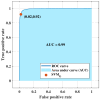
ROC curve obtained from SVMG employing principal components of real parts of S21 signals for NF-WF signal classification.

References

    1. Yedjou C.G., Sims J.N., Miele L., Noubissi F., Lowe L., Fonseca D.D., Alo R.A., Payton M., Tchounwou P.B. Breast Cancer Metastasis Drug Resistance. Springer; Basel, Switzerland: 2019. Health and racial disparity in breast cancer; pp. 31–49. - PMC - PubMed
    1. Miglioretti D.L., Lange J., Van Den Broek J.J., Lee C.I., Van Ravesteyn N.T., Ritley D., Kerlikowske K., Fenton J.J., Melnikow J., De Koning H.J., et al. Radiation-induced breast cancer incidence and mortality from digital mammography screening: A modeling study. Ann. Intern. Med. 2016;164:205–214. doi: 10.7326/M15-1241. - DOI - PMC - PubMed
    1. Seiffert K., Thoene K., Zu Eulenburg C., Behrens S., Schmalfeldt B., Becher H., Chang-Claude J., Witzel I. The effect of family history on screening procedures and prognosis in breast cancer patients-Results of a large population-based case-control study. Breast. 2021;55:98–104. doi: 10.1016/j.breast.2020.12.008. - DOI - PMC - PubMed
    1. Miller A.M., Champion V.L. Attitudes about breast cancer and mammography: Racial, income, and educational differences. Women Health. 1997;26:41–63. doi: 10.1300/J013v26n01_04. - DOI - PubMed
    1. Loving V.A., Aminololama-Shakeri S., Leung J.W. Anxiety and its association with screening mammography. J. Breast Imaging. 2021;3:266–272. doi: 10.1093/jbi/wbab024. - DOI - PubMed

Publication types