. 2023 Jan 24;120(4):e2200057120.
doi: 10.1073/pnas.2200057120. Epub 2023 Jan 17.

Intranasal delivery of full-length anti-Nogo-A antibody: A potential alternative route for therapeutic antibodies to central nervous system targets

Affiliations

Intranasal delivery of full-length anti-Nogo-A antibody: A potential alternative route for therapeutic antibodies to central nervous system targets

Daphne Correa et al. Proc Natl Acad Sci U S A. .

Abstract

Antibody delivery to the CNS remains a huge hurdle for the clinical application of antibodies targeting a CNS antigen. The blood-brain barrier and blood-CSF barrier restrict access of therapeutic antibodies to their CNS targets in a major way. The very high amounts of therapeutic antibodies that are administered systemically in recent clinical trials to reach CNS targets are barely viable cost-wise for broad, routine applications. Though global CNS delivery of antibodies can be achieved by intrathecal application, these procedures are invasive. A non-invasive method to bring antibodies into the CNS reliably and reproducibly remains an important unmet need in neurology. In the present study, we show that intranasal application of a mouse monoclonal antibody against the neurite growth-inhibiting and plasticity-restricting membrane protein Nogo-A leads to a rapid transfer of significant amounts of antibody to the brain and spinal cord in intact adult rats. Daily intranasal application for 2 wk of anti-Nogo-A antibody enhanced growth and compensatory sprouting of corticofugal projections and functional recovery in rats after large unilateral cortical strokes. These findings are a starting point for clinical translation for a less invasive route of application of therapeutic antibodies to CNS targets for many neurological indications.

Keywords: Nogo-A; antibody therapy; intranasal; neurodegeneration; stroke recovery.


Conflict of interest statement

M.E.S. is the co-founder and president of the board of NovaGo Therapeutics AG. All other authors declare that they have no competing interests.

Figures

Fig. 1.
Fig. 1.
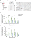
Quantification and localization of anti-Nogo-A antibody 11C7 by ELISA and immunohistochemistry in the adult mouse CNS 6 h and 24 h after a single intranasal application. (A) Concentrations of anti-Nogo-A antibody in different CNS regions 6 h (n=6) and 24 h (n=5) after a single intranasal application (500 μg/nares) as detected by capture ELISA. Statistical evaluation was done using the mixed-effects two-way ANOVA model followed by Bonferroni post hoc correction. (B) Regional distribution of anti-Nogo-A antibody 6 h after a single intranasal application at different doses: 0 μg (n=2); 10 μg (n=3); 100 μg (n=3); and 1,000 μg (n=3). One-way ANOVA followed by Bonferroni post hoc correction was used. Data are presented as mean ± SEM, asterisks indicate significances: *P < 0.05, **P < 0.01. (CL) Detection of biotinylated mouse anti-Nogo-A antibody by histochemistry in adult mouse brain 6 h after a single intranasal application (100 µg; 50 µg/nostril). (C and H) Nissl stain of the olfactory and brainstem regions. (Scale bar, 100 µm.) (E, G, J, and L) Diffuse, as well as a cellular 11C7-biotin signal is found in the granule cell layer of the olfactory bulb (E and G) and the spinal trigeminal nucleus (J and L) in the brainstem. (D, F, I, and K) Only low background staining is seen in the corresponding brain regions in the untreated mouse. (F, G, K, and L). Color-coded staining intensity of HRP–DAB-stained cryostat sections of olfactory and brainstem region. GCL: granule cell layer; Sp5: spinal trigeminal nucleus. (Scale bar, 50 µm.)
Fig. 2.
Fig. 2.
Localization of mouse anti-Nogo-A antibody 11C7 by immunohistochemistry in the adult rat brain 24 h after a single intranasal application or after three successive daily intranasal applications (72 h). (A) Regional distribution of anti-Nogo-A antibody immunoreactivity in the adult rat brain at 24 and 72 h post-intranasal application. Color coding shows the intensity of staining in the olfactory bulb, motor cortex, hippocampus, cerebellum, brainstem, and pons. Sections of untreated rat show low background staining only. (B) Mouse antibody 11C7 was detected on/in cells in many brain regions, in particular after repeated intranasal application. Examples shown are from the olfactory bulb, pyramidal cells of lamina V of the motor cortex and CA1 of the hippocampus, Purkinje cells of the cerebellum, and large neurons in the trapezoid nucleus and principal sensory trigeminal nucleus (Pr5). In white matter, e.g., corpus callosum, the typical row-forming oligodendrocytes are also labeled. All these labeled cell types are known to express Nogo-A. Double immunofluorescent labeling of the trapezoid nucleus for endogenous Nogo-A and mouse antibody 11C7 confirmed the colocalization (Lower Right). AOL: anterior olfactory nucleus, lateral; (V) Lamina V of motor cortex; CA1: cornu ammonis 1 of the hippocampus; ML, molecular layer; PCL, Purkinje cell layer; Pr5: principal sensory trigeminal nucleus; Tz: Nucleus of the trapezoid body; CC: corpus callosum. – Red channel: Nogo-A; Green channel: Mouse IgG (11C7 detection). (Scale bars, 100 µm.)
Fig. 3.
Fig. 3.
Regional tissue concentrations of anti-Nogo-A or control antibody following intranasal or intrathecal application. (A) CNS tissue concentrations of anti-Nogo-A antibody or isotype control by ELISA in intact rats after 7 d (7d) of intrathecal, lumbar pump infusion (total mount infused: 2.5 to 3 mg). Data are mean values of μg of antibody normalized to gram tissue wet weight ± SEM. (B) Mean concentrations of anti-Nogo-A and isotype control antibody in the rat serum and (C) cerebrospinal fluid (CSF) after 7 d of pump infusion. (D) Regional tissue concentrations of anti-Nogo-A antibody in the brain and spinal cord following daily intranasal application (1 mg/day) for 7 d. (E) Mean concentrations of anti-Nogo-A in the rat serum. (F) Percent of antibodies detected in the CNS (brain and spinal cord), at day 7 of the total 7 mg applied intranasally. (G) Regional CNS tissue concentrations of anti-Nogo-A antibody or isotype control in intact rats after 14 d of intranasal application (1 mg antibody/day). (H) Mean concentrations of anti-Nogo-A and isotype control in the rat serum ± SEM. (I) Percent of antibody detected in the CNS at day 14 of the total 14 mg applied via nasal route. Statistical evaluation was done using ANOVA measures followed by Šídák's multiple comparisons test and Student’s t-test; for the nonparametric test: the Kruskal–Wallis test followed by Dun's correction was used; n=3 to 5; asterisks indicate significances: *P < 0.05, **P < 0.01, ***P < 0.001, ****P < 0.0001.
Fig. 4.
Fig. 4.
Intranasal application of anti-Nogo-A antibody enhanced functional recovery. (A) Experimental timeline (d: days), adult rats received large unilateral cortical strokes followed by 14 d of intranasal application of either anti-Nogo-A antibody or control antibody. Weekly behavioral assessment was carried out starting from day 2 to day 42 poststroke. At the end of the behavioral assessment anterograde tracing of the CST fibers was carried out for neuroanatomical analysis in the cervical spinal cord. (B) Representative coronal section of a brain depicting cellular damage throughout the layers of the sensorimotor cortex 63 d poststroke. (Scale bar, 1.5 mm.) (C) Quantification of cortical stroke depth. Dotted line represents the position of ipsilesional corticospinal motor neurons (cortical layer V). (D) Success rate in the SPG task at baseline (BL; intact, trained) and after a unilateral photothrombotic stroke to the sensorimotor cortex of the preferred paw from day 2 to Day 42 postlesion of anti-Nogo-A antibody (green, n = 9) or control antibody (blue, n = 6)-treated animals. Data are presented as mean ± SEM. Statistical evaluation was carried out with two-way ANOVA repeated measure followed by Bonferroni post hoc, asterisks indicate significances: **P < 0.01 and ***P < 0.001. (E) No correlation was found between the lesion depth and the success rate on the SPG task at day 42 poststroke (P > 0.05, r = 0.13, Spearman correlation).
Fig. 5.
Fig. 5.
Intranasal application of anti-Nogo-A for 2-w. enhanced contralesional CST sprouting into the denervated hemicord after stroke. (A) Anterograde axonal tracer BDA was injected into the contralesional (intact hemisphere) sensory–motor cortex and the cervical segments C3–C4 were analyzed to quantify CST fibers in the midline and the stroke-denervated hemicord. (B) Low magnification micrograph of BDA-labeled corticospinal fibers from intact spinal hemicord (Right) reinnervating the stroke-denervated hemicord (Left; Inset) at spinal cord level C3. CST fibers were counted as intersections with vertical lines placed at the midline (M) and given distances in the gray matter (D1 to D4). (Scale bar, 100 µm.) (C) Representative micrographs showing corticospinal fibers (arrowheads) from the contralesional cortex in the denervated cervical spinal cord (C3) in anti-Nogo-A and control antibody-treated animals. (Scale bar, 100 µm.) (D and E) Number of CST collaterals projecting over the midline (M) and innervating the denervated gray matter at D1 to D4 in anti-Nogo-A and control antibody-treated animals at cervical level C3 (D) and C4 (E) normalized to 10% of BDA-positive labeled CST fibers at the brainstem level. Intranasally treated animals with anti-Nogo-A showed significantly more fibers at M, D1, and D2. To account for the variability of BDA labeling in the corticofugal pathway, we determined the number of BDA-positive fibers in the cerebral peduncle at the level of the midbrain. Cervical spinal cord fiber counts were normalized to 10% of the labeled corticofugal fibers. Data are presented as mean ± SEM, (green dots: individual animals treated with anti-Nogo-A and blue dots: control antibody); statistical evaluation was carried out with Student’s t-test (two-tailed, unpaired), asterisks indicate significances: *P < 0.05 and **P < 0.01.
Fig. 6.
Fig. 6.
Anterogradely labeled projection from the intact, contralesional forelimb motor cortex to the pons. (A) Micrograph showing cross-section at the mid-pontine level of anti-Nogo-A antibody-treated animal 63 d poststroke. In addition to the strongly labeled intact side, fibers crossing the midline and innervating the central and medial pontine nuclei of the stroke-denervated side (arrows) can be seen. (CP): cerebral peduncle; (1, 1′): Central pontine nucleus; (2, 2′): Medial pontine nucleus; (3, 3′): Lateral pontine nucleus -1; (4, 4′): Lateral pontine nucleus -2. (1-4: intact side; 1′-4′: stroke-denervated side). (Scale bar, 500 µm.) (B) Magnification of the boxed region depicted in (A), showing crossed fibers terminating and arborizing in the central and medial basilar pontine nuclei (arrows). (Scale bar, 100 µm.) (C) Qualification of projections from the intact, contralesional forelimb cortex to the stroke-denervated central, medial, and lateral basilar pontine nuclei, normalized to the number of fibers counted in 10% of the area of the midbrain cerebral peduncle. Level of significance: *P = 0.01, two-way ANOVA multiple comparisons followed by Šídák's correction. (D) Significant positive correlation of ipsilesional corticopontine projections to the central nucleus to the success rate of skilled forelimb grasping at 42 d poststroke (***P = 0.0006, r = 0.86, Pearson correlation).

Similar articles

Cited by

References

    1. Ferrero J., et al. , First-in-human, double-blind, placebo-controlled, single-dose escalation study of aducanumab (BIIB037) in mild-to-moderate Alzheimer’s disease. Alzheimer’s Dement. Transl. Res. Clin. Interv. 2, 169–176 (2016). - PMC - PubMed
    1. Ranger A., et al. , Anti-LINGO-1 has no detectable immunomodulatory effects in preclinical and phase 1 studies. Neurol. Neuroimmunol. NeuroInflammation 5, 417 (2018). - PMC - PubMed
    1. Freskgård P. O., Urich E., Antibody therapies in CNS diseases. Neuropharmacology 120, 38–55 (2017). - PubMed
    1. Kucher K., et al. , First-in-man intrathecal application of neurite growth-promoting anti-nogo- a antibodies in acute spinal cord injury. Neurorehabil. Neural Repair 32, 578–589 (2018). - PubMed
    1. Lu R. M., et al. , Development of therapeutic antibodies for the treatment of diseases. J. Biomed. Sci. 27, 1–30 (2020). - PMC - PubMed

Publication types

LinkOut - more resources