. 2023 Jan;16(1):e014067.
doi: 10.1161/CIRCIMAGING.122.014067. Epub 2023 Jan 17.

Nuclear Methods for Immune Cell Imaging: Bridging Molecular Imaging and Individualized Medicine

Affiliations

Nuclear Methods for Immune Cell Imaging: Bridging Molecular Imaging and Individualized Medicine

Gyu Seong Heo et al. Circ Cardiovasc Imaging. 2023 Jan.

Abstract

Inflammation is a key mechanistic contributor to the progression of cardiovascular disease, from atherosclerosis through ischemic injury and overt heart failure. Recent evidence has identified specific roles of immune cell subpopulations in cardiac pathogenesis that diverges between individual patients. Nuclear imaging approaches facilitate noninvasive and serial quantification of inflammation severity, offering the opportunity to predict eventual outcome, stratify patient risk, and guide novel targeted molecular therapies against specific leukocyte subpopulations. Here, we will discuss the established and emerging nuclear imaging methods to label and track exogenous and endogenous immune cells, with a particular focus on clinical situations in which targeted molecular inflammation imaging would be advantageous. The expanding options for imaging inflammation provide the foundation to bridge between molecular imaging and individual therapy.

Keywords: heart failure; inflammation; magnetic resonance imaging; myocardial infarction; positron emission tomography.


Figures
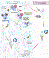
Figure 1.
Figure 1.. Schematic overview of nuclear methods for immune cell labeling.
(A) exogenous ex vivo cell labeling. (B) endogenous in vivo cell imaging. (Created with BioRender.com)
Figure 2.
Figure 2.. PET imaging of immune cells in animal models of AMI.
68Ga-Pentixafor PET images showing prognostic value of CXCR4 expression. (A) Sample cardiac short-axis (SA) PET images at 3d after MI of a surviving mouse and a mouse dying early of left ventricular (LV) rupture. (B) 68Ga-Pentixafor uptake within the infarct of surviving and LV rupture mice. (C) Correlation of 68Ga-pentixafor signal to the left ventricular ejection fraction (LVEF) at 6 weeks on univariate analysis (modified from). PET/CT images of 68Ga-DOTA-ECL1i at d3 (D) and 64Cu-DOTA-ECL1i at d4 (E) after MI show comparable uptake (F) in the infarcted region of the same mouse. (reproduced from) (G) Correlation between 68Ga-DOTA-ECL1i heart uptake at d4 and LVEF and akinetic area measured on d28 after MI (reproduced from). 18F-LW223 PET with BPTC quantification detects macrophage-driven inflammation within heart 7d after MI without need for additional perfusion scan. Representative K1 (H) and BPTC images (I) of LV of MI rat demonstrating true TSPO signal across heart. (J) BPTC values across global heart and LV anterior wall demonstrating TSPO expression within infarct (reproduced from). K1: rate constant for transfer from arterial plasma to tissues; BPTC:binding potential for impaired radiotracer transfer from plasma to tissue; A: anterior; P: posterior
Figure 3.
Figure 3.. PET imaging of immune cells in animal models of pressure overload (A), myocarditis (B), and cardiomyopathy (C).
(A) CXCR4 PET defines diffuse myocardial inflammation after pressure-overload. (left) Representative 68Ga-pentixafor and 18F-FDG images in sham and mice after transverse aortic constriction (TAC). (right) Comparison of 68Ga-pentixafor (red) uptake after TAC with sham (black). (reproduced from) (B) 18F-FDG and 18F-FOL PET images in rat with autoimmune myocarditis. PET images show focal 18F-FOL uptake (arrows) in posterior LV wall. (modified from) (C) CCR2 and CCR5 PET/CT showed detection of cardiac macrophages in a mouse model of dilated cardiomyopathy. (reproduced from) Tnnt2: Troponin T2
Figure 4.
Figure 4.. Molecular imaging of inflammation after AMI.
(A) CXCR4 PET 3d after lateral wall ST-elevation MI with low (left) or high signal (right). (B) Intensity of the early CXCR4 signal correlates LVEF at time of imaging and at 7±3 months follow-up (modified from).
Figure 5.
Figure 5.. Imaging of inflammation in cardiac sarcoidosis.
Whole body and cardiac distribution of 18F-FDG in sarcoidosis displays focal patchy pattern of uptake in left ventricle on transaxial PET/CT images (lower panel). Patient preparation with no carbohydrate high fat diet and heparin administration suppressed myocardial 18F-FDG uptake to facilitate immune cell visualization.
Figure 6.
Figure 6.. Clinical imaging of inflammation in valvular disease.
(A) Focal abnormal 18F-FDG accumulation identified around the aortic prosthetic valve (middle) despite no abnormality on computed tomographic angiography (CTA, left) or open surgical exploration (right). (B) Subsequent CTA revealed mycotic aneurysm (left) and transesophogeal echocardiography identified abscess formation (middle). Severe infection was confirmed with follow-up surgical exploration (right) (Reproduced from). RCA: right coronary artery; LCA: left coronary artery; PHV: prosthetic heart valve

References

    1. Steffens S, Nahrendorf M, Madonna R. Immune cells in cardiac homeostasis and disease: emerging insights from novel technologies. Eur Heart J. 2022;43:1533–1541. doi: 10.1093/eurheartj/ehab842 - DOI - PMC - PubMed
    1. Swirski FK, Nahrendorf M. Cardioimmunology: the immune system in cardiac homeostasis and disease. Nat Rev Immunol. 2018;18:733–744. doi: 10.1038/s41577-018-0065-8 - DOI - PubMed
    1. Bajpai G, Bredemeyer A, Li W, Zaitsev K, Koenig AL, Lokshina I, Mohan J, Ivey B, Hsiao HM, Weinheimer C, Kovacs A, Epelman S, Artyomov M, Kreisel D, Lavine KJ. Tissue Resident CCR2- and CCR2+ Cardiac Macrophages Differentially Orchestrate Monocyte Recruitment and Fate Specification Following Myocardial Injury. Circ Res. 2019;124:263–278. doi: 10.1161/circresaha.118.314028 - DOI - PMC - PubMed
    1. Ni SH, Xu JD, Sun SN, Li Y, Zhou Z, Li H, Liu X, Deng JP, Huang YS, Chen ZX, Feng WJ, Wang JJ, Xian SX, Yang ZQ, Wang S, Wang LJ, Lu L. Single-cell transcriptomic analyses of cardiac immune cells reveal that Rel-driven CD72-positive macrophages induce cardiomyocyte injury. Cardiovasc Res. 2022;118:1303–1320. doi: 10.1093/cvr/cvab193 - DOI - PubMed
    1. Lafuse WP, Wozniak DJ, Rajaram MVS. Role of Cardiac Macrophages on Cardiac Inflammation, Fibrosis and Tissue Repair. Cells. 2021;10:51. - PMC - PubMed

Publication types