. 2023 Jan 17;6(4):e202201488.
doi: 10.26508/lsa.202201488. Print 2023 Apr.

Vasopressin V1a receptor and oxytocin receptor regulate murine sperm motility differently

Affiliations

Vasopressin V1a receptor and oxytocin receptor regulate murine sperm motility differently

Hiroyoshi Tsuchiya et al. Life Sci Alliance. .

Abstract

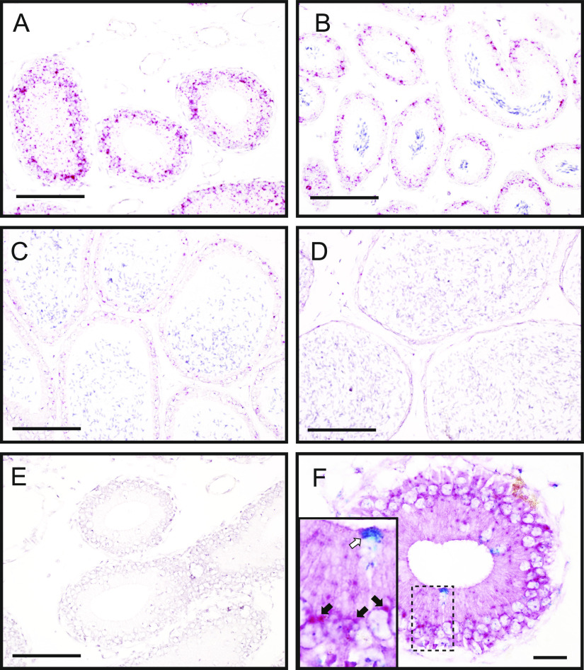
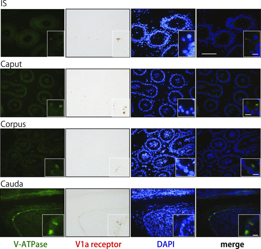
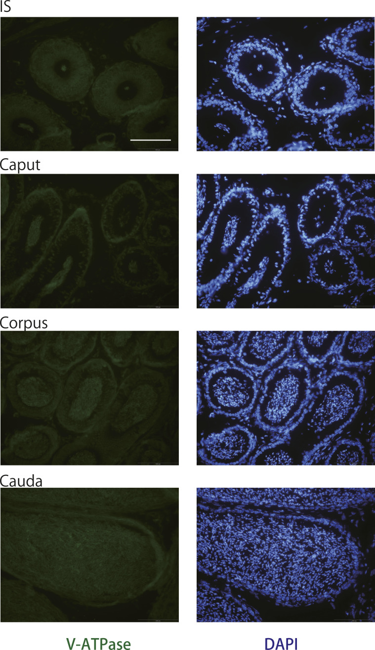
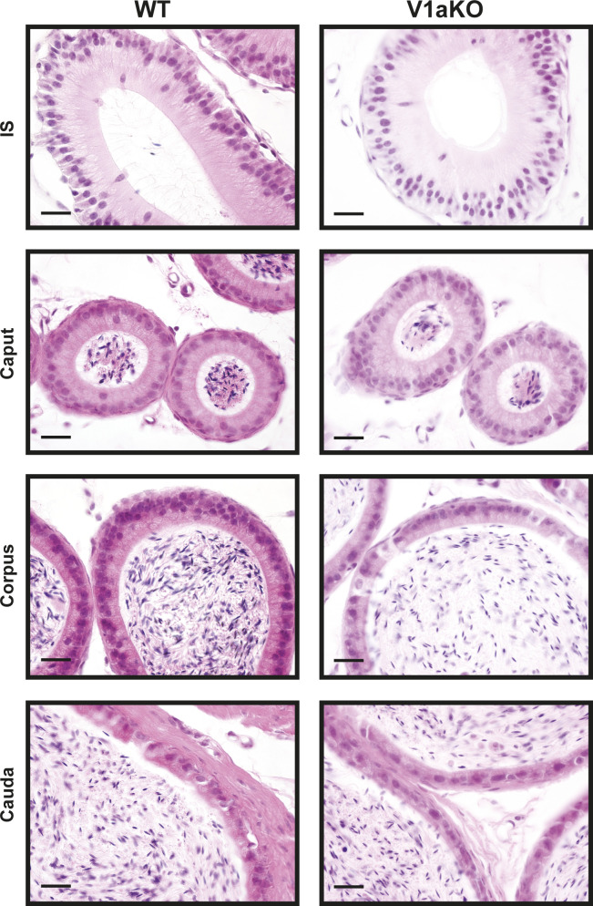
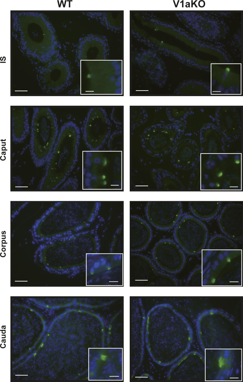
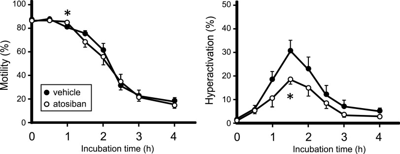
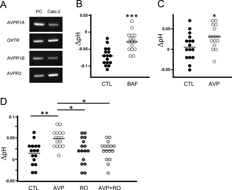
Specific receptors for the neurohypophyseal hormones, arginine vasopressin (AVP) and oxytocin, are present in the male reproductive organs. However, their exact roles remain unknown. To elucidate the physiological functions of pituitary hormones in male reproduction, this study first focused on the distribution and function of one of the AVP receptors, V1a. In situ hybridization analysis revealed high expression of the Avpr1a in Leydig cells of the testes and narrow/clear cells in the epididymis, with the expression pattern differing from that of the oxytocin receptor (OTR). Notably, persistent motility and highly proportional hyperactivation were observed in spermatozoa from V1a receptor-deficient mice. In contrast, OTR blocking by antagonist atosiban decreased hyperactivation rate. Furthermore, AVP stimulation could alter the extracellular pH mediated by the V1a receptor. The results highlight the crucial role of neurohypophyseal hormones in male reproductive physiology, with potential contradicting roles of V1a and OTR in sperm maturation. Our findings suggest that V1a receptor antagonists are potential therapeutic drugs for male infertility.


Conflict of interest statement

The authors declare that they have no conflict of interest.

Figures

Figure 1.
Figure 1.. Morphological abnormalities of the epididymis in V1a-deficient mice.
(A, B) Weights of the epididymis (A) and testes (B) from WT and V1a-deficient (V1aKO) mice were adjusted based on individual body weights (n = 8). However, the differences were not statistically significant. (C) Macroscopic morphology of the epididymis in WT and V1aKO mice. (D) Longitudinal lengths of the epididymis from WT and V1aKO mice, measured using a ruler (n = 8). (E) Thinnest parts of the epididymis at the corpus epididymis of WT and V1aKO mice were compared using various criteria. The images were stained with Mayer’s hematoxylin and eosin Y solution. (F, G, H, I) Lengths of the portions with three or fewer tubes (F), total tissue area (G), area of the single duct (H), and the inner diameter of the duct (I) (n = 10–11) were compared between WT and V1aKO mice. (J) Epididymides of WT and V1aKO were stained using the β-Galactosidase staining procedure. For comparisons between the two groups, we performed Welch’s t test. The results are expressed as mean ± SEM, ∗P < 0.05, ∗∗P < 0.01, ∗∗∗P < 0.005, n.s., not significant, versus WT.
Figure S1.
Figure S1.. Weights of tissues and the serum testosterone concentrations from WT or V1aKO male mice.
(A) Weights of the seminal vesicle, preputial gland, and prostate gland from WT or V1aKO mice were adjusted based on individual body weight (n = 10). (B) Concentrations of serum testosterone were measured using enzyme immunoassay (n = 10–13). For comparisons between the two groups, we performed Welch’s t test. The results are expressed as mean ± SEM, n.s., not significant, versus WT.
Figure S2.
Figure S2.. Hematoxylin and eosin Y staining of continuous sections of corpus epididymis.
(A, B) Epididymides were sampled from WT (A) or V1aKO (B) male mice. Each section was ∼60 μm apart. Red flame indicates the thinnest slice of the epididymis. Scale bars = 200 μm.
Figure 2.
Figure 2.. Distribution of the Avpr1a transcripts in murine tissues.
(A, B, C, D, E, F, G, H, I, J, K) In situ hybridization analyses of Avpr1a transcripts were performed in the murine testes (A, B, C), adrenal gland (D, E, F), epididymis ((G) initial segment (IS), (H) caput, (I) corpus, (J) cauda), and vas deferens (K). Arrows indicate positive brown signals for the Avpr1a transcripts. (B, E) Areas surrounded by the dotted lines (A, D), respectively. (C, F) The negative controls. (G) Inset in (G) shows an area surrounded by dotted lines of (G). Scale bars = 200 μm (A, D), 100 μm (G, H, I, J), and 20 μm (B, C, E, F, K).
Figure 3.
Figure 3.. Distribution of the Oxtr transcripts was different from that of Avpr1a in murine epididymis.
(A, B, C, D, E, F) In situ hybridization analyses of Oxtr transcripts were performed in the murine epididymis ((A, E, F) IS, (B) caput, (C) corpus, (D) cauda). (E) represents negative control. (F) shows a merged image of double staining with Avpr1a transcripts (blue; white arrow in inset) and Oxtr transcripts (red; black arrow in inset) in the IS section. Scale bars = 100 μm (A, B, C, D, E) and 20 μm (F).
Figure 4.
Figure 4.. Avpr1a signals colocalized with V-ATPase in murine epididymis.
In the merged images, red represents Avpr1a transcripts (pseudocolor) and green represents V-ATPase protein. Blue indicates the nuclei. Scale bars = 100 and 10 μm (inset).
Figure S3.
Figure S3.. Negative control of the immunohistochemical staining of V-ATPase B1 and B2 subunits in the epididymis from WT male mice.
As a negative control, epididymal sections were incubated with incubation buffer only instead of primary antibody. Blue indicates nuclei stained with DAPI. Scale bars = 100 μm.
Figure S4.
Figure S4.. Hematoxylin and eosin Y staining of epididymis from WT or V1aKO male mice.
Scale bars = 20 μm.
Figure S5.
Figure S5.. Immunohistochemical staining of V-ATPase B1 and B2 subunits (green) in the epididymis from WT or V1aKO male mice.
Blue indicates nuclei stained with DAPI. Scale bars = 50 and 10 μm (inset).
Figure 5.
Figure 5.. Increased sperm activity in V1a-deficient mice (white) than in WT (black) mice.
(A) Straight-line velocity, curvilinear velocity, average path velocity, linearity, straightness, amplitude of lateral head displacement, beat–cross frequency, and wobbler coefficient were recorded and calculated using a computer (n = 8). (B) Persistent motility and hyperactivation were measured manually (n = 8). For comparisons between the two groups, we performed Welch’s t test. The results are expressed as mean ± SEM, ∗P < 0.05, ∗∗P < 0.01, ∗∗∗P < 0.005, versus WT values.
Figure 6.
Figure 6.. Hyperactivation of the spermatozoa was up-regulated in 14-d atosiban-injected mice (white) compared with control (black).
Persistent motility and hyperactivation were manually measured. For comparisons between the two groups, we performed Welch’s t test. The results are presented as mean ± SEM, ∗P < 0.05, versus the vehicle group (n = 8).
Figure 7.
Figure 7.. AVP stimulation increased the extracellular pH, and V1a receptor antagonist blocked it.
(A) Gene expression levels of receptor subtypes of AVP and oxytocin in human renal carcinoma Caki-2 cells. Human liver, brain, and kidney were used as a positive control for AVPR1A, AVPR1B and OXTR, and AVPR2, respectively. (B) pH changes from the background buffer were plotted in the graph. (B, C) 1 μM bafilomycin A1 (BAF) or 1 μM AVP treatment has been compared with the vehicle (CTL). For comparisons between the two groups, we performed Welch’s t test. The bars represent median, ∗P < 0.05, ∗∗∗P < 0.005, versus the CTL group (n = 16). (D) pH changes induced by AVP with or without V1a antagonist RO5028442 (RO) were plotted. For comparisons between multiple groups, Bartlett’s test followed by one-way ANOVA and Tukey’s test were combined. The bars represent mean, ∗P < 0.05, ∗∗P < 0.01, versus the CTL group (n = 16).

Similar articles

Cited by

References

    1. Agmo A, Andersson R, Johansson C (1978) Effect of oxytocin on sperm numbers in spontaneous rat ejaculates. Biol Reprod 18: 346–349. 10.1095/biolreprod18.3.346 - DOI - PubMed
    1. Akerlund M, Bossmar T, Brouard R, Kostrzewska A, Laudanski T, Lemancewicz A, Serradeil-Le Gal C, Steinwall M (1999) Receptor binding of oxytocin and vasopressin antagonists and inhibitory effects on isolated myometrium from preterm and term pregnant women. Br J Obstet Gynaecol 106: 1047–1053. 10.1111/j.1471-0528.1999.tb08112.x - DOI - PubMed
    1. Al-bataineh MM, Gong F, Marciszyn AL, Myerburg MM, Pastor-Soler NM (2014) Regulation of proximal tubule vacuolar h(+)-atpase by pka and amp-activated protein kinase. Am J Physiol Renal Physiol 306: F981–F995. 10.1152/ajprenal.00362.2013 - DOI - PMC - PubMed
    1. Al-Bataineh MM, Alzamora R, Ohmi K, Ho PY, Marciszyn AL, Gong F, Li H, Hallows KR, Pastor-Soler NM (2016) Aurora kinase a activates the vacuolar h+-atpase (v-atpase) in kidney carcinoma cells. Am J Physiol Renal Physiol 310: F1216–F1228. 10.1152/ajprenal.00061.2016 - DOI - PMC - PubMed
    1. Aoyagi T, Birumachi Ji, Hiroyama M, Fujiwara Y, Sanbe A, Yamauchi J, Tanoue A (2007) Alteration of glucose homeostasis in v1a vasopressin receptor-deficient mice. Endocrinology 148: 2075–2084. 10.1210/en.2006-1315 - DOI - PubMed

Publication types