. 2023 Jan;13(1):e1129.
doi: 10.1002/ctm2.1129.

LncRNA BC promotes lung adenocarcinoma progression by modulating IMPAD1 alternative splicing

Affiliations

LncRNA BC promotes lung adenocarcinoma progression by modulating IMPAD1 alternative splicing

Qi Wen Chen et al. Clin Transl Med. 2023 Jan.

Abstract

Background: The therapeutic value of targeted therapies in patients with lung cancer is reduced when tumours acquire secondary resistance after an initial period of successful treatment. However, the molecular events behind the resistance to targeted therapies in lung cancer remain largely unknown.

Aims: To discover the important role and mechanism of lncRNA BC in promoting tumor metastasis and influencing clinical prognosis of LUAD.

Materials & methods: Microarrays were used to screen a comprehensive set of lncRNAs with differential expression profiles in lung cancer cells. The functional role and mechanism of lncRNA were further investigated by gain- and loss-of-function assays. RNA pull-down, protein assays, and mass spectrometry were used to identify proteins that interacted with lncRNA. TaqMan PCR was used to measure lncRNA in lung adenocarcinoma and adjacent nontumor tissues from 428 patients. The clinical significance of lncRNA identified was statistically confirmed in this cohort of patients.

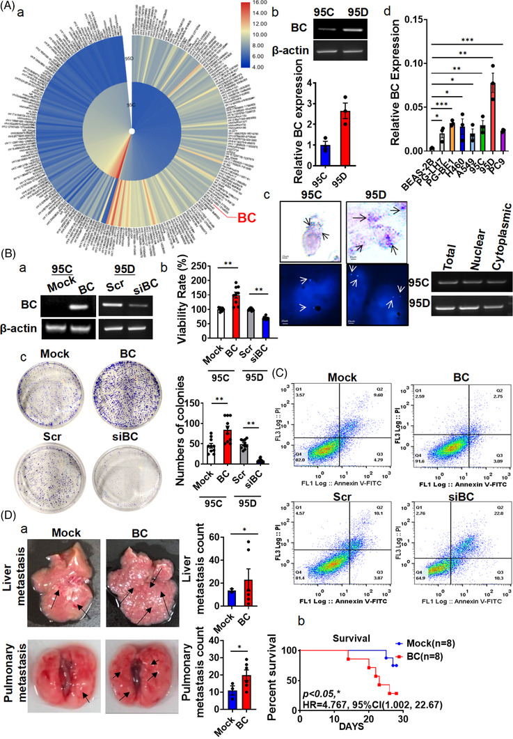
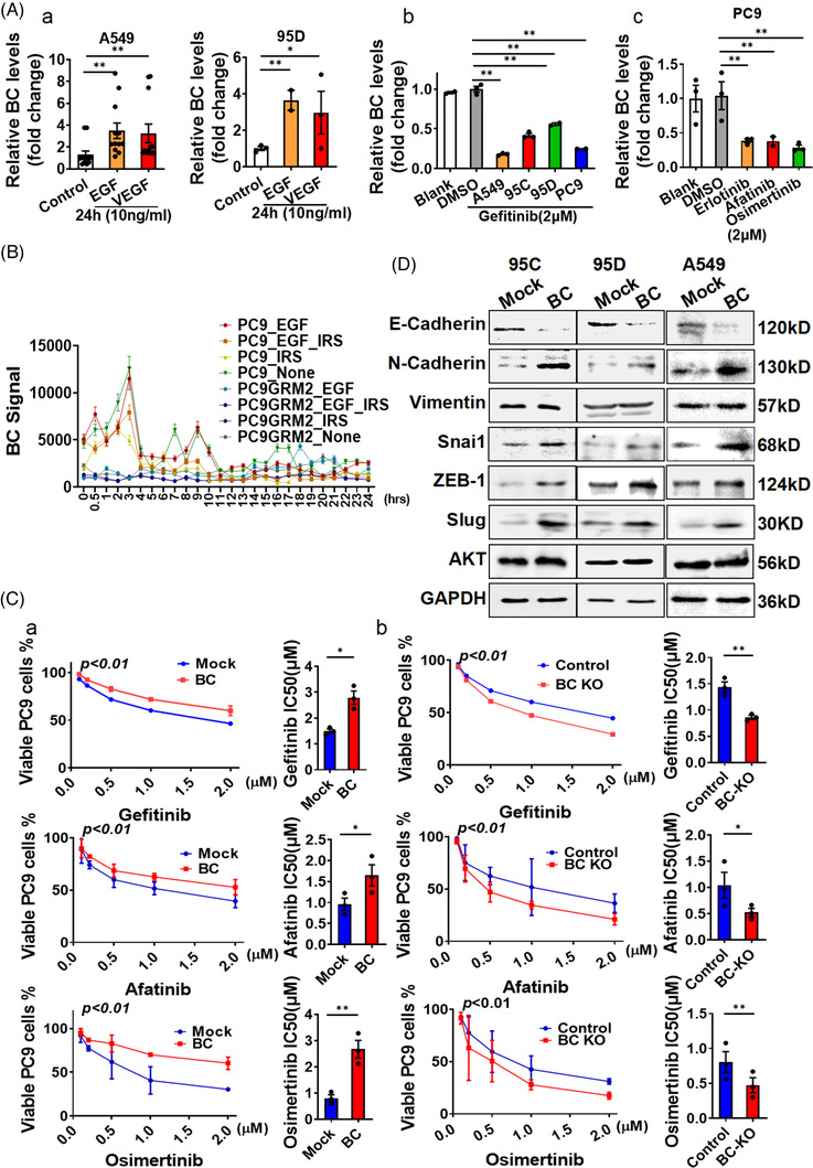
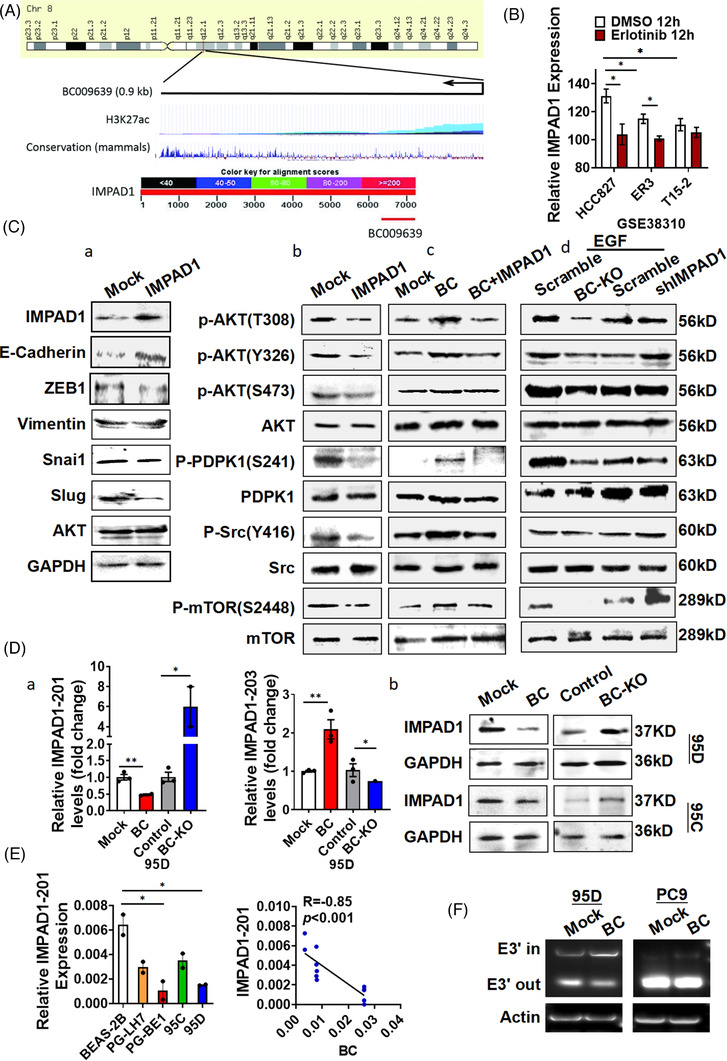
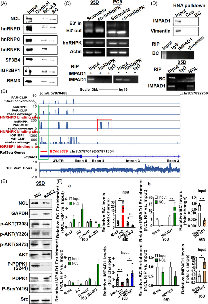
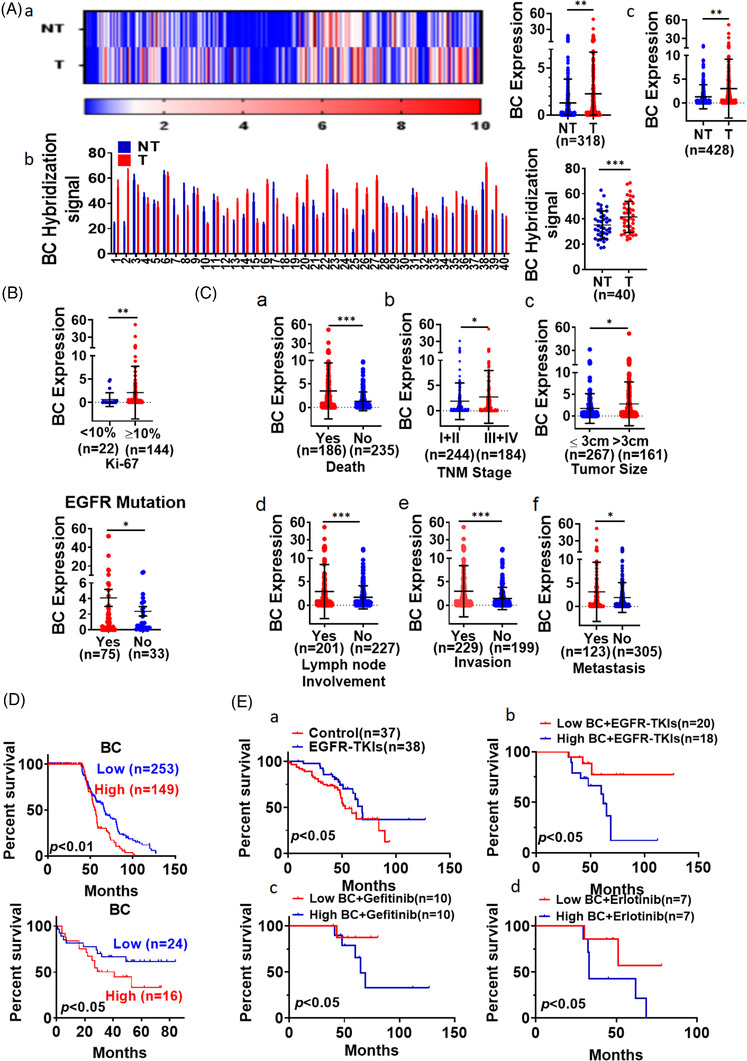
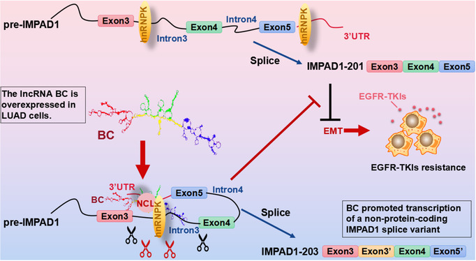
Results: In this study, we show that the long non-coding RNA BC009639 (BC) is involved in acquired resistance to EGFR-targeted therapies. Among the 235 long non-coding RNAs that were differentially expressed in lung cancer cell lines, with different metastatic potentials, BC promoted growth, invasion, metastasis, and resistance to EGFR-tyrosine kinase inhibitors (EGFR-TKIs), both in vitro and in vivo. BC was highly expressed in 428 patients with lung adenocarcinoma (LUAD) and high BC expression correlated with reduced efficacy of EGFR-TKI therapy. To uncover the molecular mechanism of BC-mediated EGFR-TKI resistance in lung cancer, we screened and identified nucleolin and hnRNPK that interact with BC. BC formed the splicing complex with nucleolin and hnRNPK to facilitate the production of a non-protein-coding inositol monophosphatase domain containing 1 (IMPAD1) splice variant, instead of the protein-coding variant. The BC-mediated alternative splicing (AS) of IMPAD1 resulted in the induction of the epithelial-mesenchymal transition and resistance to EGFR-TKI in lung cancer. High BC expression correlated with clinical progress and poor survival among 402 patients with LUAD.

Disscussion: Through alternative splicing, BC boosted the non-coding IMPAD1-203 transcript variant while suppressing the IMPAD1-201 variant. In order to control the processing of pre-mRNA, BC not only attracted RNA binding proteins (NCL, IGF2BP1) or splicing factors (hnRNPK), but also controlled the formation of the splicing-regulator complex by creating RNA-RNA-duplexes.

Conclusion: Our results reveal an important role for BC in mediating resistance to EGFR-targeted therapy in LUAD through IMPAD1 AS and in implication for the targeted therapy resistance.

Keywords: EGFR-TKI resistance; alternative splicing; inositol monophosphatase domain containing 1; lung adenocarcinoma; non-coding RNA.


Conflict of interest statement

The authors have declared that no conflicts of interest and no competing interest exist.

Figures

FIGURE 1
FIGURE 1
BC overexpressed in lung adenocarcinoma (LUAD) cells, promoted lung cancer metastasis. (A) (a) Hierarchical clustering of 235 differentially expressed long non‐coding RNAs (lncRNAs) (|fold change| > 3) was performed using microarray data. Red, high expression; blue, low expression. (b) The expression of BC in 95C and 95D cells was analysed by RT‐PCR. (c) BC in nuclear and cytoplasmic fractions were detected by in situ hybridization, fluorescence in situ hybridization (left) and RT‐PCR (right). Arrows indicate positive hybridization sites. (d) BC expression was measured by RT‐qPCR in BEAS‐2B, PG‐LH7, PG‐BE1, H460, A549, PC9, 95C and 95D cells. (B) (a) Overexpression and knock‐down of BC were validated by RT‐PCR. (b) The viability of 95D cells was analysed by the MTT assay after BC overexpression or knock‐down. (c) Colony formation assays were performed with BC‐overexpressing and BC‐silenced cells. Quantitative analysis is shown on the right. (C) Flow cytometry was performed to measure cell cycle progression after ectopic expression or silencing of BC. (D) (a) Representative images of livers and lungs of nude mice, 4 weeks after tail‐vein injection (n = 8). Arrows indicate metastatic foci. Liver and pulmonary metastasis foci were quantitatively analysed. (b) The death rate of both groups is summarized. Data are represented as mean ± SEM of at least three independent experiments. *p < .05, **p < .01
FIGURE 2
FIGURE 2
BC enhanced resistance to EGFR‐tyrosine kinase inhibitors (EGFR‐TKIs) by activating the epithelial–mesenchymal transition. (A) (a) BC levels were determined by RT‐qPCR in A549 and 95D cells treated with EGF or VEGF. (b) BC levels were determined by RT‐qPCR in A549, 95C, 95D and PC9 cells treated with 2 μm/L gefitinib (24 h), vehicle or blank control. (c) BC expression was measured in PC9 cells treated with 2 μm/L erlotinib, afatinib or osimertinib. (B) BC expression was measured in gefitinib‐sensitive PC9 cells and gefitinib‐resistant PC9G cells from public microarray dataset GSE34228. IRS, gefitinib‐treatment. PC9GRM2, gefitinib‐resistance cell line. (C) (a and b) Viability of (a) BC‐overexpressing and (b) BC knockout PC9 cells was measured by the MTT assay after treatment with various concentrations of gefitinib (upper), afatinib (middle) or osimertinib (bottom) for 24 h. The IC50 of the EGFR‐TKIs was also measured. (D) The expression of cadherin, vimentin, Snai1, Slug, ZEB‐1 and AKT were determined by Western blot in 95C, 95D and A549 cells with BC overexpression. Data are represented as mean ± SEM of at least three independent experiments. *p < .05, **p < .01
FIGURE 3
FIGURE 3
BC enhanced the epithelial–mesenchymal transition by regulating inositol monophosphatase domain containing 1 (IMPAD1) alternative splicing. (A) Schematic representation of BC and UCSC Genome Browser tracks depicting H3K27ac chromatin immunoprecipitation (ChIP)‐seq coverage (upper) and mammalian conservation in human cell lines (middle). Schematic diagram of the complementation between the IMAPD1 nucleotide sequence and BC determined by BLAST (lower). (B) IMPAD1 expression analysis of cells resistant or sensitive to erlotinib was performed using public microarray dataset GSE38310. (C) (a) IMPAD1, cadherin, vimentin, Snai1, ZEB‐1, Slug and AKT were detected by Western analysis in 95D and A549 cells overexpressing IMPAD1; (b) phosphorylation of AKT, PDPK1, Src and mTOR in 95D cells overexpressing IMPAD1 was measured; (c and d) phosphorylation levels of AKT, PDPK1, Src and mTOR were determined by Western blot in (c) 95D cells with BC and IMPAD1 co‐transfection and (d) 95D cells with BC knockout or IMPAD1 knock‐down after 10 ng/ml EGF treatment. (D) (a) The IMPAD1‐201 and IMPAD1‐203 splice variants were detected by RT‐qPCR in 95D cells after BC overexpression or knock‐down; (b) IMPAD1 expression was detected by Western analysis in 95D and 95C cells with BC overexpression or knockout. (E) IMPAD1‐201 expression was measured by RT‐qPCR in BEAS‐2B, PG‐LH7, PG‐BE1, 95C and 95D cells (left). Pearson correlation between BC and IMPAD1‐201 expression levels is shown (right). (F) The splicing of IMPAD1‐203 exon 3′ in 95D and PC9 cells was detected by PCR. Data are represented as mean ± SEM of at least three independent experiments. *p < .05, **p < .01
FIGURE 4
FIGURE 4
BC regulates inositol monophosphatase domain containing 1 (IMPAD1) alternative splicing via an interaction with hnRNPK and NCL. (A) Binding of BC to NCL, heterogeneous nuclear ribonucleoprotein (hnRNP), IGF2BP1, SF3B4 and RBM3 was analysed by BC RNA pull‐down assay. (B) Genome browser views of hnRNPD, hnRNPK and IGF2BP2 PAR‐CLIP binding sites and sequence conservation across vertebrates in genomic regions spanning IMPAD1 and BC. The aligned PAR‐CLIP reads are highlighted in BC‐aligned regions and IMPAD1 intron 3 with frame. (C) Splicing of IMPAD1‐203 exon 3′ was detected by PCR in 95D and PC9 cells following hnRNPK knock‐down (upper). RNA immunoprecipitation using the hnRNPK antibody was performed in 95D cells with IMPAD1 overexpression (lower). IgG‐bound RNA was used as a negative control. (D) IMPAD1 or vimentin binding to BC was analysed by BC RNA pull‐down and RNA immunoprecipitation (top, middle). Binding of NCL to BC and IMPAD1 mRNA were detected by RNA immunoprecipitation (bottom). (E) NCL, PDPK1, Src, AKT and their phosphorylation were detected by Western analysis in 95D cells with NCL knock‐down. (F) RNA immunoprecipitation and RT‐qPCR analysis were performed to measure BC and IMPAD1 mRNA binding to NCL protein in 95D cells with BC overexpression or knock‐down (a) or in 95D cells with IMPAD1 overexpression (b). Data are represented as mean ± SEM of at least three independent experiments. *p < .05, **p < .01
FIGURE 5
FIGURE 5
BC was highly expressed in lung adenocarcinoma (LUAD) tissues. (A) (a) Differential BC expression between LUAD (T) tissues and adjacent non‐tumour (NT) tissues from 318 patients was determined by RT‐qPCR; (b) BC expression was measured by in situ hybridization in LUAD tissues and adjacent non‐tumour tissues from 40 patients; (c) BC expression analysis in the combined cohort (n = 428). (B) BC levels in LUAD tissues were compared between patients with different positive rate of Ki‐67 staining (upper). BC levels were measured in lung cancer tissues with and without EGFR mutation from 428 patients with LUAD (lower). (C) BC levels in LUAD tissues were compared between patients with different (a) survival, (b) TNM stage, (c) tumour size, (d) lymph node involvement, (e) invasion and (f) remote metastasis. Data are representative of at least three independent experiments. *p < .05, **p < .01. (D) Overall survival among 402 patients with LUAD was analysed according to BC expression levels measured by RT‐qPCR (upper). Kaplan–Meier analysis of 40 patients with LUAD showed differential survival corresponding to BC in situ hybridization signal levels (lower). (E) Kaplan–Meier analysis of overall survival of patients with LUAD that received EGFR‐tyrosine kinase inhibitor (EGFR‐TKI) treatment and conventional chemotherapy (a); Kaplan–Meier analysis of overall survival of patients with LUAD that received EGFR‐TKI treatment based on BC expression (b); overall survival analysis of patients who received treatment with gefitinib (c) or erlotinib (d) based on BC expression
FIGURE 6
FIGURE 6
Long non‐coding RNA (LncRNA) BC mediates the resistance to EGFR‐tyrosine kinase inhibitor (EGFR‐TKI) in lung cancer cells via regulating inositol monophosphatase domain containing 1 (IMPAD1) alternative splicing and epithelial–mesenchymal transition (EMT) process.

References

    1. Reck M, Heigener DF, Mok T, Soria JC, Rabe KF. Management of non‐small‐cell lung cancer: recent developments. Lancet. 2013;382(9893):709‐719. 10.1016/S0140-6736(13)61502-0 - DOI - PubMed
    1. Bray F, Ferlay J, Soerjomataram I, Siegel RL, Torre LA, Jemal A. Global cancer statistics 2018: gLOBOCAN estimates of incidence and mortality worldwide for 36 cancers in 185 countries. CA Cancer J Clin. 2018;68(6):394‐424. 10.3322/caac.21492 - DOI - PubMed
    1. Kaira K, Takahashi T, Murakami H, et al. The role of betaIII‐tubulin in non‐small cell lung cancer patients treated by taxane‐based chemotherapy. Int J Clin Oncol. 2013;18(3):371‐379. 10.1007/s10147-012-0386-8 - DOI - PubMed
    1. Verdecchia A, Francisci S, Brenner H, et al. Recent cancer survival in Europe: a 2000–02 period analysis of EUROCARE‐4 data. Lancet Oncol. 2007;8(9):784‐796. 10.1016/S1470-2045(07)70246-2 - DOI - PubMed
    1. Mayekar MK, Bivona TG. Current landscape of targeted therapy in lung cancer. Clin Pharmacol Ther. 2017;102(5):757‐764. 10.1002/cpt.810 - DOI - PubMed

Publication types