Review
. 2023 Jan 17;8(1):39.
doi: 10.1038/s41392-022-01298-z.

Nucleic acid drug vectors for diagnosis and treatment of brain diseases

Affiliations
Review

Nucleic acid drug vectors for diagnosis and treatment of brain diseases

Zhi-Guo Lu et al. Signal Transduct Target Ther. .

Abstract

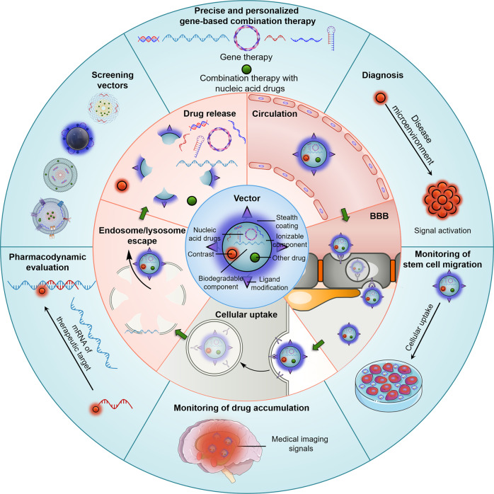
Nucleic acid drugs have the advantages of rich target selection, simple in design, good and enduring effect. They have been demonstrated to have irreplaceable superiority in brain disease treatment, while vectors are a decisive factor in therapeutic efficacy. Strict physiological barriers, such as degradation and clearance in circulation, blood-brain barrier, cellular uptake, endosome/lysosome barriers, release, obstruct the delivery of nucleic acid drugs to the brain by the vectors. Nucleic acid drugs against a single target are inefficient in treating brain diseases of complex pathogenesis. Differences between individual patients lead to severe uncertainties in brain disease treatment with nucleic acid drugs. In this Review, we briefly summarize the classification of nucleic acid drugs. Next, we discuss physiological barriers during drug delivery and universal coping strategies and introduce the application methods of these universal strategies to nucleic acid drug vectors. Subsequently, we explore nucleic acid drug-based multidrug regimens for the combination treatment of brain diseases and the construction of the corresponding vectors. In the following, we address the feasibility of patient stratification and personalized therapy through diagnostic information from medical imaging and the manner of introducing contrast agents into vectors. Finally, we take a perspective on the future feasibility and remaining challenges of vector-based integrated diagnosis and gene therapy for brain diseases.


Conflict of interest statement

The authors declare no competing interests.

Figures

Fig. 1
Fig. 1
Mechanisms of nucleic acid drug-based combination therapy and medical imaging contrast-based diagnosis for precise and personalized treatment of brain diseases. The inner ring shows a conventional contrast-labeled nucleic acid drug vector. The middle ring demonstrates the biological barriers that nucleic acid drug vectors face for delivery to the brain. The outer ring illustrates how medical imaging-based diagnostics contribute to precise and personalized therapy
Fig. 2
Fig. 2
Schematic illustration of the biological barriers to systemic delivery of nucleic acid drugs for brain diseases and strategies to overcome these barriers. a Degradation and clearance in circulation. b BBB. c Cellular uptake. d Endosome/lysosome barriers. e Nucleic acid drug release
Fig. 3
Fig. 3
Schematic illustration of classes of nucleic acid drug vectors. PNPs have the advantages of low synthesis difficulty, high chemical structure tunability, simultaneous multifunctionality, and the disadvantages of the tendency to agglomerate and potential toxicity. Lipid-based nanoparticles have the advantage of simple preparation, structural tunability, and interaction with bio-membranes and the disadvantage of low drug loading efficiency and tendency to be cleared by the liver. EVs are natural vectors for cell-to-cell delivery of biomolecules and combine certain therapeutic properties. However, EVs are difficult to isolate, extract, drug load, and modify with low yields and suffer from heterogeneity. Inorganic nanoparticle-based nucleic acid drug vectors have the advantages of homogeneous particle size, shape, surface potentials, highly tunable size and shape, and possible medical imaging signals and the disadvantages of toxicity and solubility. Nucleic acid drug conjugates (not shown) have the advantage of almost absolute homogeneity, high stability, low non-drug component content, and less restricted administration routes and the disadvantage of less functionality and low endosome/lysosome escape efficiency
Fig. 4
Fig. 4
Chemical structures of polymers and polymer derivatives used for nucleic acid drug delivery. Chitosan; pHis, poly (L-histidine); branched PEI, polyethyleneimine; PEI-PEG, polyethyleneimine-g-poly(ethylene glycol); PLO, poly-L-ornithine; PAA, poly(amidoamine); PBAE, poly(β-amino esters); OEI, oligoethylenimine; TBD-PEG-N3, 1,1,2,2-tetraphenylethene-benzo[c][1,2,5] thiadiazole-2-(diphenyl methylene) malononitrile-polyethylene glycol-azide; Thiolated dextrin; PVBLG-8, poly(γ-(4-(((2-(piperidin-1-yl)ethyl)amino)methyl)benzyl-L-glutamate); PQDEA, poly(N-(2-(acryloyloxy)ethyl)-N-(p-acetyloxyphenyl)-N,N-diethylammonium chloride); PADDAC, poly(N-(2-(acryloyloxy)ethyl)-N-(p-(2,4-dinitrophenoxy)benzyl)-N,N-diethylammonium chloride); B-PDEAEA, poly((2-acryloyl)ethyl(p-boronic acid benzyl)diethylammonium bromide); PDMAEMA, poly(dimethylaminoethyl methacrylate); PdXYP, polydixylitol-polyethyleneimine; T-PA-G-SA, Tet-1-poly(2-(dimethylamino)ethyl acrylate)-b-poly(2-aminoethyl methacrylate-GFLG-salsalate); PCB-Se-Se-Sim, polycarboxybetaine-co-poly(polycarboxybetaine-Se-Se-simvastatin); cRGD-PEO-b-P(CL-g-DP), cRGD-poly(ethylene oxide)-b-poly(ɛ-caprolactone-g-N,N-dimethyldipropylenetriamine); P2-PEG-HSA-AA, poly(ethylene glycol)-human serum albumin-cis-aconitic anhydride; DOPA-PLys-PEG, dopamine-polylysine-b-poly(ethylene glycol); Glu-PEG-PLys(MPA/IM), glucosyl-poly(ethylene glycol)-b-poly(L-lysine modified with 3-mercaptopropyl amidine and 2-thiolaneimine); mPEG-SS-PLys, poly(ethylene glycol)-b-poly(L-lysine) bearing a disulfide linkage; PEG-b-PMPMC-g-PTX, poly(ethylene glycol)-b-poly(5-mthyl-5-propargyl-1,3-dioxan-2-one)-g-paclitaxel; p(OEGMA-DMAEMA)-b-p((MAVE)-(MAVE-PDP)), poly(oligo(ethylene glycol) monomethyl ether methacrylate-dimethylaminoethyl methacrylate)-b-poly((2-(vinyloxy)ethyl methacrylate)-((2 R)-N-((2 R,5 R)-5-benzyl-11,17-dimethyl-3,6,16-trioxo-1-phenyl-10,12-dioxa-4,7-diazaoctadec-17-en-2-yl)-3-phenyl-2-(2-(pyren-2-yl)acetamido)propanamide)); PCB-P(DPA-co-DMA)-PG, Poly(carboxybetaine)-b-poly(dimethylaminoethyl methacrylate-co-diisopropylethyl methacrylate)-b-poly(aminoethyl methacrylate); mPEG-bPEI-PAsp(DIP-BzA), monomethoxy-poly(ethylene glycol)-b-branched polyethyleneimine-b-poly(N-(N′,N′-diisopropylaminoethyl)-co-benzylamino)aspartamide; PEG-b-P(GuF), poly(ethylene glycol)-b-poly((N-(3-methacrylamidopropyl) guanidinium-co-2,2,3,3-tetrafluoropropyl methacrylate)
Fig. 5
Fig. 5
Chemical structures of lipids and lipid derivatives used for nucleic acid drug delivery. DSPE-PCB, 1,2-distearoyl-sn-glycero-3-phosphoethanolamine-polycarboxybetaine; DSPE-PEG, 1,2-distearoyl-sn-glycero-3-phosphoethanolamine-poly(ethylene glycol); DPPC, 1,2-dipalmitoyl-sn-glycero-3-phosphocholine; DOPC, 1,2-dioleoyl-sn-glycero-3-phosphocholine; POPC, 1-palmitoyl-2-oleoyl-glycero-3-phosphocholine; DOPE, 1,2-dioleoyl-sn-glycero-3-phosphoethanolamine; DSPC, 1,2-distearoyl-sn-glycero-3-phosphocholine; DPPE-PEG, 1,2-dipalmitoyl-sn-glycero-3-phosphoethanolamine-poly(ethylene glycol); DOPG, 1,2-dioleoyl-sn-glycero-3-phospho-(1’-rac-glycerol); DMG-PEG, 1,2-dimyristoyl-rac-glycero-3-methoxy-poly(ethylene glycol); POPG, 1-palmitoyl-2-oleoyl-sn-glycero-3-phospho-(1’-rac-glycerol) (sodium salt); DLin-MC3-DMA, (6Z,9Z,28Z,31Z)-heptatriaconta-6,9,28,31-tetraen-19-yl 4-(dimethylamino)butanoate; C16-PEG2000-Ceramide, N-palmitoyl-sphingosine-1-(succinyl(methoxy(polyethylene glycol)2000)); X-ULFA-1, 4-(dimethylamino)-N’,N’-di((9Z,12Z)-octadeca-9,12-dien-1-yl)butanehydrazide; X-ULFA-2, 4-((di((9Z,12Z)-octadeca-9,12-dien-1-yl)amino)oxy)-N,N-dimethyl-4-oxobutan-1-amine; X-ULFA-3, 2-(di((9Z,12Z)-octadeca-9,12-dien-1-yl)amino)ethyl 3-(dimethylamino)propanoate; X-ULFA-4, 2-(di((9Z,12Z)-octadeca-9,12-dien-1-yl)amino)ethyl 3-(4-methylpiperazin-1-yl)propanoate; X-O14B, bis(2-(decyldisulfaneyl)ethyl) 3,3’-((3-(bis(2-hydroxyethyl)amino)propyl)azanediyl)dipropionate; ssPalmO-X, ((disulfanediylbis(propane-3,1-diyl))bis(piperidine-1,4-diyl))bis(ethane-2,1-diyl) (3E,3’E,5E,5’E,7E,7’E,9E,9’E)-bis(4,8-dimethyl-10-(2,6,6-trimethylcyclohex-1-en-1-yl)deca-3,5,7,9-tetraenoate); ssPalmO-Phe, ((((((disulfanediylbis(ethane-2,1-diyl))bis(piperidine-1,4-diyl))bis(ethane-2,1-diyl))bis(oxy))bis(2-oxoethane-2,1-diyl))bis(4,1-phenylene))bis(methylene) dioleate; X-OCholB, bis(2-((2-((((17-isopentyl-10,13-dimethyl-2,3,4,7,8,9,10,11,12,13,14,15,16,17-tetradecahydro-1H-cyclopenta[a]phenanthren-3-yl)oxy)carbonyl)amino)ethyl)disulfaneyl)ethyl) 3,3’-((3-(pyrrolidin-1-yl)propyl)azanediyl)dipropionate; DOTAP, 1,2-dioleoyl-3-trimethylammonium-propane (chloride salt); DPTAP, 1,2-dipalmitoyl-3-trimethylammonium-propane (chloride salt); DOTMA, 1,2-di-O-octadecenyl-3-trimethylammonium propane (chloride salt)
Fig. 6
Fig. 6
Schematic illustration of co-loading and release of multi drugs. a The degradation of PNPs promotes the co-release of nucleic acid drugs and chemical drugs. b The covalent bonds linking the chemical drugs and PNPs were cleaved to mediate the pioneering release of the chemical drugs. The chemical drug release further mediates the degradation of PNPs and the release of nucleic acid drugs. c The charge transition of the vector surface mediates the release of nucleic acid drugs. The nano-enzyme drugs do not need to be released. d The charge transition of the vector surface mediates the pioneering release of nucleic acid drugs. The covalent bonds linking the chemical drugs and vectors were cleaved to mediate the subsequent release of the chemical drugs. e The degradation of the lipid shells promotes the PNP release. The charge transition of the cationic outer layer of PNPs mediates the pioneering release of nucleic acid drugs. The disease microenvironment mediates the conversion of the inner core of PNPs from hydrophobic to hydrophilic, thus facilitating the degradation of PNPs and the subsequent release of the chemical agent. f Degradation of the lipid outer membrane mediates the pioneering release of the chemical agent. The charge transition of the cationic outer layer of PNPs mediates the subsequent release of nucleic acid drugs. g The exosome outer membrane fuses with the cell membrane to deliver the PNPs directly to the cytoplasm. The disease microenvironment mediates the charge transition in the outer layer and the hydrophile transition in the hydrophobic inner core of PNPs, facilitating the co-release of chemical and nucleic acid drugs. h The exosome outer membrane fuses with the cell membrane to deliver the endogenous nucleic acid drugs and PNPs directly to the cytoplasm. The hydrophile transition in the hydrophobic inner core of PNPs mediates the subsequent release of the chemical drugs
Fig. 7
Fig. 7
Schematic illustration of contrast addition methods and diagnostic applications. Contrast-labeled nucleic acid drug vectors are theoretically equipped for real-time monitoring of drug accumulation in the lesion. a, b T2-weighted MRI contrast agents (SPIONs) are encapsulated in the cores of multidrug-loaded PNPs by hydrophobic interactions. c T1-weighted MRI contrast agents (Mn(II) chelates) are used to cross-link cationic polymers into PNPs, thereby loading nucleic acid drugs. d The unactivated T1-weighted MRI contrast agents (Mn(III)) and the less sensitive T1-weighted MRI contrast agents (Mn(IV)) are adsorbed on albumin to form nanoparticles, thereby loading nucleic acid drugs. e Chemical drugs, nucleic acid drugs, and T2-weighted MRI contrast agents (SPIONs) are co-loaded in LPNPs. The contrast agents are encapsulated in the cores of PNPs by hydrophobic interactions. f Chemical drugs, nucleic acid drugs, and T2-weighted MRI contrast agents (SPIONs) are co-loaded in LPNPs. The contrast agents are encapsulated in the cores of PNPs by hydrophobic interactions. g T1-weighted MRI contrasts (Gd(III) chelates)-modified lipids are synthesized to construct cationic lipid-based nanoparticles to load nucleic acid drugs. h Chemical drugs, endogenous nucleic acid drugs, and T2-weighted MRI contrast agents (SPIONs) are co-loaded in exosome/polymer hybrid nanoparticles. The contrast agents are encapsulated in the cores of PNPs by hydrophobic interactions. i Nucleic acid drugs and T1-weighted MRI contrasts (Gd(III) chelates) are co-loaded on organic-inorganic hybrid nanoparticles via electrostatic interactions and covalent bonds, respectively. j T2-weighted MRI contrast agents (SPIONs) are developed as vectors based on organic-inorganic hybrid nanoparticles, thus loading chemical drugs and nucleic acid drugs via covalent bonds and electrostatic interactions, respectively. k CT contrast agents (AuNPs) are developed as vectors based on organic-inorganic hybrid nanoparticles, thus loading chemical drugs and nucleic acid drugs via covalent bonds and electrostatic interactions, respectively. l Radionuclides (18F-flumazenil, 125I or 99mTc) are directly conjugated to nucleic acid drugs for nuclear medicine imaging (PET, SPECT/CT, or SPECT). m-p Diagnostic applications of the contrast-labeled nucleic acid drug vectors. m Monitoring of the migration of transplanted stem cells in vivo through the uptake of contrast-labeled nucleic acid drug vectors. n Pharmacodynamic evaluation by specific binding of contrast-labeled nucleic acid drug vectors to therapeutic targets. o The migration of transplanted stem cells in vivo can be monitored in real-time after the uptake of contrast-labeled nucleic acid drug vectors. o and p Disease diagnosis via disease microenvironment-activated signals from contrast agents in nucleic acid drug vectors

References

    1. Whitehead KA, Langer R, Anderson DG. Knocking down barriers: advances in siRNA delivery. Nat. Rev. Drug Discov. 2009;8:129–138. doi: 10.1038/nrd2742. - DOI - PMC - PubMed
    1. Hou X, Zaks T, Langer R, Dong Y. Lipid nanoparticles for mRNA delivery. Nat. Rev. Mater. 2021;6:1078–1094. doi: 10.1038/s41578-021-00358-0. - DOI - PMC - PubMed
    1. Katti A, Diaz BJ, Caragine CM, Sanjana NE, Dow LE. CRISPR in cancer biology and therapy. Nat. Rev. Cancer. 2022;22:259–279. doi: 10.1038/s41568-022-00441-w. - DOI - PubMed
    1. Carthew RW, Sontheimer EJ. Origins and mechanisms of miRNAs and siRNAs. Cell. 2009;136:642–655. doi: 10.1016/j.cell.2009.01.035. - DOI - PMC - PubMed
    1. Li Y, et al. Strategies and materials of “SMART” non-viral vectors: overcoming the barriers for brain gene therapy. Nano Today. 2020;35:101006. doi: 10.1016/j.nantod.2020.101006. - DOI

Publication types