. 2023 Jan 17;13(1):862.
doi: 10.1038/s41598-023-27712-2.

A preclinical study comparing single- and double-root 3D-printed Ti-6Al-4V implants

Affiliations

A preclinical study comparing single- and double-root 3D-printed Ti-6Al-4V implants

Inna Chung et al. Sci Rep. .

Abstract

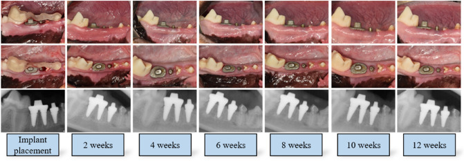
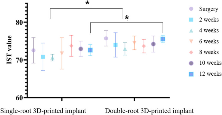
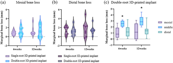
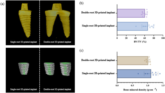
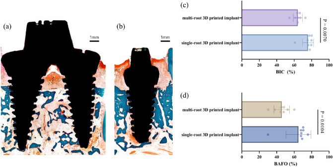
Recently, double-root implants have been investigated using 3D-printed technology. Here, we investigated damping capacity, microcomputed tomographic (micro-CT) and histological analyses of double-root 3D-printed implants compared with single-root 3D printed implants. Single- and double-root 3D-printed implants were fabricated and placed at both sides of mandibular third and fourth premolars in four beagle dogs. The damping capacity was measured, and periapical X-rays were taken every 2 weeks for 12 weeks. The bone volume/tissue volume (BV/TV) and bone mineral density (BMD) around the implants were measured with micro-CT. Bone-to-implant contact (BIC) and bone area fraction occupancy (BAFO) were measured in histological samples. The implant stability values between the groups were not significantly different, except at 4 and 12 weeks. The marginal bone changes were similar at the mesial and distal areas between the groups. The BV/TV and BMD values of the double-root 3D-printed implants showed no statistical difference through micro-CT analysis, but the double-root 3D-printed implants showed lower BIC and BAFO values through histomorphometric analysis compared to the single-root 3D-printed implants. Compared to single-root implants, 3D-printed double-root implants demonstrated comparable stability and bone remodeling around the fixtures, but the statistically significant bone loss in the furcation area remains problematic.


Conflict of interest statement

The authors declare no competing interests.

Figures

Figure 1
Figure 1
Clinical and radiographic photograph of single- and double-root 3D-printed implant. All protective caps were removed 2 weeks after implant placement for plaque control and implant stability measurements. There were no clinical signs of peri-implant inflammation, including redness, spontaneous bleeding, swelling, or ulceration. The clinical and radiographic photos were taken at the time of implant placement and 2, 4, 6, 8, 10, 12 weeks after implant placement.
Figure 2
Figure 2
Implant stability test (IST) values of single- and double-root 3D-printed implants. The IST values were significantly greater for the double-root 3D-printed implants at 4 and 12 weeks than for the single-root 3D-printed implants, as shown by Sidak’s multiple comparison test (p = 0.0143, p = 0.0320). There were no significant differences within the group during each time point.
Figure 3
Figure 3
Radiographic changes (a) mesial bone loss in single- and double-root 3D-printed implant at 6 and 12 weeks (b) distal bone loss in single- and double-root 3D-printed implant at 6 and 12 weeks (c) Mesial, middle, and distal bone loss in double-root 3D-printed implant at 6 and 12 weeks. Asterisks (*) indicate statistically significant difference in marginal bone loss in the middle area compared with mesial and distal areas at 6 weeks and 12 weeks. 3D: 3-dimensional.
Figure 4
Figure 4
Representative micro-computed tomography (a) and analysis (b, c). (a) Gray and green areas indicate the VOI and 3D-printed implants, respectively. BMD and BV/TV were measured in the gray area. (b) The BV/TV of single-root and multi-root 3D printed implant were 67.11 ± 13.05% and 60.76 ± 5.43%, respectively, showing no statistically significant difference. (c) The bone mineral density of single-root and multi-root 3D printed implant were 1.11 ± 0.23 g mm−3 and 1.02 ± 0.08 g mm−3, respectively, showing no statistically significant difference. CT: computed tomography, VOI: volume of interest, 3D: 3-dimensional, BMD: bone mineral density, BV/TV: bone volume/tissue volume.
Figure 5
Figure 5
Representative of histologic view (a, b) and analysis (b, d). (a)Histomorphometric view of double-root 3D-printed implant. Note the bone resorption in the furcation area. (b) Histomorphometric view of single-root 3D-printed implant (c) The BIC in single-root and multi-root 3D-printed implants had statistically significant difference (75.87% ± 6.32% and 64.18 ± 5.23%, respectively, p = 0.0070) (d) The BAFO was statistically significantly different (p = 0.0104) in single-root and multi-root 3D-printed implants (64.88 ± 14.37% and 45.81 ± 9.01%, respectively). 3D: 3-dimensional, BIC: bone-to-implant contact, BAFO: bone area fraction occupancy.

References

    1. Yu NH, et al. Estimation and change of edentulism among the Korean population: Korea national health and nutrition examination survey 2007–2018. Epidemiol. Health. 2021;43:e2021020. doi: 10.4178/epih.e2021020. - DOI - PMC - PubMed
    1. Buser D, et al. 10-year survival and success rates of 511 titanium implants with a sandblasted and acid-etched surface: A retrospective study in 303 partially edentulous patients. Clin. Implant Dent. Relat. Res. 2012;14(6):839–851. doi: 10.1111/j.1708-8208.2012.00456.x. - DOI - PubMed
    1. Degidi M, Nardi D, Piattelli A. 10-year follow-up of immediately loaded implants with TiUnite porous anodized surface. Clin. Implant Dent. Relat. Res. 2012;14(6):828–838. doi: 10.1111/j.1708-8208.2012.00446.x. - DOI - PubMed
    1. Fischer K, Stenberg T. Prospective 10-year cohort study based on a randomized controlled trial (RCT) on implant-supported full-arch maxillary prostheses. Part 1: Sandblasted and acid-etched implants and mucosal tissue. Clin. Implant Dent. Relat. Res. 2012;14(6):808–815. doi: 10.1111/j.1708-8208.2011.00389.x. - DOI - PubMed
    1. Zhang XX, et al. Long-term outcomes of early loading of straumann implant-supported fixed segmented bridgeworks in Edentulous Maxillae: A 10-year prospective study. Clin. Implant Dent. Relat. Res. 2016;18(6):1227–1237. doi: 10.1111/cid.12420. - DOI - PubMed

Publication types