. 2023 Jan 17;13(1):884.
doi: 10.1038/s41598-023-27894-9.

Comparing the accuracy of ultrasound-based measurements of the cervical vagus nerve

Affiliations

Comparing the accuracy of ultrasound-based measurements of the cervical vagus nerve

Johann Dörschner et al. Sci Rep. .

Abstract

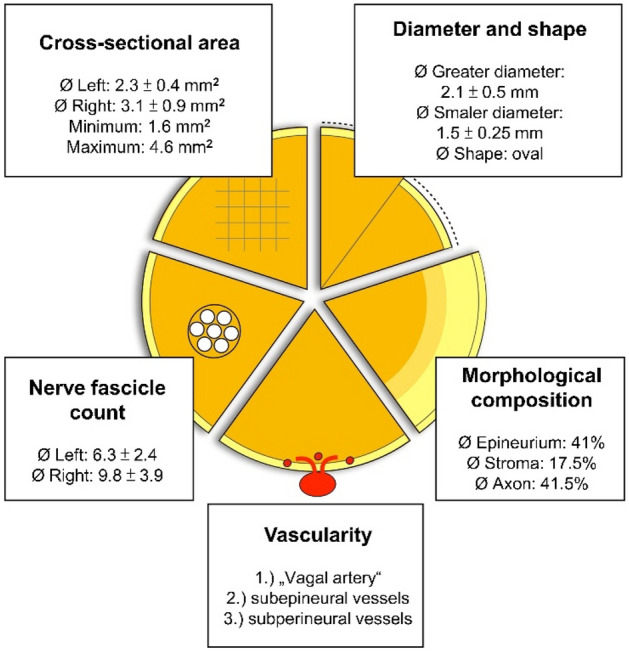
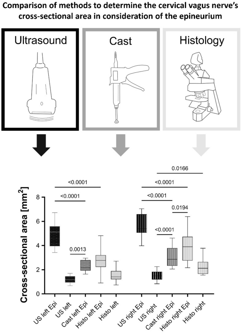
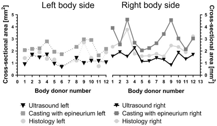
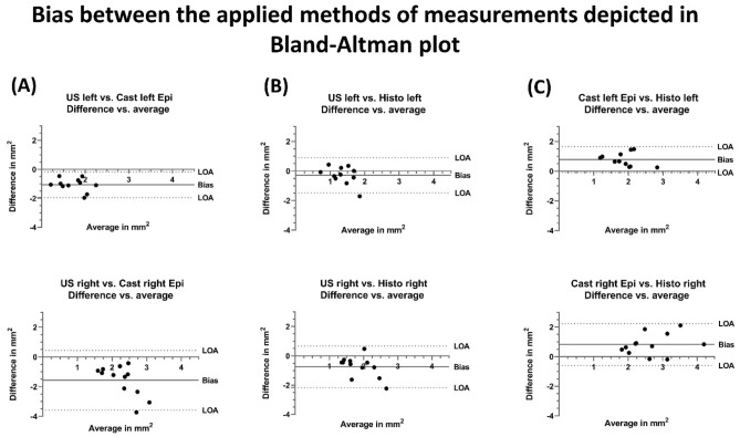
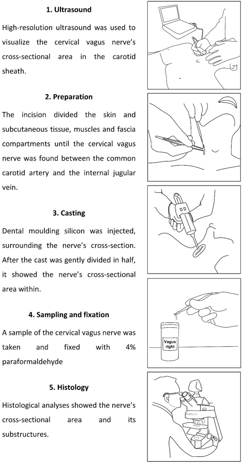
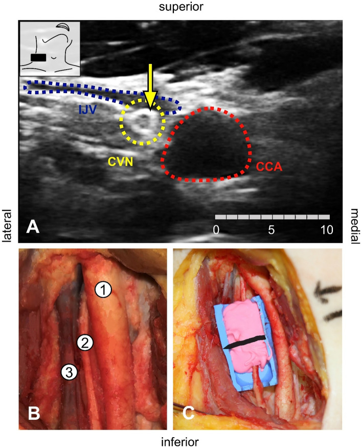
Vagus nerve stimulation (VNS) has become a promising therapy especially for drug resistant epilepsy and other pathologies. Side effects or missing therapeutic success are observed due to cuff electrodes that are too narrow or too wide. Preoperative high-resolution ultrasound is used to evaluate the size of the cervical vagus nerve (CVN) to estimate the size of cuff electrodes for VNS. It remains unclear how precise ultrasound reflects the CVN dimensions, which has been the objective of this study. CVN cross-sections and diameters were investigated in 23 sides from 12 bodies, using ultrasound, histology, and CVN casting in situ as a reference. Morphometric data were obtained including fascicle count and nerve composition in histology. CVN yielded significant side-, age-, and BMI-related differences. CVN cross-sections were smaller in ultrasound when compared to casting and histology (1.5 ± 0.4 vs. 3.1 ± 0.9 vs. 2.3 ± 0.7 mm2). With the given setting in ultrasound, CVN cross-sections were consistently underestimated when compared to casting. Ultrasound-based cross-section measurements are related to a biased estimation of CVN size. A factor to correct for method related differences may help to adjust for accurate cuff electrode sizes for patient needs and to reduce undesired effects and potentially material consumption.


Conflict of interest statement

The authors declare no competing interests.

Figures

Figure 1
Figure 1
Overview on morphological findings derived from casting (cross-sectional area, diameter, and shape) and histology (nerve fascicle count, vascularity, and morphological composition; results with standard deviation; for diameter, shape, and morphological composition the average of both body sides is given).
Figure 2
Figure 2
Histology sample of the cervical vagus nerve stained with hematoxylin–eosin. Nerve fascicles surrounded by perineurium and epineurium can be seen (A). Vascular supply with the “vagal artery” laying external to the epineurium (B), subperineural vessels within the perineurium (C), and minor vessels within the axon tissue within the nerve’s fascicles (D) are shown. Pictures of (BD) are magnifications of (A) in given resolution.
Figure 3
Figure 3
Box plots comparing different methods to determine the cervical vagus nerve’s cross-sectional area in consideration of the epineurium. Dotted Boxes indicate the left side, striped boxes the right side. The boxes show the 25th, 50th and 75th percentile, whiskers the minima and maxima. The solid line marks the median. Black—ultrasound (US), darker grey—cast, lighter grey—histology (Histo); Epi: Measurements contain the epineurium.
Figure 4
Figure 4
Determining the cervical vagus nerve cross-sectional area with ultrasound, cast, and histology. Interindividual differences donor- and side-depended were detected. Black—ultrasound darker grey—casting, lighter grey—histology. Dotted lines indicate the left and full lines the right body side.
Figure 5
Figure 5
Bland–Altman plots showing the differences of measurement between the applied methods for cross-sectional area detection. Most values lied within the 95% confidence interval (LOA) yielding that differences are distributed within close borders. Ultrasound (US) and casting (Cast) with epineurium (Epi) showed negative bias (A), ultrasound versus histology (Histo) likewise a negative bias (B), and casting with epineurium versus histology a positive bias for both body sides (C). LOA Limits of agreement.
Figure 6
Figure 6
Five steps showing the workflow of analyzing the cervical vagus nerve’s cross-sectional area.
Figure 7
Figure 7
(A) The cervical vagus nerve (CVN), the internal jugular vein (IJV), and the common carotid artery (CCA) are shown using high-resolution ultrasound. The yellow arrow points out the nerve’s hyperechoic epineurium. The nerve’s cross-sectional area is measured within this structure (scale in mm). (B) Preparation of the common carotid artery (1), the cervical vagus nerve (2), and the internal jugular vein (3). Most often the nerve is found in between the great vessels. (C) Casting of the cervical vagus nerve with pink-colored dental molding silicon. The blue mold provides sufficient casting by separating the nerve from surrounding tissue and moisture. The black line marks the level of examination.

References

    1. Coady MA, Adler F, Davila JJ, Gahtan V. Nonrecurrent laryngeal nerve during carotid artery surgery: Case report and literature review. J. Vasc. Surg. 2000;32:192–196. doi: 10.1067/mva.2000.105680. - DOI - PubMed
    1. Elliott RE, et al. Vagus nerve stimulation for children with treatment-resistant epilepsy: A consecutive series of 141 cases. J. Neurosurg. Pediatr. 2011;7:491–500. doi: 10.3171/2011.2.PEDS10505. - DOI - PubMed
    1. Englot DJ, Chang EF, Auguste KI. Vagus nerve stimulation for epilepsy: A meta-analysis of efficacy and predictors of response. J. Neurosurg. 2011;115:1248–1255. doi: 10.3171/2011.7.JNS11977. - DOI - PubMed
    1. Crumrine PK. Vagal nerve stimulation in children. Semin. Pediatr. Neurol. 2000;7:216–223. doi: 10.1053/spen.2000.9218. - DOI - PubMed
    1. Hirsch M, Reinacher P, Schulze-Bonhage A. Vagusnervstimulation in der Behandlung von Epilepsiepatienten. Akt. Neurol. 2016;43:501–510. doi: 10.1055/s-0042-116022. - DOI