. 2023 Jan 17;6(1):60.
doi: 10.1038/s42003-022-04385-7.

Tumor-specific intracellular delivery: peptide-guided transport of a catalytic toxin

Affiliations

Tumor-specific intracellular delivery: peptide-guided transport of a catalytic toxin

Curtis A Allred et al. Commun Biol. .

Abstract

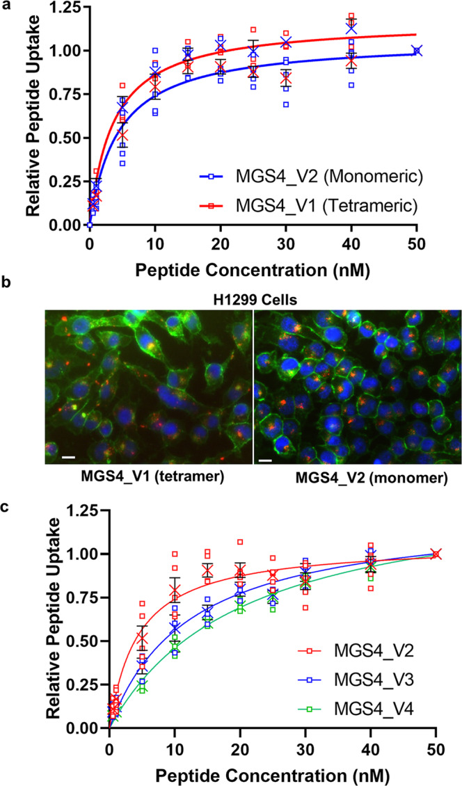
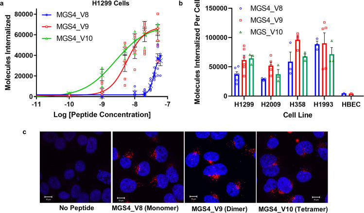
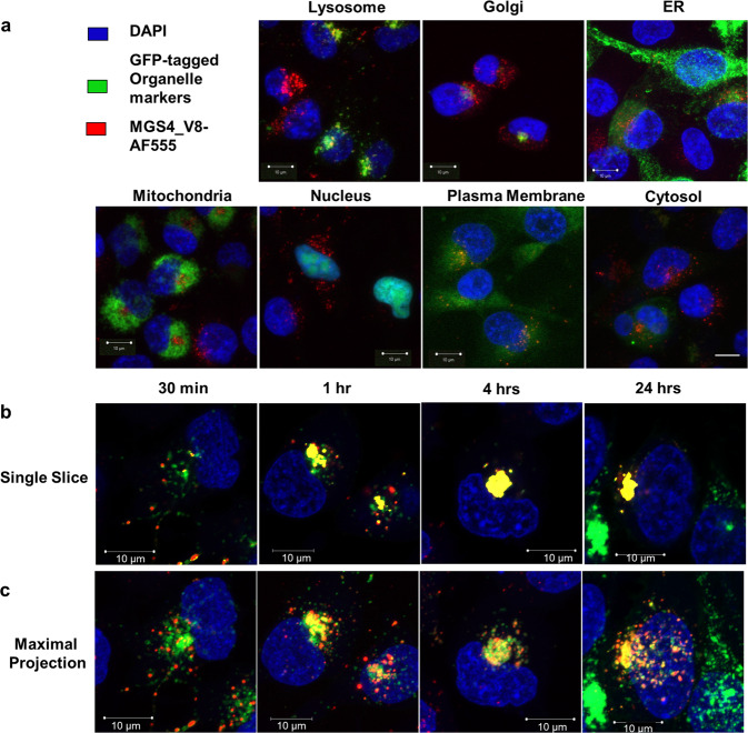
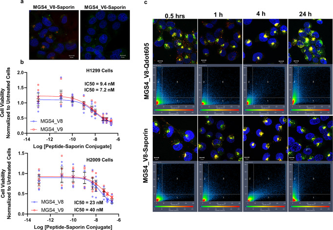
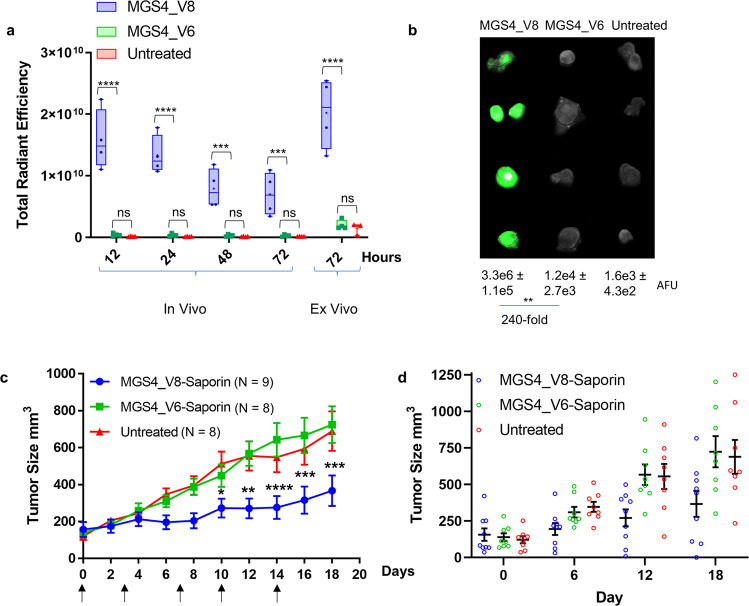
There continues to be a need for cancer-specific ligands that can deliver a wide variety of therapeutic cargos. Ligands demonstrating both tumor-specificity and the ability to mediate efficient cellular uptake of a therapeutic are critical to expand targeted therapies. We previously reported the selection of a peptide from a peptide library using a non-small cell lung cancer (NSCLC) cell line as the target. Here we optimize our lead peptide by a series of chemical modifications including truncations, N-terminal capping, and changes in valency. The resultant 10 amino acid peptide has an affinity of <40 nM on four different NSCLC cell lines as a monomer and is stable in human serum for >48 h. The peptide rapidly internalizes upon cell binding and traffics to the lysosome. The peptide homes to a tumor in an animal model and is retained up to 72 h. Importantly, we demonstrate that the peptide can deliver the cytotoxic protein saporin specifically to cancer cells in vitro and in vivo, resulting in an effective anticancer agent.


Conflict of interest statement

The authors declare no competing interests.

Figures

Fig. 1
Fig. 1. Monomeric and tetrameric MGS4 have similar binding, internalization and subcellular location.
a Binding and internalization of tetrameric MGS4_V1 and monomeric MGS4_V2 on live H1299 cells in culture. Cells were incubated with the peptide conjugated to streptavidin-phycoerythrin for 1 h at 37 °C. Non-internalized peptide was removed and the cells were analyzed by flow cytometry. b H1299 cells were incubated with MGS4_V1 or MGS4_V2 conjugated to streptavidin-Alexa Fluor 555 (red) for 1 h, washed, fixed and counterstained with WGA-Alexa Fluor 488 (green, cell membrane) and DAPI (blue, nuclei) and analyzed by fluorescence microscopy. The scale bar represents 20 µm. MGS4_V1 and MGS4_V2 internalize to a similar degree and localize to a similar destination. c Truncated monomeric MGS4 peptides have similar EC50 as the parental full-length peptide. Individual measurements are shown. The mean is shown as an “X” and black error bars represent standard error for a minimum of three experimental replicates (SEM). All original binding data and nonlinear regression analysis of the data are included in the supplementary materials.
Fig. 2
Fig. 2. Increased valency of truncated MGS4_V8 decreases EC50 but does not impact absolute update of the peptide or subcellular location.
a MGS4_V8, MGS4_V9, or MGS4_V10 were conjugated with streptavidin-Alexa Fluor 647 and H1299 cells were incubated with the labeled conjugate for 1 h. Non-internalized peptide was removed, and the mean number of peptides internalized per cell was determined to calculate the EC50. Individual measurements are shown. The mean is shown as an “X”. Error bars, in black, represent standard error measurements and are below the height of the symbols in some cases. b The average number of peptide molecules per cell at 50 nM at 1 h was determined on four NSCLC cell lines and one normal human bronchial epithelial cell line (HBEC). Error bars represent SEM and individual data points are shown. c H1299 cells were incubated with 50 nM peptide-Streptavidin-Qdot605 for 1 h, removed, and replaced with normal growth media. After 24 h, cells were fixed, and counterstained with DAPI (blue). Representative maximally projected z-stacks for each group reveal no apparent difference in peptide internalization or localization. The scale bar represents 10 µm.
Fig. 3
Fig. 3. MGS4_V8 traffics to and accumulates in lysosomes over time.
a H1299 cells were labelled with GFP-tagged organelle-specific proteins for ER, Golgi, lysosome, mitochondria, nucleus, plasma membrane, and cytosol (green). Cells were incubated with 50 nM MGS4_V8-Streptavidin Alexa Fluor 555 (red) for 1 h at 37 °C, then fixed and counterstained with DAPI (blue). MGS4_V8 colocalizes with lysosomes observed as yellow puncta indicated by the red arrows. No significant colocalization is observed with other subcellular organelles. b Lysosome labelled H1299 cells (green) were incubated with 50 nM MGS4_V8-Streptavidin Alexa Fluor 555 (red) for 0.5, 1, 4, or 24 h, then washed, fixed, and counterstained with DAPI (Blue). Representative single z-slice images are shown. Peptide-filled vesicles can be seen trafficking to lysosomes at 30 min, with many already colocalizing by 1 h. Most peptide is found within lysosomes by 4 h and retained there at 24 h. c Maximally projected, compressed z-stacks from images in panel b. The scale bar in all images represents 10 µm.
Fig. 4
Fig. 4. MGS4_V8 effectively delivers active saporin to cancer cells.
a H1299 cells were incubated biotinylated MGS4_V8 conjugated to streptavidin-saporin for 1 h, then washed, fixed and counterstained with an anti-saporin antibody (red), WGA-AF488 (green) and DAPI (blue). MGS4_V8 successfully delivers saporin into cancer cells while the control peptide, MGS4_V6 cannot. b MGS4_V8 and MGS4_V9 saporin-conjugates were serially diluted and incubated with H1299 and H2009 cells for 6 h after which, the MGS4-saporin conjugates were removed and complete growth media returned to the wells. At 72 h, viability was measured. IC50 values are provided in the inset. Individual measurements are shown. The mean is shown as an “X”. Error bars, in black, represent standard error measurements. Non-linear regression analysis is included in the supplementary material. c Colocalization time course as before, comparing MGS4_V8-streptavidin-Qdot trafficking to MGS4_V8-saporin trafficking. Pixels are plotted based on intensity in the red channel (x-axis) and the green channel (y-axis). Box 1 represents the population of saporin or Qdots not colocalized with the lysosome. Conversely, box 2 represents lysosomal staining not associated with Qdots or saporin signal. Box 3 contains colocalized pixels, which are falsely colored yellow and represent saporin or Qdots colocalized within the lysosomal compartment. A subpopulation of saporin containing vesicles remain distinct from lysosomes (box 1). The scale bar in all images represents 10 µm.
Fig. 5
Fig. 5. MGS4_V8 homes to xenograft tumor after systemic delivery and can deliver saporin resulting in reduced tumor growth.
H2009 tumor bearing nude mice (N = 4) were injected I.V. with MGS4_V8 or MGS4_V6 conjugated to NIR dye Alexa Fluor 750. At 12, 24, 48, and 72 h postinjection, mice were anesthetized and imaged on an IVIS® (Perkin Elmer) to measure total radiant efficiency in each tumor. MGS4_V8 accumulates in tumor 25–39 fold better than control peptide, MGS4_V6. MGS4_V6 accumulation is statistically no different than untreated tumors. Ex vivo NIR imaging of the tumors at the end of the experiment mirrors the data observed in the living animals. Whiskers represent min-to-max values, 25th to 75th percentile are represented by the box, the line shows the median, the + symbol represents the mean, and individual data is shown as a dot. Data for individual animals is included in Supplementary Table 8. b Excised tumors from the previous experiment were fixed in PBS + 4% formaldehyde and then imaged again together on a LI-COR® Odyssey. Arbitrary fluorescence units (AFU) were determined for each tumor and the average and SEM are shown below the image. The mean fluorescent intensity is 240-fold greater for MGS4_V8-Alexa Fluor 750 conjugate than the MGS4_V6 control conjugate. c Saporin was conjugated to either targeting MGS4_V8 or nontargeting MGS4_V6 and 7.5 µg of the conjugate was injected I.V. into mice bearing subcutaneous tumors. Animals were dosed 2 times per week for 2.5 weeks (indicated by arrows). Tumors were measured every other day. MGS4_V8-saporin clearly slows tumor growth, while nontargeted saporin has no effect compared to untreated animals. Error bars represent SEM, *p-value < 0.05, **p-value < 0.01, ***p-value < 0.001, ****p-value < 0.0001 (two-way ANOVA). d Tumor size (mm3) are shown for individual animals at days 0, 6, 12, and 18. Mean value is represented by the horizontal line and the error bars represent standard error. There is no statistical difference between MGS4_V6-Saporin and untreated at any day. At days 12 and 18 MGS4_V8-Saporin is statistically different than untreated (p-values 0.0099 and 0.0029, respectively) and MGS4_V6-Saporin (p-values 0.0069 and 0.0009, respectively).

Similar articles

Cited by

References

    1. American Cancer Society: Cancer Facts & Figures 2022, https://www.cancer.org/research/cancer-facts-statistics/all-cancer-facts....
    1. König D, Savic Prince S, Rothschild SI. Targeted therapy in advanced and metastatic non-small cell lung cancer. an update on treatment of the most important actionable oncogenic driver alterations. Cancers. 2021;13:804–841. doi: 10.3390/cancers13040804. - DOI - PMC - PubMed
    1. Verma S, et al. Trastuzumab emtansine for HER2-positive advanced breast cancer. N. Engl. J. Med. 2012;367:1783–1791. doi: 10.1056/NEJMoa1209124. - DOI - PMC - PubMed
    1. Younes A, et al. Results of a pivotal phase II study of brentuximab vedotin for patients with relapsed or refractory Hodgkin’s lymphoma. J. Clin. Oncol. 2012;30:2183–2189. doi: 10.1200/JCO.2011.38.0410. - DOI - PMC - PubMed
    1. Pro B, et al. Brentuximab Vedotin (SGN-35) in Patients With Relapsed or Refractory Systemic Anaplastic Large-Cell Lymphoma: Results of a Phase II Study. J. Clin. Oncol. 2012;30:2190–2196. doi: 10.1200/JCO.2011.38.0402. - DOI - PubMed

Publication types

MeSH terms