. 2023 Mar;33(3):201-214.
doi: 10.1038/s41422-022-00775-y. Epub 2023 Jan 17.

SARS-CoV-2 Z-RNA activates the ZBP1-RIPK3 pathway to promote virus-induced inflammatory responses

Affiliations

SARS-CoV-2 Z-RNA activates the ZBP1-RIPK3 pathway to promote virus-induced inflammatory responses

Shufen Li et al. Cell Res. 2023 Mar.

Abstract

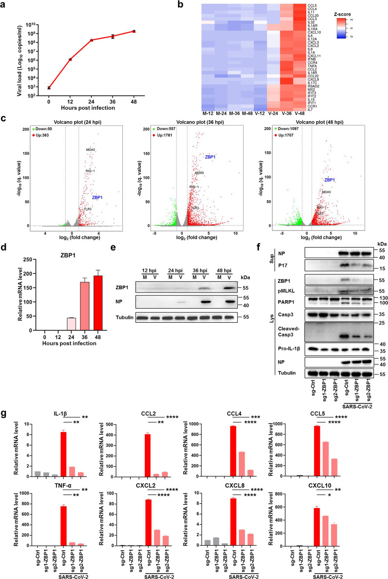
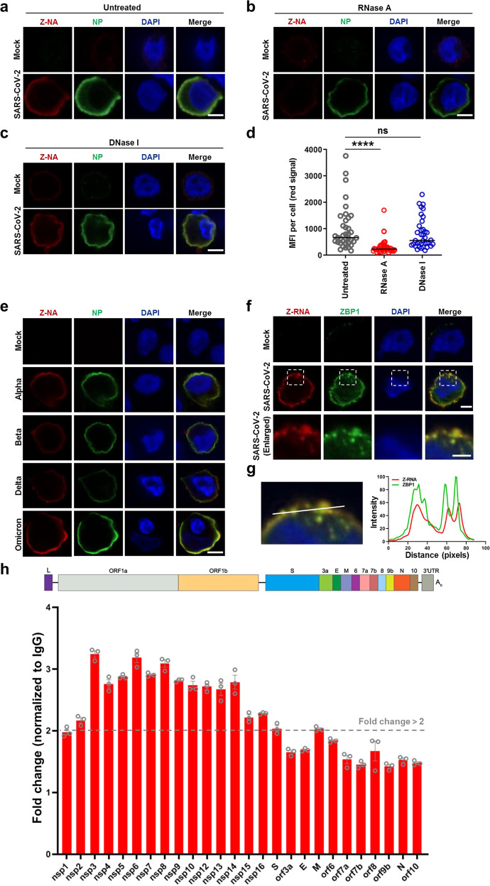
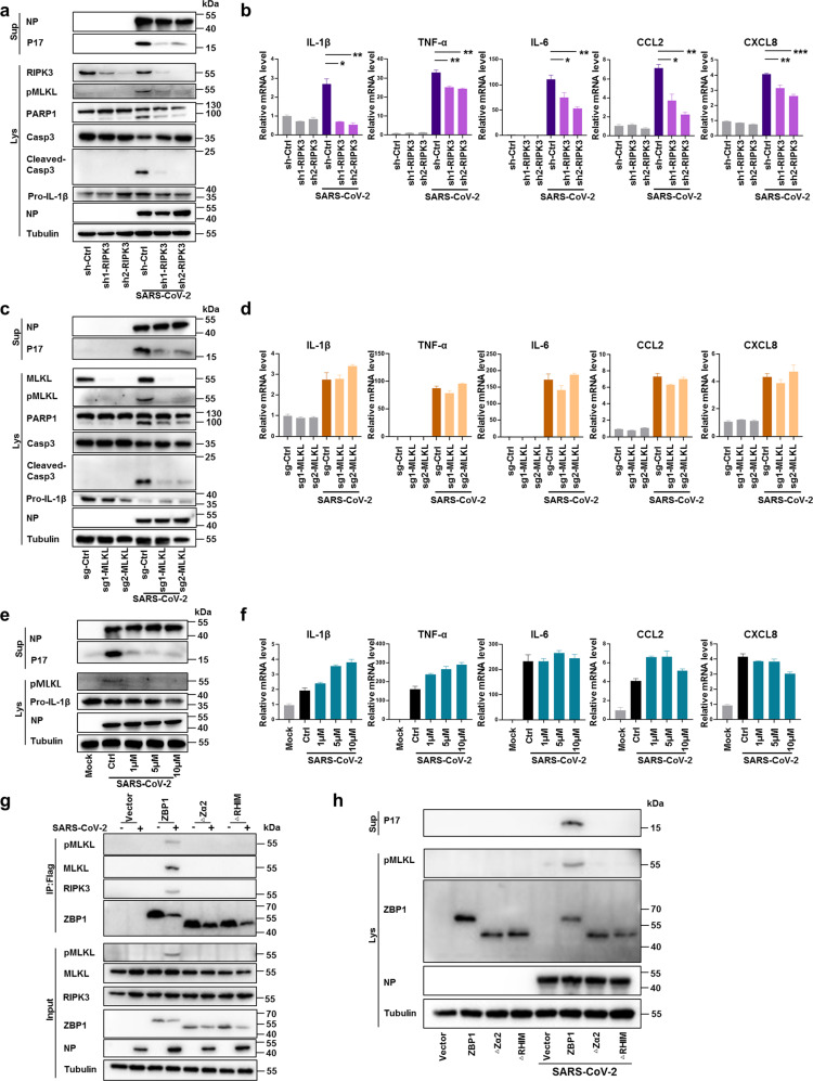
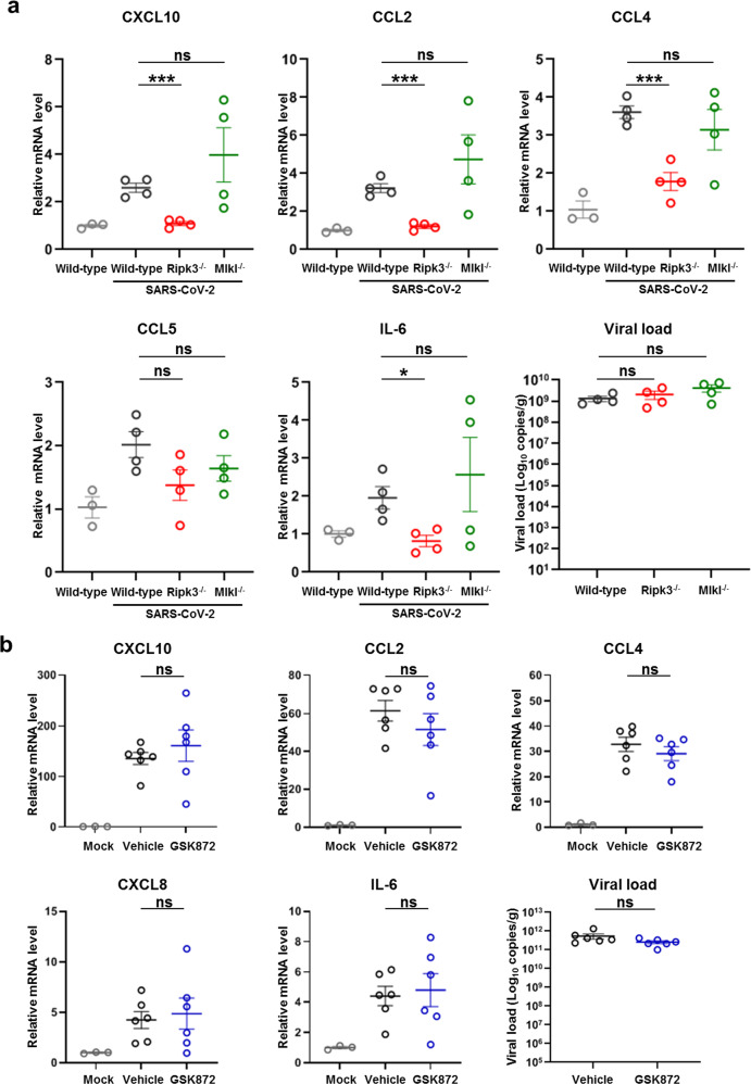
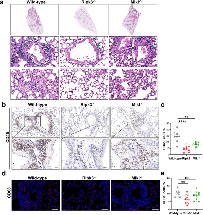
SARS-CoV-2 infection can trigger strong inflammatory responses and cause severe lung damage in COVID-19 patients with critical illness. However, the molecular mechanisms by which the infection induces excessive inflammatory responses are not fully understood. Here, we report that SARS-CoV-2 infection results in the formation of viral Z-RNA in the cytoplasm of infected cells and thereby activates the ZBP1-RIPK3 pathway. Pharmacological inhibition of RIPK3 by GSK872 or genetic deletion of MLKL reduced SARS-CoV-2-induced IL-1β release. ZBP1 or RIPK3 deficiency leads to reduced production of both inflammatory cytokines and chemokines during SARS-CoV-2 infection both in vitro and in vivo. Furthermore, deletion of ZBP1 or RIPK3 alleviated SARS-CoV-2 infection-induced immune cell infiltration and lung damage in infected mouse models. These results suggest that the ZBP1-RIPK3 pathway plays a critical role in SARS-CoV-2-induced inflammatory responses and lung damage. Our study provides novel insights into how SARS-CoV-2 infection triggers inflammatory responses and lung pathology, and implicates the therapeutic potential of targeting ZBP1-RIPK3 axis in treating COVID-19.


Conflict of interest statement

The authors declare no competing interests.

Figures

Fig. 1
Fig. 1. SARS-CoV-2 infection upregulates ZBP1 to stimulate inflammatory signaling and necroptosis.
ae Calu-3 cells were infected with SARS-CoV-2 (MOI = 0.1) or mock treated, cells and supernatants were harvested at the indicated times. Extracellular vRNA levels were determined with quantitative RT-PCR (qRT-PCR) (a). b, c Transcriptomic analysis of infected cells harvested at the indicated times. Heat map profiling expression of inflammation regulated genes that were significantly upregulated upon SARS-CoV-2 infection (b). M, mock, V, virus infection. Volcano plot indicating differentially expressed genes. Red and green indicated up- and downregulated genes, respectively (c). The transcription (d) and expression (e) level of ZBP1 was determined by qRT-PCR and western blot assay. f, g Calu-3 cells stably expressing non-targeting sgRNA (scramble) or sgRNAs against ZBP1 were infected with SARS-CoV-2 (MOI = 0.1). Cells and supernatants were harvested at 48 hpi. NP and P17 levels in supernatants and expression levels of ZBP1, pMLKL, PARP1, Casp3, cleaved-Casp3, pro-IL-1β or NP in cell lysates were measured by western blot assay (f). Total RNA was extracted and relative mRNA level of indicated cytokine and chemokine genes were detected by qRT-PCR (g). Data shown are means ± SEM. Statistical significance was analyzed by Student’s t-test. *P < 0.05; **P < 0.01; ***P < 0.001; ****P < 0.0001.
Fig. 2
Fig. 2. Formation of Z-RNA during SARS-CoV-2 infection.
ad SARS-CoV-2 original strain (MOI = 0.1) infected or uninfected (mock) Calu-3 cells were fixed and permeabilized at 48 hpi, and then either untreated (a) or treated with RNase A (b) or DNase I (c). Cells were co-stained with antibodies against Z-NA (red) and viral antigen NP (green). Nuclei were stained by DAPI (blue). d Quantification of the median florescence intensity (MFI) of Z-NA staining in SARS-CoV-2-infected cells in ac. Comparison of median values between two groups was analyzed by Mann–Whitney test. ****P < 0.0001; ns, no significance. Scale bar, 10 μm. e Calu-3 cells were infected with SARS-CoV-2 Alpha, Beta, Delta and Omicron strains (MOI = 0.1) for 48 h, and stained with antibodies against Z-NA (red) and NP (green). Scale bar, 10 μm. f SARS-CoV-2 original strain infected Calu-3 cells were co-stained with Z-NA and ZBP1 antibodies. Scale bars, 10 μm and 5 μm in enlarged image. g Quantitative analysis of the co-localization between Z-RNA and ZBP1 was performed with ImageJ. h Identification of viral derived Z-RNA. Calu-3 cells infected with SARS-CoV-2 (MOI = 0.1) for 48 h were lysed and incubated with Z-RNA antibody or isotype mouse IgG for immunoprecipitation. Enriched RNA was subjected to qRT-PCR analysis.
Fig. 3
Fig. 3. ZBP1 promotes SARS-CoV-2-induced inflammatory signaling and lung damage.
aj Zbp1−/− and wild-type mice were divided into three groups: Ad5-hACE2 non-transduced and non-infected (n = 3), transduced but non-infected (n = 3), transduced and infected (n = 6). Mice were intranasally transduced with Ad5-hACE2 (2.5 × 108 PFU) and infected with SARS-CoV-2 (1 × 105 TCID50) at 5 days post transduction. Lung samples were harvested at 2 dpi. a Relative level of indicated genes was analyzed with qRT-PCR and normalized to non-transduced and uninfected wild-type controls. Data shown are means ± SEM. Statistical significance was analyzed by Student’s t-test. **P < 0.01; ns, no significance. b The histopathological changes in lung were evaluated by H&E staining. Scale bars, 1000 μm and 50 μm in enlarged image. Inflammatory cell infiltration (green arrow) and alveolar septa expansion (red arrow) were observed in the lung sections. Immunostaining of CD45 (c), CD68 (e), MPO (g) and CD3 (i) representing the infiltration of leukocytes, macrophages, neutrophils and T cells, respectively. c, i Scale bars, 50 μm and 10 μm in the enlarged image. e, g Scale bars, 50 μm. d, f, h, j Quantitative analysis of positive cells was performed with ImageJ for c, e, g, i, respectively. Data shown are means ± SEM. Statistical significance was analyzed by Student’s t-test. **P < 0.01; ****P < 0.0001.
Fig. 4
Fig. 4. RIPK3 regulates SARS-CoV-2-induced necroptosis and inflammatory signaling.
a, b Calu-3 cells stably expressing non-targeting shRNA (scramble) or shRNAs against RIPK3 were infected with SARS-CoV-2 at MOI of 0.1. Cells and supernatants were harvested at 48 hpi. a NP and P17 levels in supernatants and expression levels of RIPK3, pMLKL, PARP1, Casp3, cleaved-Casp3, Pro-IL-1β or NP in cell lysates were determined by western blot assay. b Relative mRNA level of indicated cytokine and chemokine genes was detected by qRT-PCR. c, d Calu-3 cells stably expressing non-targeting sgRNA (scramble) or sgRNAs against MLKL were infected with SARS-CoV-2 (MOI = 0.1). Cells and supernatants were harvested at 48 hpi. c NP and P17 levels in supernatants and expression levels of MLKL, pMLKL, PARP1, Casp3, cleaved-Casp3, Pro-IL-1β or NP in cell lysates were determined by western blot assay. d Relative mRNA level of indicated cytokine and chemokine genes was detected by qRT-PCR. e, f Calu-3 cells were pre-treated with GSK872 at concentration of 1, 5 or 10 μM followed by infection with SARS-CoV-2 (MOI = 0.1) for 48 h. e NP and P17 level in the supernatants, pMLKL, pro-IL-1β or NP in cell lysates were measured by western blot assay. f Relative mRNA level of indicated cytokine and chemokine genes was determined by qRT-PCR. g, h ZBP1 knockout Calu-3 cells reconstituted with FLAG-tagged full-length ZBP1or truncation mutants deleted of Zα2 domain (ZBP1-∆Zα2) or RHIM domain (ZBP1-∆RHIM) were infected with SARS-CoV-2 (MOI = 0.1) or mock treated. g Cell lysates were harvested at 72 hpi and immunoprecipitated with anti-Flag magnetic beads, and detected by western blot assay. h Cell lysates and supernatant were harvested at 48 hpi. IL-1β P17 levels in the supernatants and the expression level of pMLKL, ZBP1or NP in the cell lysates were determined by western blot analysis. Data shown are means ± SEM. Statistical significance was analyzed by Student’s t-test. *P < 0.05; **P < 0.01; ***P < 0.001.
Fig. 5
Fig. 5. The scaffolding function of RIPK3 is critical for SARS-CoV-2-triggered inflammatory chemokine production in the lung.
a Ripk3−/−, Mlkl−/− or wild-type C57BL/6 mice (n = 4) were intranasally transduced with Ad5-hACE2 (2.5 × 108 PFU) and infected with SARS-CoV-2 (1 × 105 TCID50) at 5 days post transduction. Ad5-hACE2 transduced but uninfected wild-type C57BL/6 mice (n = 3) were used as the control. Lung samples were harvested at 2 dpi. Relative mRNA level of indicated genes was analyzed with qRT-PCR and normalized to samples from transduced but uninfected wild-type mice. b The effect of GSK872 on SARS-CoV-2-induced upregulation of cytokines and chemokines in the lung. BALB/c mice were intranasally infected with the mouse-adapted virus (WBP-1, 1 × 104 TCID50) and treated with vehicle (n = 6) or GSK872 (10 mg/kg/d) (n = 6) by intraperitoneal injection. Mice were euthanized at 5 dpi and lung samples were harvested for qRT-PCR determination. Data shown are means ± SEM. Statistical significance was analyzed by Student’s t-test. *P < 0.05; ***P < 0.001; ns, no significance.
Fig. 6
Fig. 6. RIPK3 contributes to SARS-CoV-2-induced lung damage and immune cell infiltration into the lung.
ae Ad5-hACE2 (2.5 × 108 PFU) transduced Ripk3−/−, Mlkl−/− or wild-type C57BL/6 mice were infected with SARS-CoV-2 (1 × 105 TCID50). Lung samples were harvested at 2 dpi. a The histopathological changes were evaluated by H&E staining. Inflammatory cell infiltration (green arrow) and alveolar septa expansion (red arrow) were indicated. Scale bars, 1000 μm in the original image and 50 μm in the enlarged image. bj Immunostaining of CD45 (b), CD68 (d), CD3 (f) and MPO (h) representing the infiltration of leukocytes, macrophages, T cells and neutrophils respectively. b, f Scale bars, 50 μm and 10 μm in the enlarged image. d, h Scale bars, 50 μm. c, e, g, i Quantitative analysis of positive cells performed with ImageJ for b, d, f, h, respectively. Data shown are means ± SEM. Statistical significance was analyzed by Student’s t-test. *P < 0.05; **P < 0.01; ***P < 0.001; **** P < 0.0001; ns, no significance. j Co-staining of CD8 (red) and CXCR3 (green) representing the recruitment of CD8+CXCR3+ T cells in the lung section of SARS-CoV-2-infected wild-type and Ripk3−/− mice. Scale bars, 10 μm.

Comment in

References

    1. Hu B, Guo H, Zhou P, Shi ZL. Characteristics of SARS-CoV-2 and COVID-19. Nat. Rev. Microbiol. 2021;19:141–154. doi: 10.1038/s41579-020-00459-7. - DOI - PMC - PubMed
    1. Wiersinga WJ, Rhodes A, Cheng AC, Peacock SJ, Prescott HC. Pathophysiology, transmission, diagnosis, and treatment of Coronavirus disease 2019 (COVID-19): a review. JAMA. 2020;324:782–793. doi: 10.1001/jama.2020.12839. - DOI - PubMed
    1. Li H, et al. SARS-CoV-2 and viral sepsis: observations and hypotheses. Lancet. 2020;395:1517–1520. doi: 10.1016/S0140-6736(20)30920-X. - DOI - PMC - PubMed
    1. Tay MZ, Poh CM, Renia L, MacAry PA, Ng LFP. The trinity of COVID-19: immunity, inflammation and intervention. Nat. Rev. Immunol. 2020;20:363–374. doi: 10.1038/s41577-020-0311-8. - DOI - PMC - PubMed
    1. Mehta P, et al. COVID-19: consider cytokine storm syndromes and immunosuppression. Lancet. 2020;395:1033–1034. doi: 10.1016/S0140-6736(20)30628-0. - DOI - PMC - PubMed

Publication types