. 2023 Mar;597(5):672-681.
doi: 10.1002/1873-3468.14571. Epub 2023 Jan 17.

The proline-rich domain of fission yeast WASp (Wsp1p) interacts with actin filaments and inhibits actin polymerization

Affiliations

The proline-rich domain of fission yeast WASp (Wsp1p) interacts with actin filaments and inhibits actin polymerization

Aaron D Rosenbloom et al. FEBS Lett. 2023 Mar.

Abstract

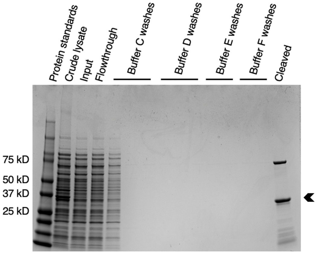
Members of the Wiskott-Aldrich Syndrome protein (WASp) family activate Arp2/3 complex (actin-related proteins 2 and 3 complex) to form actin filament branches. The proline-rich domain (PRD) of WASp contributes to branching nucleation, and the PRD of budding yeast Las17 binds actin filaments [Urbanek AN et al. (2013) Curr Biol 23, 196-203]. Biochemical assays showed the recombinant PRD of fission yeast Schizosaccharomyces pombe Wsp1p binds actin filaments with micromolar affinity. Recombinant PRDs of both Wsp1p and Las17p slowed the elongation of actin filaments by Mg-ATP-actin monomers by half and slowed the spontaneous polymerization of Mg-ATP-actin monomers modestly. The affinity of PRDs of WASp-family proteins for actin filaments is high enough to contribute to the reported stimulation of actin filament branching by Arp2/3 complex.

Keywords: Arp2/3 complex; Wiskott-Aldrich syndrome protein (WASp); actin.


Conflict of interest statement

Competing interests: The authors declare that no competing interests exist.

Figures

Fig. 1.
Fig. 1.
SDS-PAGE and Coomassie brilliant blue staining of samples from each step of the purification of Wsp1p-PRD. Left gel: lane 1, protein standards; lane 2, crude lysate; lane 3 clarified sample of crude lysate applied to TALON resin; lane 4, unbound flowthrough from TALON resin; lanes 5-7, successive washes of TALON resin with Buffer A (480 mM NaCl, 3 mM KCl, 10 mM phosphate, 10 mM imidazole pH 7.4); lanes 8-11, elution of bound proteins from TALON resin with elution buffer (Buffer A with an additional 240 mM imidazole pH 7.4). Middle gel: lane 1, protein standards; lane 2, combined fractions eluted from TALON resin and applied to glutathione Sepharose 4B beads; lane 3, unbound flowthrough from glutathione Sepharose 4B beads; lanes 4-6, successive washes of the glutathione Sepharose 4B beads with Buffer B (480 mM NaCl, 3 mM KCl, and 10 mM phosphate at a pH 7.4); lanes 7-9, washes of glutathione Sepharose 4B beads with KMEID (50 mM KCl, 1 mM MgCl2, 1 mM EGTA, 10 mM imidazole pH 7.0, 2 mM DTT); lane 10, pure Wsp1p-PRD cleaved from glutathione Sepharose 4B beads. Right hand gel shows two lanes of purified Wsp1p-PRD from another preparation. The arrowhead marks Wsp1p-PRD.
Fig. 2.
Fig. 2.
Coomassie brilliant blue stained SDS-PAGE gel of purification steps of S. cerevisiae Las17-PRD. Lane 1, protein standards; lane 2, crude lysate; lane 3, clarified lysate applied to glutathione Sepharose 4B beads; lane 4, unbound flowthrough from glutathione Sepharose 4B beads; lanes 5-7, washes of glutathione Sepharose 4B beads with Buffer C (10 mM phosphate buffer pH 7.4, 480 mM NaCl, 3 mM KCl, and 0.1% Tween-20); lanes 8-10, washes of glutathione Sepharose 4B beads with Buffer D (10 mM phosphate buffer pH 7.4, 140 mM NaCl, and 3 mM KCl); lanes 11-12, washes of glutathione Sepharose 4B beads with Buffer E (50 mM Tris-HCl, 400 mM NaCl, 1 mM EDTA, 1 mM DTT, and brought to a pH of 7 at room temperature); lanes 13-14, washes of glutathione Sepharose 4B beads with Buffer F (50 mM Tris-HCl, 150 mM NaCl, 1 mM EDTA, 1 mM DTT brought to pH 7 at room temperature); lane 15, protein cleaved from glutathione Sepharose 4B beads. Mass spectrometry of tryptic digests identified the main band (arrowhead) as Las17-PRD, the upper band as E. coli DnaK, and the minor lower molecular weight bands as fragments of Las17-PRD, likely due to ribosome stalling.
Fig. 3.
Fig. 3.
Interaction of Alexa-488-labeled Wsp1p-PRD for actin filaments. (A-B) Measurement of affinity by a pelleting assay. A range of concentrations of actin filaments were incubated with 1 μM of Alexa-488-labeled Wsp1p-PRD in 50 mM KCl, 1 mM MgCl2, 1 mM EGTA, 10 mM imidazole at pH 7.0 for 30 min at 25°C followed by ultracentrifugation at 240,000 g for 30 min. The fraction bound was calculated from the fluorescence of the supernatant. (A) Fraction of Alexa488-Wsp1p-PRD bound vs. actin filament concentration. Points are the mean with standard deviation as the error (n = 5-6). The solid line is the fit to the data of a binding isotherm with a Kd of 6.1 ± 0.6 μM. (B) Fraction of Wsp1p-PRD-Alexa488 bound vs. actin filament concentration. Points are the mean with the range as the error (n = 2-3). The solid line is the fit to the data of a binding isotherm with a Kd of 5 ± 1 μM. (C-D) Fluorescence anisotropy of Alexa488 bound to the N- or C-terminus of Wsp1p-PRD. A range of concentrations of actin filaments were incubated with 20 nM of Alexa488-Wsp1p-PRD or 100 nM of Wsp1p-PRD-Alexa 488 in 50 mM KCl, 1 mM MgCl2, 1 mM EGTA, and 10 mM imidazole at pH 7.0 at 25°C. Each sample was excited with polarized light, and the anisotropy was calculated based on the polarized components of the emission. (C) Fluorescence anisotropy of Alexa488-Wsp1p-PRD vs. concentration of actin filaments. Points are the mean ± standard deviation (n = 5-6). (D) Fluorescence anisotropy of Wsp1p-PRD-Alexa488 vs. concentration of actin filaments. Points are values from one experiment.
Fig. 4.
Fig. 4.
Time course of the spontaneous polymerization of four concentrations of Mg-ATP-actin monomers (10% pyrenyl-actin) alone or with 2.5 μM or 5 μM (A) Wsp1p-PRD or (B) Las17-PRD. Conditions: 50 mM KCl, 1 mM MgCl2, 1 mM EGTA, 10 mM imidazole pH 7.0, 0.6 mM Tris, 0.15 mM DTT, 60 μM ATP, 30 μM CaCl2 and 0.00005% Antifoam 204.
Fig. 5.
Fig. 5.
Effect of PRDs on the time course of elongation of actin filaments from preformed seeds (0.5 μm polymerized actin, 0.3 nM ends). Conditions: 2 μM 10% Mg-ATP-pyrenyl-actin monomers with either no PRD (black), 5 μM Wsp1p-PRD (green), or 5 μM of Las17-PRD (pink). For comparison, 2.5 μM of Mg-ATP-actin with 10% labeled actin were polymerized without preformed filaments or PRD (grey). (A) Examples of time courses corrected for photobleaching. (B) Initial parts of the polymerization curves from A after the initial dead time.

References

    1. Urbanek AN, Smith AP, Allwood EG, Booth WI, Ayscough KR. A novel actin-binding motif in Las17/WASP nucleates actin filaments independently of Arp2/3. Curr Biol. 2013;23(3):196–203. - PubMed
    1. Pollard TD, Cooper JA. Actin, a central player in cell shape and movement. Science. 2009;326(5957):1208–12. - PMC - PubMed
    1. Rosenbloom AD, Kovar EW, Kovar DR, Loew LM, Pollard TD. Mechanism of actin filament nucleation. Biophys J. 2021;120(20):4399–417. - PMC - PubMed
    1. Pollard TD. Regulation of actin filament assembly by Arp2/3 complex and formins. Annu Rev Biophys Biomol Struct. 2007;36:451–77. - PubMed
    1. Machesky LM, Atkinson SJ, Ampe C, Vandekerckhove J, Pollard TD. Purification of a Cortical Complex Containing 2 Unconventional Actins from Acanthamoeba by Affinity-Chromatography on Profilin-Agarose. J Cell Biol. 1994;127(1):107–15. - PMC - PubMed

Publication types

MeSH terms