. 2023 Feb 20;6(2):483-493.
doi: 10.1021/acsabm.2c00759. Epub 2023 Jan 18.

Pd-Based Hybrid Nanoparticles As Multimodal Theranostic Nanomedicine

Affiliations

Pd-Based Hybrid Nanoparticles As Multimodal Theranostic Nanomedicine

Alberto Bellissima et al. ACS Appl Bio Mater. .

Abstract

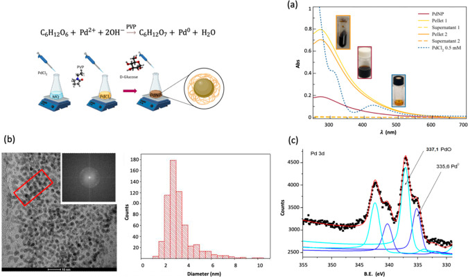
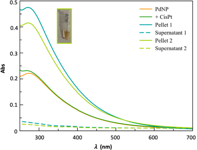
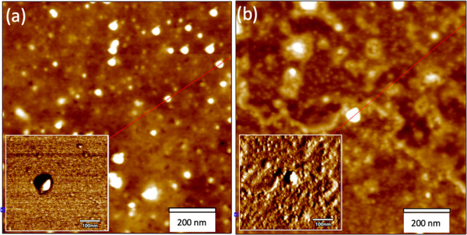
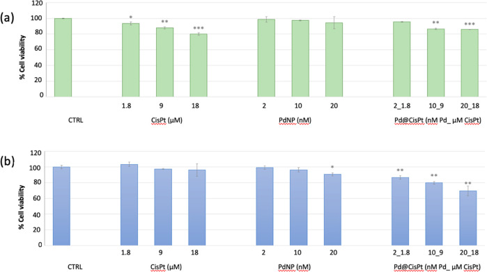
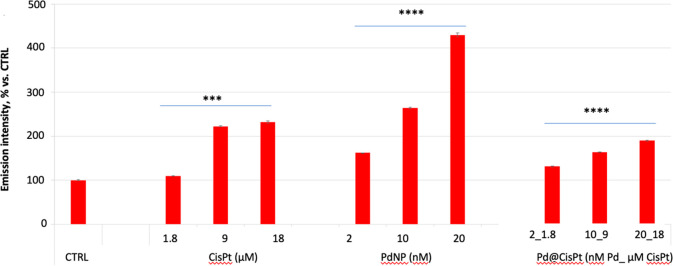
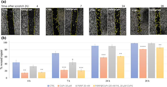
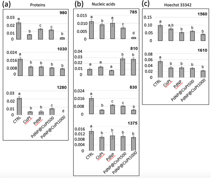
A nanodelivery system based on palladium nanoparticles (PdNP) and cisplatin (CisPt) was developed by physisorption of the drug onto the PdNP synthesized via a green redox process, using d-glucose and polyvinylpyrrolidone (PVP) as reducing and stabilizing/capping agents, respectively. UV-vis analysis and H2-evolution measurements were carried out to prove the nanoparticles' capability to act as bimodal theranostic nanomedicine, i.e., having both plasmonic and photocatalytic properties. XPS, XRD, and TEM allowed light to be shed on the chemical composition and morphology of the PdNP. The analysis of the UV-visible spectra evidenced plasmonic peak changes for the hybrid nanoparticle-drug assembly (Pd@CisPt), which pointed to a significant interaction of CisPt with the NP surface. The drug loading was quantitatively estimated by ICP-OES measurements, while DLS and AFM confirmed the strong association of the drug with the nanoparticle surface. The test of SOD-like activity in a cell-free environment proved the maintenance of the antioxidant capability of PdNP also in the Pd@CisPt systems. Finally, Pd@CisPt tested in prostate cancer cells (PC-3 line) unveiled the antitumoral action of the developed nanomedicine, related to reactive oxygen species (ROS) generation, with a condition of protein misfolding/unfolding and DNA damage, as evidenced by cytotoxicity and MitoSOX assays, as well as Raman microspectroscopy, respectively. Cell imaging by confocal microscopy evidenced cellular uptake of the nanoparticles, as well as dynamic processes of copper ion accumulation at the level of subcellular compartments. Finally, cell migration studies upon treatment with Pd@CisPt evidenced a tunable response between the inhibitory effect of CisPt and the enhanced rate of cell migration for the metal NP alone, which pointed out the promising potential of the developed theranostic nanomedicine in tissue regeneration.

Keywords: multimodal platform; photocatalytic activity; plasmonics; prostate cancer cell targeting.


Conflict of interest statement

The authors declare no competing financial interest.

Figures

Figure 1
Figure 1
(a) UV–visible spectra of PdNP (wine solid line) before and after the two centrifugation steps in 10 mM PBS at room temperature (1 min, 8000 rpm) for the resuspended pellets 1 (yellow, solid line) and 2 (orange, solid line), respectively; the supernatants spectra (dashed lines) collected after the two centrifugation steps, as well as the reference spectrum of palladium chloride (blue, dotted line) are also included. (b) TEM image of PVP-PdNP, size distribution (>600 particles have been measured), and 2D-FFT image of the area in the red box. (c) XPS spectrum of Pd 3d core-level doublets for PVP-stabilized PdNPs. Pd is present in two oxidation forms: Pd3/2 and Pd5/2 doublet of Pd0 (blue lines) and Pd3/2 and Pd5/2 doublet of Pd2+ (cyan lines).
Figure 2
Figure 2
UV–visible spectra of PdNP (1.58 × 10⁻⁷ mol/L, 1.8 × 10¹² NP/mL; orange solid line) before and after the addition of 2.4 mM CisPt (green solid line). The dark-green and light-green solid curves refer to the first and second pellets, respectively, while the dashed lines refer to the supernatants, obtained after two centrifugation steps in 10 mM PBS at room temperature (1 min, 8000 rpm). All spectra were recorded by diluting the mother solution five times in 10 mM PBS and using a quartz cuvette (optical path length = 0.1 cm).
Figure 3
Figure 3
AFM images recorded in AC mode in air of height (z scale = 10 nm) and phase (insets) for (a) PdNP and (b) Pd@CisPt.
Figure 4
Figure 4
Cell viability measured by (a) nuclear detection (imaging kit Blue/Green) and (b) mitochondrial succinate dehydrogenase activity (MTT assay) in PC-3 cells p.19 treated for 24 h with Pd@CiSt at increasing concentrations. Negative (untreated cells) and positive (i.e., cells treated with cisplatin or PdNP) controls are included. The data are expressed as the average percentage ± SD of three different experiments. Pairwise Student’s t test: *p < 0.05; **p < 0.01; ***p < 0.001 vs Ctrl.
Figure 5
Figure 5
MitoSOX assay’s results for the level of mitochondrial ROS in PC-3. Cells were incubated for 24 h with Pd@CisPt at increasing concentrations. Negative (untreated cells) and positive (i.e., cells treated with cisplatin or PdNP) controls are included. Data are expressed as the MitoSOX ratio with respect to DCF emission intensities (average percentage ± SD of three different experiments). Pairwise Student’s t test: ***p < 0.001; ****p < 0.0001 vs Ctrl.
Figure 6
Figure 6
(a) Representative micrographs of PC-3 cells, in the absence (negative control) or in the presence of Pd@CisPt (20 nM nanoparticles, 18 μM drug) hybrid collected at different times (t = 4,7,24, 28 h) after scratching (t = 0). (b) Quantitative analysis of cell migration (wound edge advancement in percent vs time). Means ± SEM values from three independent experiments. Pairwise Student’s T: *p < 0.05, **p < 0.01, ****p < 0.0001 vs Ctrl.
Figure 7
Figure 7
Univariate statistical analysis of peak height values calculated for the control, CisPt, PdNP, Pd@CisPt500, and Pd@CisPt1000 groups and related to (a) proteins (980 cm–1, 1030 cm–1, and 1280 cm–1), (b) nucleic acids (785 cm–1, 810 cm–1, 830 cm–1, and 1375 cm–1), and (c) Hoechst 33342 dye (1560 and 1610 cm–1). Values are reported as mean ± SD. Significant differences between experimental groups were determined by means of a factorial analysis of variance (one-way ANOVA), followed by Tukey’s multiple comparisons test, by the statistical software Prism6 (Graphpad Software, Inc. USA). Statistical significance was set at p < 0.05. Different letters over box charts indicate statistically significant differences among the above-defined experimental groups.

References

    1. Sultan M. H.; Moni S. S.; Madkhali O. A.; Bakkari M. A.; Alshahrani S.; Alqahtani S. S.; Alhakamy N. A.; Mohan S.; Ghazwani M.; Bukhary H. A.; et al.Characterization of cisplatin-loaded chitosan nanoparticles and rituximab-linked surfaces as target-specific injectable nano-formulations for combating cancer. Sci. Rep. 2022, 12, 468, 10.1038/s41598-021-04427-w. - DOI - PMC - PubMed
    1. Chen H.; Zhao Y. Applications of Light-Responsive Systems for Cancer Theranostics. ACS Appl. Mater. Interfaces 2018, 10 (25), 21021–21034. 10.1021/acsami.8b01114. - DOI - PubMed
    1. Ghosh S. Cisplatin: The first metal based anticancer drug. Bioorganic Chemistry 2019, 88, 102925.10.1016/j.bioorg.2019.102925. - DOI - PubMed
    1. Siddik Z. H. Cisplatin: mode of cytotoxic action and molecular basis of resistance. Oncogene 2003, 22 (47), 7265–7279. 10.1038/sj.onc.1206933. - DOI - PubMed
    1. Zhu Z.; Wang Z.; Zhang C.; Wang Y.; Zhang H.; Gan Z.; Guo Z.; Wang X. Mitochondrion-targeted platinum complexes suppressing lung cancer through multiple pathways involving energy metabolism. Chemical Science 2019, 10 (10), 3089–3095. 10.1039/C8SC04871A. - DOI - PMC - PubMed

Publication types

MeSH terms