. 2023 Jul 6;25(7):1249-1261.
doi: 10.1093/neuonc/noad018.

Lysine-specific histone demethylase 1A (KDM1A/LSD1) inhibition attenuates DNA double-strand break repair and augments the efficacy of temozolomide in glioblastoma

Affiliations

Lysine-specific histone demethylase 1A (KDM1A/LSD1) inhibition attenuates DNA double-strand break repair and augments the efficacy of temozolomide in glioblastoma

Salvador Alejo et al. Neuro Oncol. .

Abstract

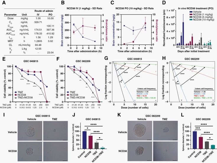
Background: Efficient DNA repair in response to standard chemo and radiation therapies often contributes to glioblastoma (GBM) therapy resistance. Understanding the mechanisms of therapy resistance and identifying the drugs that enhance the therapeutic efficacy of standard therapies may extend the survival of GBM patients. In this study, we investigated the role of KDM1A/LSD1 in DNA double-strand break (DSB) repair and a combination of KDM1A inhibitor and temozolomide (TMZ) in vitro and in vivo using patient-derived glioma stem cells (GSCs).

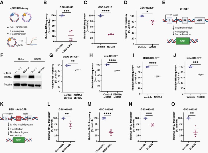
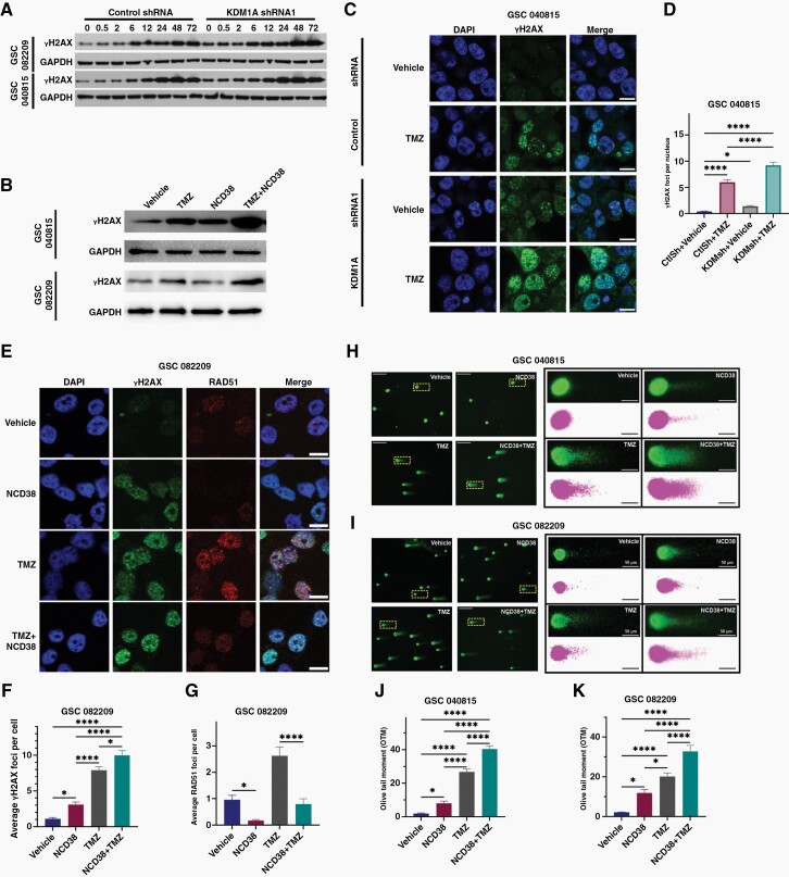
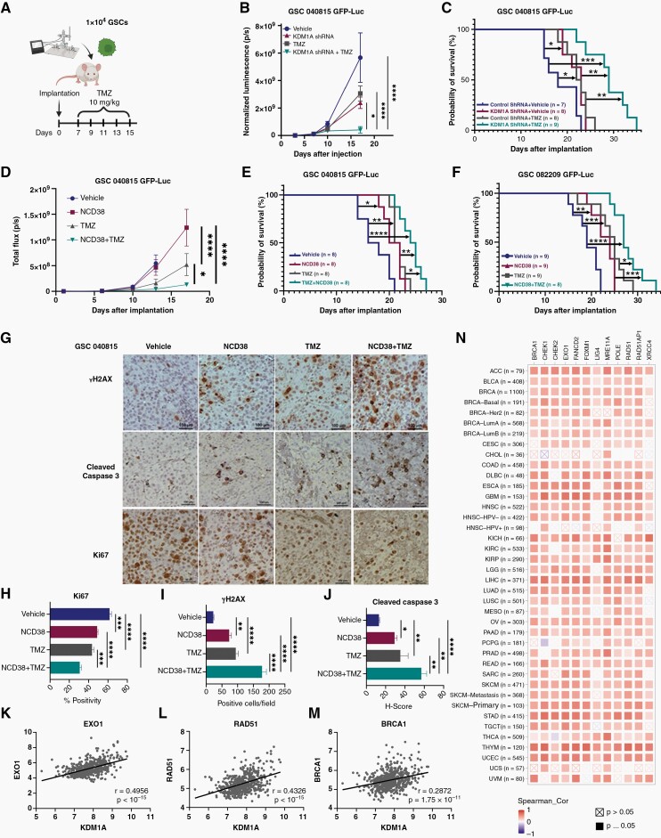
Methods: Brain bioavailability of the KDM1A inhibitor (NCD38) was established using LS-MS/MS. The effect of a combination of KDM1A knockdown or inhibition with TMZ was studied using cell viability and self-renewal assays. Mechanistic studies were conducted using CUT&Tag-seq, RNA-seq, RT-qPCR, western blot, homologous recombination (HR) and non-homologous end joining (NHEJ) reporter, immunofluorescence, and comet assays. Orthotopic murine models were used to study efficacy in vivo.

Results: TCGA analysis showed KDM1A is highly expressed in TMZ-treated GBM patients. Knockdown or knockout or inhibition of KDM1A enhanced TMZ efficacy in reducing the viability and self-renewal of GSCs. Pharmacokinetic studies established that NCD38 readily crosses the blood-brain barrier. CUT&Tag-seq studies showed that KDM1A is enriched at the promoters of DNA repair genes and RNA-seq studies confirmed that KDM1A inhibition reduced their expression. Knockdown or inhibition of KDM1A attenuated HR and NHEJ-mediated DNA repair capacity and enhanced TMZ-mediated DNA damage. A combination of KDM1A knockdown or inhibition and TMZ treatment significantly enhanced the survival of tumor-bearing mice.

Conclusions: Our results provide evidence that KDM1A inhibition sensitizes GBM to TMZ via attenuation of DNA DSB repair pathways.

Keywords: DNA repair; KDM1A/LSD1; glioblastoma; glioma stem cells; temozolomide.


Conflict of interest statement

None.

Figures

Figure 1.
Figure 1.
KDM1A knockdown sensitized Glioma stem cells (GSCs) to TMZ treatment. (A) Box plot of KDM1A was generated using a response based on overall survival at 16 months after radiation and TMZ treatment from ROC plotter datasets. (B) KDM1A expression was examined in primary versus recurrent GBM TCGA data sets. (C–D) Control and KDM1A knockdown GSCs were treated with either vehicle or TMZ for 5 days and cell viability was determined using CellTiter-Glo assays. (E–H) Neurosphere formation of control and KDM1A knockdown GSCs following TMZ treatment was determined. (I–J) Self-renewal ability of control and KDM1A knockdown GSCs following TMZ treatment was determined by extreme limiting dilution assays. *p < .05, **p < .01, ***p < .001, ****p < .0001, by t-test or one-way ANOVA.
Figure 2.
Figure 2.
NCD38 is a brain-permeable KDM1A inhibitor and synergistically enhanced TMZ-mediated reduction in cell viability and self-renewal of Glioma stem cells (GSCs). (A) Plasma pharmacokinetic parameters of NCD38 following intravenous (IV) and peroral administration in male SD rats. Blood-brain distribution of NCD38 after IV dose of 1mg/kg (n = 3) and peroral dose of 10mg/kg (n = 3) was determined by LC-MS/MS analysis. Individual plasma concentration vs. time profile of NCD38 after IV (B) and peroral (C) administration to male SD rats. (D) U251-GSCs tumor-bearing mice were treated with vehicle or different doses of NCD38. Tumor growth rate in terms of luciferase intensity was measured using Xenogen IVIS (n = 6). (E–F) GSCs were treated with NCD38 or TMZ alone and in combination for 5 days and the cell viability was determined by Cell Titer Glo assay. (G–H) GSCs were treated with NCD38 or TMZ alone or in combination and their self-renewal ability was determined by extreme limiting dilution assays. (I–L), Effect of NCD38 or TMZ alone or in combination on neurosphere formation was determined. *p < .05, **p < .01, ***p < .001, ****p < .0001, by Student’s t-test or one-way ANOVA.
Figure 3.
Figure 3.
CUT&Tag and RNA-seq analysis identified KDM1A inhibition suppress DNA repair genes expression. (A) Chipseeker annotation of KDM1A peaks to identify their chromosomal locations. (B) Functional enrichment of KDM1A binding regions in gene ontology-BP pathways through GREAT online platform. (C) GSC082209 were treated with either vehicle or NCD38 (5 μM) for 24 h and subjected to RNA sequencing. Top gene ontology terms of differentially expressed genes were shown. (D) Heat map showing downregulation of DNA damage response genes in NCD38 treated group. (E) Volcano plots comparing the gene expression levels for the NCD38 versus vehicle-treated GSCs. (F–G) GSEA testing correlation of NCD38 regulated genes with signatures of homologous recombination and non-homologous end joining gene sets. (H) Western blotting confirms the downregulation of DNA repair genes following NCD38 treatment. (I) Heat map showing downregulation of genes in the combination group.
Figure 4.
Figure 4.
KDM1A inhibition reduces both homologous recombination (HR) and non-homologous end-joining (NHEJ) repair of Glioma stem cells (GSCs). (A) schematic of qPCR-based HR reporter assay. (B–D) Effect of KDM1A-KO or NCD38 on the HR repair activity of GSCs was measured. (E) Schematic of DR-GFP reporter assay. (F) KDM1A knockdown in HeLa and U2OS cells was confirmed using western blotting. (G–J) Effect of KDM1A knockdown or NCD38 treatment (72 h) on the HR activity of U2OS and HeLa cells that stably express DR-GFP reporter plasmids was determined using flow cytometry. (K) Schematic of NHEJ reporter assay. (L–M) Effect of KDM1A knockdown on NHEJ repair activity of GSCs in terms of GFP+ cells were measured using flow cytometry. (N–O) NHEJ repair activity of GSCs was determined after 72 h treatment of NCD38. *p < .05, **p < .01, ***p < .001, ****p < . 0001, by Student’s t-test.
Figure 5.
Figure 5.
KDM1A inhibition enhances TMZ-mediated DNA damage in Glioma stem cells (GSCs). (A–B) Control or KDM1A knockdown or NCD38-treated GSCs were treated with TMZ and the level of γH2AX protein was determined using western blotting. (C–D) Control and KDM1A knockdown GSCs were treated with TMZ for 48 h and the γH2AX foci formation was measured using immunofluorescence. (E–G) GSCs were treated with either NCD38 or TMZ alone or in combination for 48 h and the γH2AX and RAD51 foci formation was determined using immunofluorescence. (H–K) GSCs treated with vehicle or NCD38 for 24 h were subjected to alkaline comet assay. *p < .05, **p < .01, ***p < .001, ****p < .0001, by one-way ANOVA.
Figure 6.
Figure 6.
KDM1A knockdown or inhibition in combination with TMZ enhances the survival of tumor-bearing mice. (A) GSCs that stably express control or KDM1A shRNA were labeled with GFP-Luc and implanted intracranially into NOD-SCID mice. (B) Mice were treated with vehicle or TMZ and tumor growth in terms of luciferase activity was monitored using Xenogen IVIS. (C) Survival plotted using Kaplan–Meier curves. (D–F) GSC040815 and GSC082209 implanted mice were treated with vehicle, NCD38, TMZ, and NCD38+TMZ. Survival was plotted using Kaplan–Meier curves. *p < .05, **p < .01, ***p < .001, ****p < .0001 by log-rank test. (G–J) Tumor tissues collected from GSC040815 groups were subjected to IHC staining for Ki67, γH2AX, and Cleaved Caspase3. *p < .05, **p < .01, ***p < .001, ****p < .0001 by one-way ANOVA. (K–M) Scatter plots from TIMER2.0 database illustrate expression level correlations between KDM1A and EXO1, RAD51, and BRCA1 in GBM TCGA data sets. Pearson correlation r and P-value computed for each dataset. (N) Heatmap from TIMER2.0 database showing the expression level correlations between KDM1A and DNA repair genes in pan-cancers.

Similar articles

Cited by

References

    1. Omuro A, DeAngelis LM. Glioblastoma and other malignant gliomas: a clinical review. JAMA. 2013;310(17):1842–1850. - PubMed
    1. Johnson DR, O’Neill BP. Glioblastoma survival in the United States before and during the temozolomide era. J Neurooncol. 2012;107(2):359–364. - PubMed
    1. Ostrom QT, Patil N, Cioffi G, Waite K, Kruchko C, Barnholtz-Sloan JS. CBTRUS statistical report: primary brain and other central nervous system tumors diagnosed in the United States in 2013-2017. Neuro Oncol. 2020;22(12 Suppl 2):iv1–iv96. - PMC - PubMed
    1. Bahadur S, Sahu AK, Baghel P, Saha S. Current promising treatment strategy for glioblastoma multiform: a review. Oncol Rev. 2019;13(2):417. - PMC - PubMed
    1. Cloughesy TF, Cavenee WK, Mischel PS. Glioblastoma: from molecular pathology to targeted treatment. Annu Rev Pathol. 2014;9:1–25. - PubMed

Publication types

MeSH terms