. 2023 Jan 18;9(3):eade4077.
doi: 10.1126/sciadv.ade4077. Epub 2023 Jan 18.

The structure of DarB in complex with RelNTD reveals nonribosomal activation of Rel stringent factors

Affiliations

The structure of DarB in complex with RelNTD reveals nonribosomal activation of Rel stringent factors

Andres Ainelo et al. Sci Adv. .

Abstract

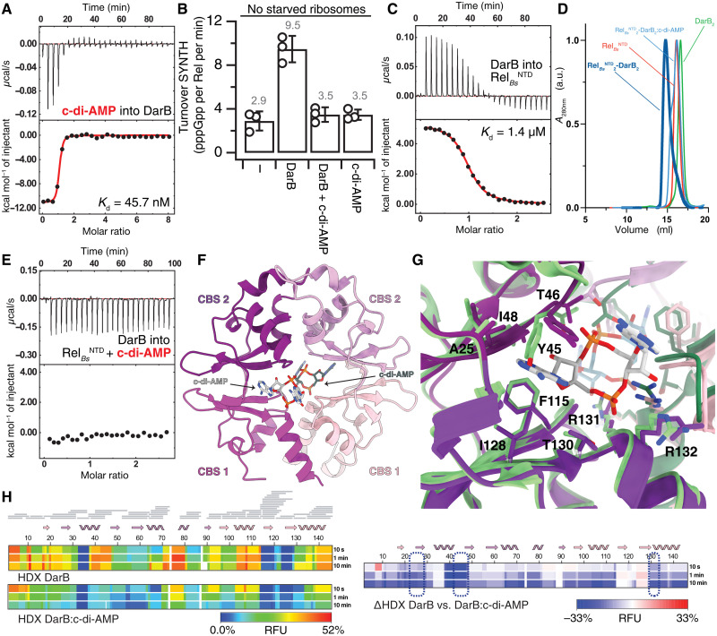
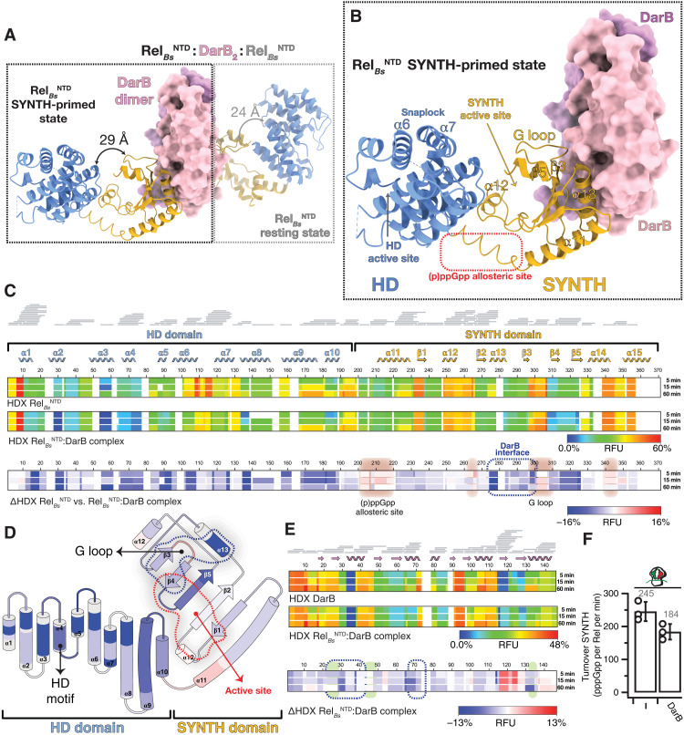
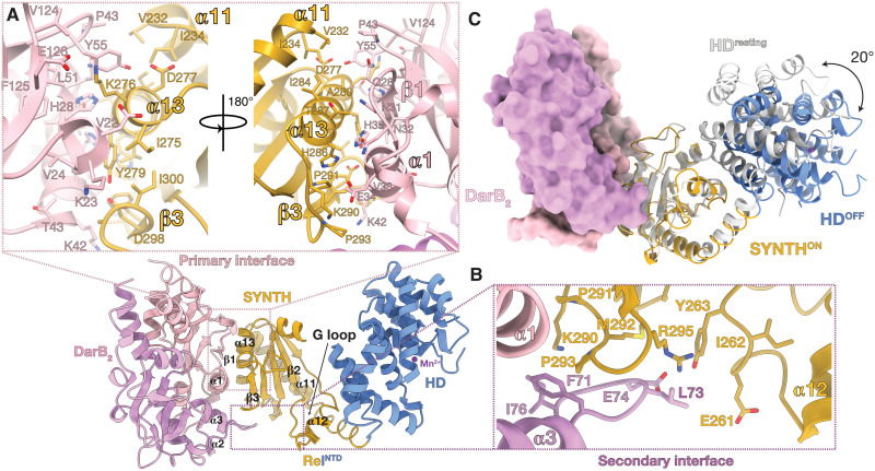
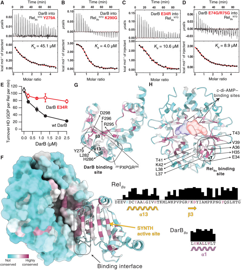
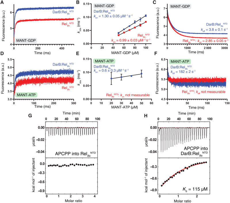
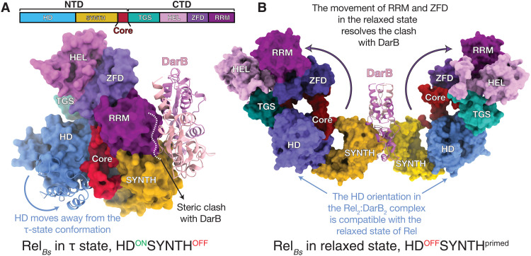
Rel stringent factors are bifunctional ribosome-associated enzymes that catalyze both synthesis and hydrolysis of the alarmones (p)ppGpp. Besides the allosteric control by starved ribosomes and (p)ppGpp, Rel is regulated by various protein factors depending on specific stress conditions, including the c-di-AMP-binding protein DarB. However, how these effector proteins control Rel remains unknown. We have determined the crystal structure of the DarB2:RelNTD2 complex, uncovering that DarB directly engages the SYNTH domain of Rel to stimulate (p)ppGpp synthesis. This association with DarB promotes a SYNTH-primed conformation of the N-terminal domain region, markedly increasing the affinity of Rel for ATP while switching off the hydrolase activity of the enzyme. Binding to c-di-AMP rigidifies DarB, imposing an entropic penalty that precludes DarB-mediated control of Rel during normal growth. Our experiments provide the basis for understanding a previously unknown mechanism of allosteric regulation of Rel stringent factors independent of amino acid starvation.


Figures

Fig. 1.
Fig. 1.. DarB:c-di-AMP conformational interplay.
(A) Binding of c-di-AMP to DarB monitored by ITC. (B) Effect of c-di-AMP on DarB and its Rel-dependent activation of pppGpp synthesis. RelBs (250 nM) was incubated with ATP and GTP, in the absence or presence of saturating DarB (10-fold excess), c-di-AMP–saturated DarB, or c-di-AMP. (C) Binding of Rel to DarB monitored by ITC. (D) Analytical size exclusion chromatography of the RelBsNTD2-DarB2 complex (in dark blue), RelNTD (in red), DarB (in green), and the RelBsNTD2-DarB2 complex incubated with c-di-AMP (in light blue). The experiment confirms that the presence of c-di-AMP is sufficient to disrupt the complex. (E) Titration of DarB into c-di-AMP + Rel monitored by ITC. (F) Structure of DarB bound to c-di-AMP. Individual CBS domains of the tandem are labeled. Only one of the two c-di-AMP molecules is shown in the figure for clarity. (G) Structural details of the c-di-AMP–binding sites of DarB in the c-di-AMP–bound complex are shown in violet and of apo-DarB in green. Residues involved in the dinucleotide coordination are labeled. (H) Heatmaps representing the HDX of DarB (top) and DarB:c-di-AMP complex (center) and the ΔHDX (bottom). Residues involved in the binding to c-di-AMP are outlined by a dashed blue line. RFU, relative fractional uptake; a.u., arbitrary units; A280nm, absorbance at 280 nm.
Fig. 2.
Fig. 2.. Structure of the RelBsNTD2:DarB heterotetrameric complex.
(A) Crystal structure of DarB2:RelBsNTD2 heterotetrameric complex with the disc-shaped DarB dimer located at the center of the complex (colored in pink and lilac) and the two RelBsNTD bound at both sides of the DarB dimer. For each RelBsNTD molecule of the complex, the HD domain is colored in light blue, and the SYNTH domain is in yellow. In the nonsymmetrical hetero-complex, the RelBsNTD in the SYNTH-primed state (left, outlined with a black dashed line) is observed in a more open and less structured conformation than the resting RelBsNTD molecule (right, outlined with a light gray dashed line). The relative HD-to-SYNTH distance in each RelBsNTD monomer is indicated. (B) Details of RelBsNTD in the SYNTH-primed state highlighting key structural elements. (C) Heatmaps showing the HDX signal kinetics of RelBsNTD (top) and RelBsNTD as part of the DarB2:RelBsNTD2 complex (center) and the ΔHDX (bottom). Both catalytic domain of RelBs and all the secondary structural elements of the NTD are shown in the figure. Residues involved in the binding interface with DarB are outlined by a dashed blue line, and the regions with increased deuterium uptake, which include the G loop that becomes exposed upon binding to DarB and the alarmone allosteric site, are shaded in red. (D) Topology representation of RelBsNTD colored as a function of the ΔHDX. (E) Heatmaps representing the HDX of DarB (top) and DarB as part of the DarB2:RelBsNTD2 complex (center) and the ΔHDX (bottom). Residues involved in the binding interface with RelBsNTD are indicated by a dashed blue line, and those involved in the binding to c-di-AMP are shaded in green. (F) Effect of DarB on the SYNTH activity of RelBs in the presence or absence of “starved” ribosomes.
Fig. 3.
Fig. 3.. DarB interacts with RelBs via the SYNTH domain.
(A) RelBs:DarB primary interaction interface involving α13 and β3 from RelBs and α1, α2, β1, and β2 from DarB. (B) RelBs:DarB secondary interaction interface formed between the α13/β3 connecting loop from RelBs adjacent to the SYNTH active site and the α2/α3 loop of DarB. (C) Superposition of RelBsNTD in the SYNTH-primed state (colored as per Fig. 2C) onto RelBsNTD in the resting state shown in light gray (PDB ID 6YXA). The 20° movement of the HD domain away from the SYNTH domain observed in RelBsNTD in complex with DarB compared with the resting RelBs (PDB ID 6YXA) is indicated with a black arrow.
Fig. 4.
Fig. 4.. Biophysical and biochemical interrogation of the RelBs:DarB binding interface.
Effect of the Y279A (A) and K290G (B) substitutions in RelBsNTD to the interaction with DarB, monitored by ITC. Effect of the substitutions to the primary E34R (C) or secondary E74G/R75G (D) DarB interfaces, to the binding to RelBsNTD, monitored by ITC. (E) Effect of an E34R substitution on DarB to the activity of DarB monitored as a function of the hydrolase activity of RelBs. (F) DarB-SYNTH interface color-coded by the conservation score of each amino acid calculated by ConSurf. Residues involved in the primary interface are shown in the conservation bar plots to the right and colored on the basis of their individual conservation profile. The strictly conserved Y of the G loop of Rel is shown in italic. Structural elements of SYNTH (G) and DarB (H) colored as in (F) underline the strong conservation of the binding interface. The contact regions between both proteins are outlined by dashed black lines with the residues directly involved in the primary binding interface and the PXPGR motif highlighted in (G) and (H) and the location of the c-di-AMP shown as a surface in (H).
Fig. 5.
Fig. 5.. Kinetics and thermodynamics of nucleotide binding to RelBs in the presence and absence of DarB.
(A) Kinetics of MANT-GDP binding to RelBsNTD (in red) and DarB:RelBsNTD (in blue) monitored by stopped flow. The interaction was measured by FRET excitation of MANT fluorescence upon mixing 10 μM protein with increasing concentrations of MANT-GDP (B). (C) Kinetics of MANT-GDP dissociation from RelBsNTD (in red) and DarB:RelBsNTD (in blue). (D) Kinetics of MANT-ATP binding to RelBsNTD (in red) and DarB:RelBsNTD (in blue). The interaction was measured as in (A) by FRET excitation of MANT-ATP fluorescence upon mixing 10 μM protein with increasing concentrations of MANT-ATP (E). (F) Kinetics of MANT-ATP dissociation from RelBsNTD (in red) and DarB:RelBsNTD (in blue). In both cases, the dissociation was monitored upon rapid mixing with an excess (2 mM) of unlabeled GDP or ATP. Binding of APCPP to RelBsNTD (G) and DarB:RelBsNTD (H) monitored by ITC.
Fig. 6.
Fig. 6.. DarB as a conformational selector of RelBs.
(A) Surface representation of an idealized (unrealistic) model of full-length RelBs in the hydrolase-compatible τ state superimposed on the crystal structure of the DarB:RelBsNTD complex (used only to illustrate why access to the τ state is sterically blocked). Comparison of the two structures shows that the closed HD-active τ state is not compatible with the binding of DarB due to the sterical clash of DarB with the RRM and ZFD domains and the closing of the SYNTH active site by HD. (B) Model of the full-length RelBs:DarB heterotetrameric complex in the HDOFF SYNTHprimed relaxed state. The rearrangement of RRM and ZFD domains allows for binding of DarB that, in turn, precludes the recoil of the CTD and corresponding inactivation of the HD. In both (A) and (B), Rel models were based on the structures of A. baumannii SpoT in the τ and relaxed states (10).

References

    1. S. E. Irving, N. R. Choudhury, R. M. Corrigan, The stringent response and physiological roles of (pp)pGpp in bacteria. Nat. Rev. Microbiol. 19, 256–271 (2021). - PubMed
    1. G. Bange, D. E. Brodersen, A. Liuzzi, W. Steinchen, Two P or not two P: Understanding regulation by the bacterial second messengers (p)ppGpp. Annu. Rev. Microbiol. 75, 383–406 (2021). - PubMed
    1. B. W. Anderson, D. K. Fung, J. D. Wang, Regulatory themes and variations by the stress-signaling nucleotide alarmones (p)ppGpp in bacteria. Annu. Rev. Genet. 55, 115–133 (2021). - PubMed
    1. G. C. Atkinson, T. Tenson, V. Hauryliuk, The RelA/SpoT homolog (RSH) superfamily: Distribution and functional evolution of ppGpp synthetases and hydrolases across the tree of life. PLOS ONE 6, e23479 (2011). - PMC - PubMed
    1. G. Mittenhuber, Comparative genomics and evolution of genes encoding bacterial (p)ppGpp synthetases/hydrolases (the Rel, RelA and SpoT proteins). J. Mol. Microbiol. Biotechnol. 3, 585–600 (2001). - PubMed