. 2023 Mar 1;43(9):1492-1508.
doi: 10.1523/JNEUROSCI.1276-22.2023. Epub 2023 Jan 18.

AAV Vector Mediated Delivery of NG2 Function Neutralizing Antibody and Neurotrophin NT-3 Improves Synaptic Transmission, Locomotion, and Urinary Tract Function after Spinal Cord Contusion Injury in Adult Rats

Affiliations

AAV Vector Mediated Delivery of NG2 Function Neutralizing Antibody and Neurotrophin NT-3 Improves Synaptic Transmission, Locomotion, and Urinary Tract Function after Spinal Cord Contusion Injury in Adult Rats

Hayk A Petrosyan et al. J Neurosci. .

Abstract

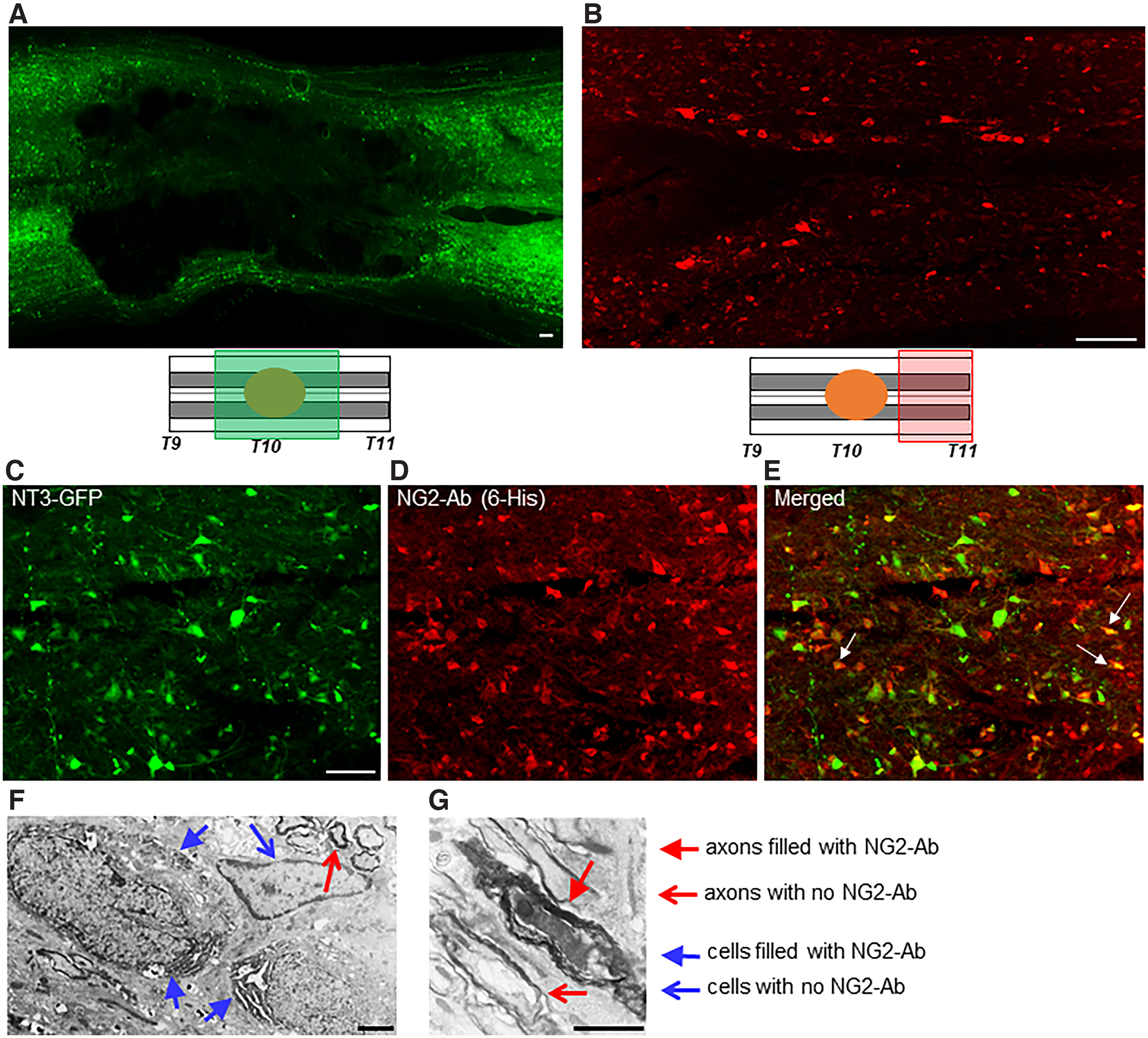
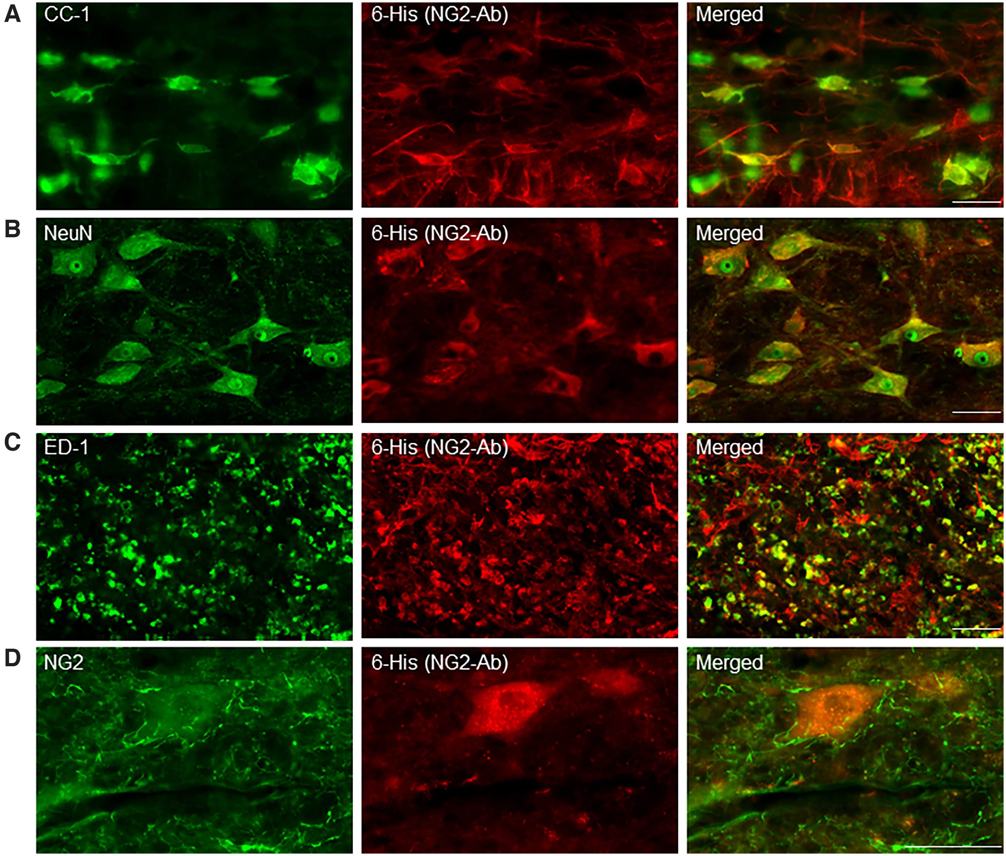
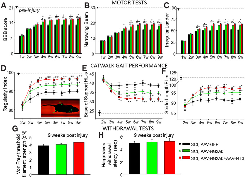
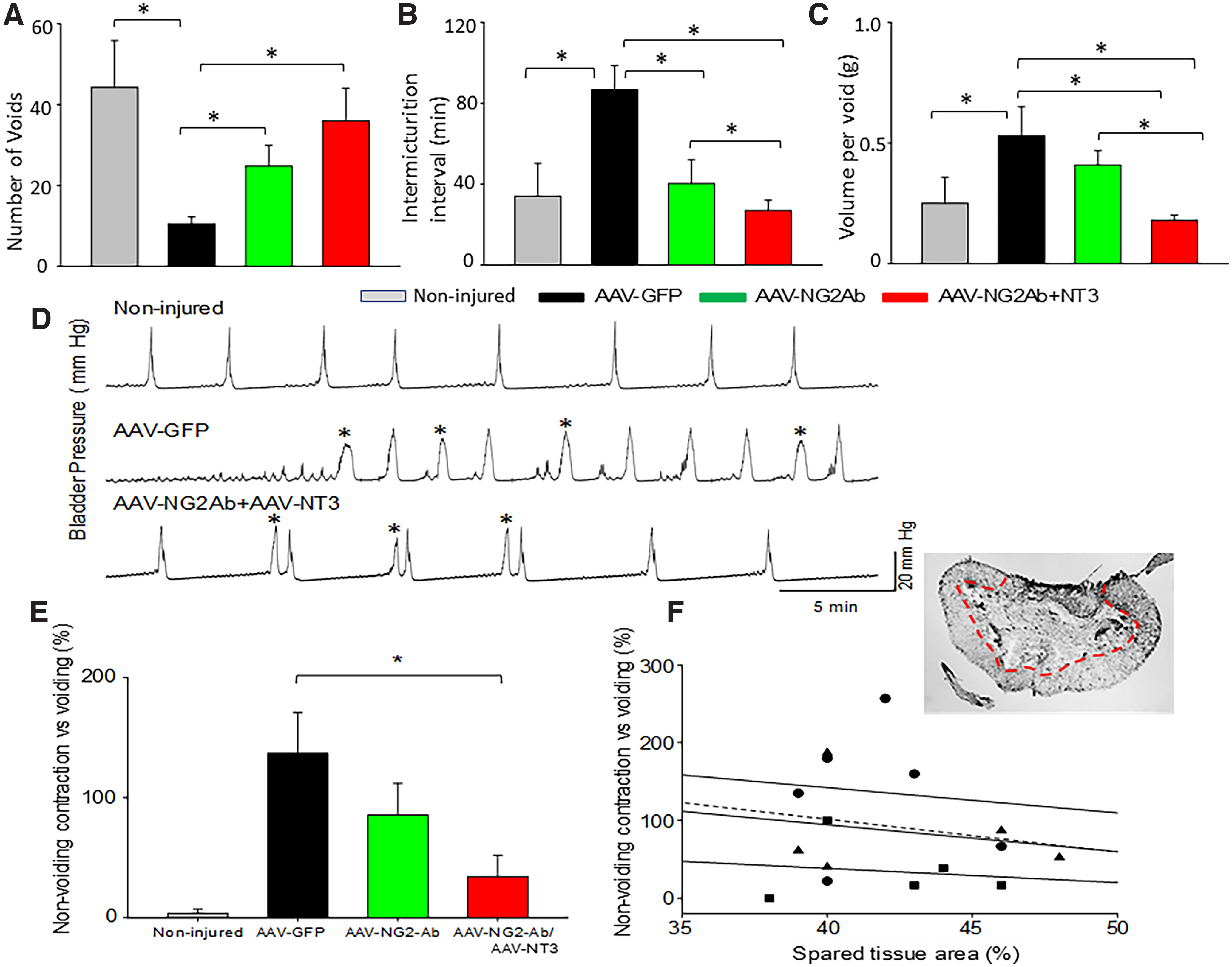
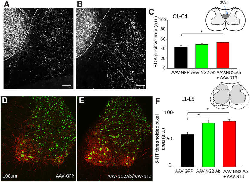
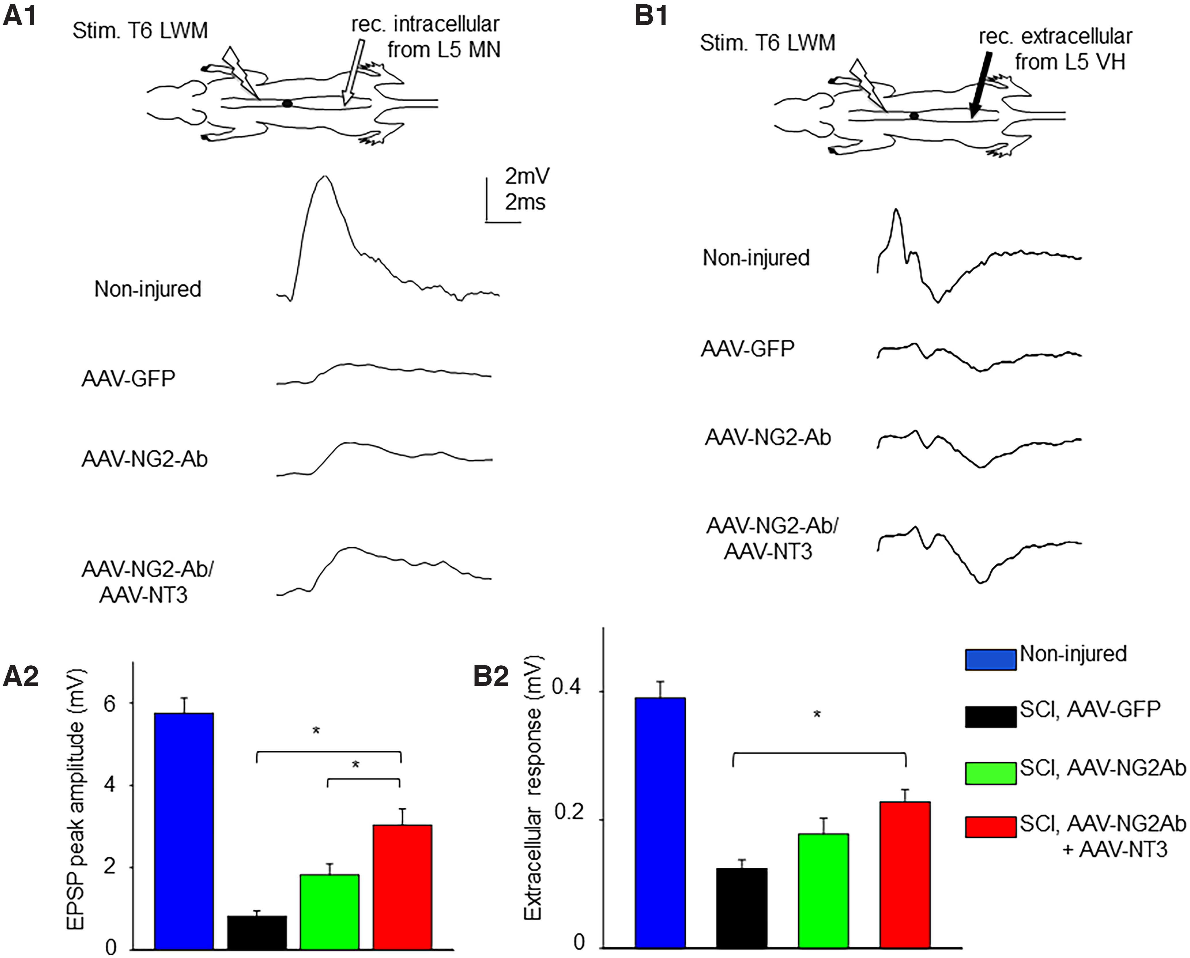
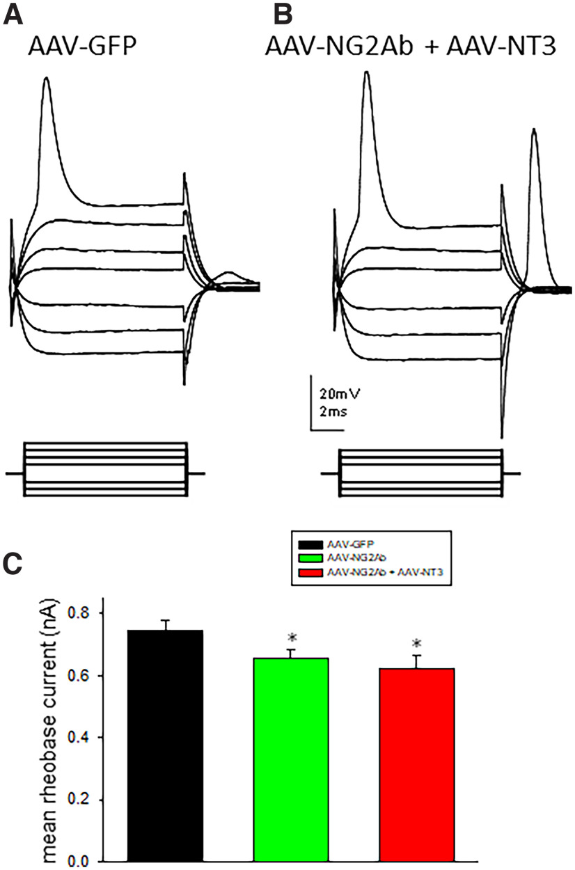
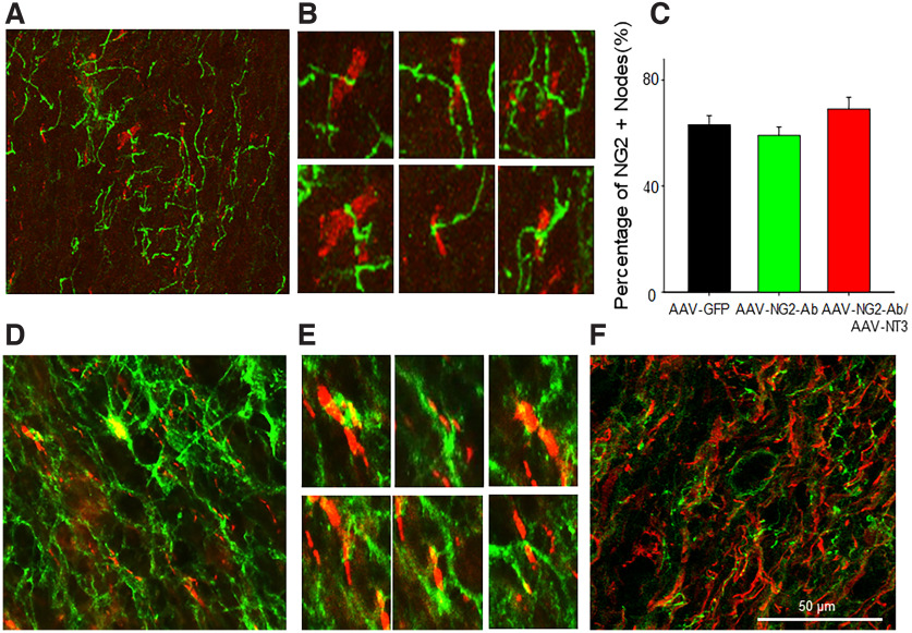
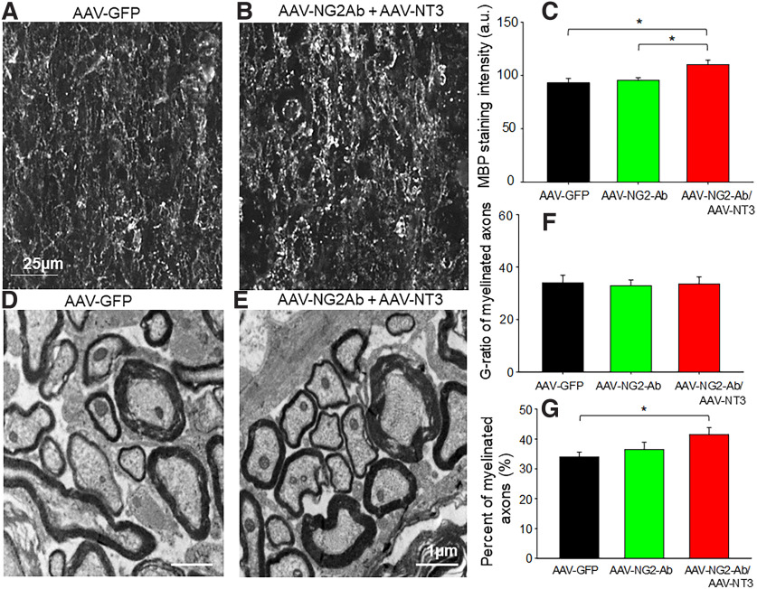
NG2 is a structurally unique transmembrane chondroitin sulfate proteoglycan (CSPG). Its role in damaged spinal cord is dual. NG2 is considered one of key inhibitory factors restricting axonal growth following spinal injury. Additionally, we have recently detected its novel function as a blocker of axonal conduction. Some studies, however, indicate the importance of NG2 presence in the formation of synaptic contacts. We hypothesized that the optimal treatment would be neutralization of inhibitory functions of NG2 without its physical removal. Acute intraspinal injections of anti-NG2 monoclonal antibodies reportedly prevented an acute block of axonal conduction by exogenous NG2. For prolonged delivery of NG2 function neutralizing antibody, we have developed a novel gene therapy: adeno-associated vector (AAV) construct expressing recombinant single-chain variable fragment anti-NG2 antibody (AAV-NG2Ab). We examined effects of AAV-NG2Ab alone or in combination with neurotrophin NT-3 in adult female rats with thoracic T10 contusion injuries. A battery of behavioral tests was used to evaluate locomotor function. In vivo single-cell electrophysiology was used to evaluate synaptic transmission. Lower urinary tract function was assessed during the survival period using metabolic chambers. Terminal cystometry, with acquisition of external urethral sphincter activity and bladder pressure, was used to evaluate bladder function. Both the AAV-NG2Ab and AAV-NG2Ab combined with AAV-NT3 treatment groups demonstrated significant improvements in transmission, locomotion, and bladder function compared with the control (AAV-GFP) group. These functional improvements associated with improved remyelination and plasticity of 5-HT fibers. The best results were observed in the group that received combinational AAV-NG2Ab+AAV-NT3 treatment.SIGNIFICANCE STATEMENT We recently demonstrated beneficial, but transient, effects of neutralization of the NG2 proteoglycan using monoclonal antibodies delivered intrathecally via osmotic mini-pumps after spinal cord injury. Currently, we have developed a novel gene therapy tool for prolonged and clinically relevant delivery of a recombinant single-chain variable fragment anti-NG2 antibody: AAV-rh10 serotype expressing scFv-NG2 (AAV-NG2Ab). Here, we examined effects of AAV-NG2Ab combined with transgene delivery of Neurotrophin-3 (AAV-NT3) in adult rats with thoracic contusion injuries. The AAV-NG2Ab and AAV-NG2Ab+AAV-NT3 treatment groups demonstrated significant improvements of locomotor function and lower urinary tract function. Beneficial effects of this novel gene therapy on locomotion and bladder function associated with improved transmission to motoneurons and plasticity of axons in damaged spinal cord.

Keywords: Bladder Function; Locomotion; NG2; Proteoglycan; SCI; Transmission.


Conflict of interest statement

The authors declare no competing financial interests.

Figures

Figure 1.
Figure 1.
Robust transduction of spinal cord tissue following intraspinal injections of AAV-NT3 and AAV-NG2Ab. A, B, Horizontal sections of T9-T11 spinal segments. GFP signal was used to visualize AAV-mediated delivery of NT3 coexpressing GFP (A, green) and immunostaining with anti-6-His to detect 6-His-tagged NG2-Ab (B, red). Diagrams represent the area where high-power images were taken. C–E, High-magnification images of the gray matter close to injury epicenter demonstrating GFP signal (C) and anti-6-His-stained sections (D) represent many transduced cells with NT3-GFP and NG2Ab. E, Merged image represents colocalization of NT3 and NG2-Ab in many cells (arrows). F, G, Electron microscope images confirm expression of NG2-Ab in cells (F) and axons (G) in AAV-NG2Ab-injected rats. Arrowheads point to cells and fiber with no 6-His signal. Filled arrowheads point to cells and a fiber transduced with AAV-NG2Ab. Scale bars, 2 µm.
Figure 2.
Figure 2.
Transduction of different types of cells following intraspinal injections of AAV-NG2Ab in injured spinal cord. Images from T9-T11 horizontal sections of T10 contused spinal cord taken in the LWM caudal to injury at depth corresponding to VLF. Double immunostaining with anti-6-His (red, to detect AAV-mediated delivery of NG2Ab) and oligodendrocyte marker CC-1 (A), neuronal marker NeuN (B), and macrophage marker ED-1 (C). D, Double immunostaining with NG2 marker (green) and anti-6-His (red) to visualize AAV-mediated delivery of NG2Ab in the vicinity of NG2-positive processes. Merged images represent colocalization of anti-6-His with CC1, NeuN, ED-1, and NG2 signals, indicating AAV-NG2Ab-mediated transduction of oligodendrocytes, neurons, macrophages/microglia, and NG2-positive processes, respectively.
Figure 3.
Figure 3.
Treatment with AAV-NG2Ab and AAV-NT3 improved locomotor function after contusion SCI assessed by BBB, Narrowing Beam, Irregular Ladder, and CatWalk gait analysis. A–F, Summary of results demonstrating significantly better locomotor performance evident in (A) BBB score, (B) longer distance passed in Narrowing Beam, (C) fewer number of slips in Irregular Ladder, (D) higher regularity index, (E) smaller base of support, and (F) longer stride length. Dotted line indicates pre-injury scoring for each motor test. Data are mean ± SEM (n = 12 rats per group). Pre-injury scoring for each motor test is indicated with the dotted line; data presented as mean ± SEM (n = 12 rats per group). *p < 0.05 statistical difference between control (AAV-GFP) group and treatment (AAV-NG2Ab alone, or AAV-NG2Ab + AAV-NT3) groups; **p < 0.05 statistical difference between AAV-NG2Ab alone and AAV-NG2Ab + AAV-NT3 groups (D, E). Withdrawal reflex, measured as (G) von Frey threshold filament strength and (H) Hargreaves withdrawal latency, was not significantly affected by these treatments.
Figure 4.
Figure 4.
Treatment with AAV-NG2Ab and AAV-NT3 improved bladder function after contusion SCI. A–C, Bladder function was assessed during the survival period using metabolic chambers to collect and quantify overnight urine production. Summary of results demonstrating improved bladder function evident in significant (A) increased number of voids, (B) decreased intermicturition intervals, and (C) decreased volume per void in treated rats compared with rats that received control treatment (n = 8 rats per group). D, E, Terminal cystometry electrophysiology recordings performed after 6 weeks after injury, with simultaneous acquisition of EUS EMG activity. D, Representative traces of bladder pressure recordings demonstrating less nonvoiding contractions (asterisks) in treated rats compared with control treated rats. E, Summary of results demonstrating significantly decreased number of nonvoiding contractions, in AAV-NG2Ab- and AAV-NT3-treated rats compared with control treated animals (n = 5 randomly selected rats per group). F, Correlation between the spared tissue at the injury epicenter (outlined by dotted line on the image of the cryosection) and changes in voiding between groups (circles represent AAV-GFP; triangles represent AAV-NG2Ab; squares represent AAV-NG2Ab+AAV-NT3); analysis revealed an absence of correlation. Dotted line indicates correlation analysis for the combined data from all groups between spared tissue and nonvoiding contractions. Data are mean ± SEM. *p < 0.05.
Figure 5.
Figure 5.
Treatment with AAV-NG2-Ab and AAV-NT3 following thoracic contusion promotes anatomic plasticity in damaged spinal cord. Treatment resulted in sprouting of corticospinal tract fibers in cervical spinal cord (A–C) and plasticity of serotonergic fibers in lumbar spinal cord (D–F). A, B, Representative images of BDA-labeled corticospinal tract fibers of animals from AAV-GFP control group and AAV-NG2-Ab+AAV-NT3 group, respectively. C, Summary of results demonstrating significantly larger BDA-positive area in gray matter of AAV-NG2AB+AAV-NT3 group in C1-C4 levels compared with AAV-GFP or AAV-NG2Ab only groups. D, E, Representative images of 5-HT (red, to identify 5-HT-positive fibers) and Neu-N (green, to identify neurons) immunoreactivity in L5 segment (corresponding to position of recording electrodes in electrophysiology experiments) in animals from AAV-GFP control group and AAV-NG2-Ab+AAV-NT3 group, respectively. F, Summary of quantitative analyses of thresholded pixel area of 5-HT-positive fibers revealed a significant increase in 5-HT-positive fibers in intermedial and ventral (outlined by dotted line) gray matter of AAV-NG2Ab and AAV-NG2AB+AAV-NT3 group in L1-L5 levels. Data are mean ± SEM (n = 5 rats/group). *p < 0.05.
Figure 6.
Figure 6.
AAV-NG2Ab combined with AAV-NT3 improved transmission to lumbar motoneurons in chronically contused rats. A, B, Representative traces of intracellular (A1) and extracellular (B1) responses (10 averaged consecutive responses) recorded from L5 ventral horn and evoked by electric stimulation of LWM at T6 demonstrating larger amplitude in rats from AAV-NG2Ab group and AAV-NG2AB+AAV-NT3 group compared with control treated rats. A2, Summary of results demonstrating significant facilitation of intracellular responses recorded from L5 ventral horn neurons in AAV-NG2Ab-treated and AAV-NG2Ab+AAV-NT-3-treated rats compared with control AAV-GFP-treated rats. B2, Summary of results demonstrating similar significant facilitation of extracellular responses in AAV-NG2Ab-treated and AAV-NG2Ab+AAV-NT-3-treated rats compared with control AAV-GFP-treated rats. Diagram represents position of stimulation and recording electrodes. Data are mean ± SEM. *p < 0.05. In the same graph (A2), we have compared effects of AAV-NG2Ab and AAV-NT3 on transmission with the results of our previous study where we have examined the effects of intraspinal injections of ChABC (adopted from Hunanyan et al., 2013).
Figure 7.
Figure 7.
AAV-NG2Ab combined with AAV-NT3 treatment improved axonal excitability. A, B, Representative traces of intra-axonal recording performed from individual axons in L1-L2 LWM, recorded from AAV-GFP-anesthetized (A) and AAV-NG2AB+AAV-NT3-anesthetized (B) rats, respectively, demonstrating increased excitability (lower rheobase current) and absence of changes in the input resistance of axons in rats from AAV-NG2AB+AAV-NT3 group compared with control contused treated with AAV-GFP group rats. C, Summary of results demonstrating significantly lower rheobase current in both AAV-NG2Ab (n = 26 axons) and AAV-NG2AB+AAV-NT3 (n = 22 axons) groups compared with AAV-GFP group (n = 24 axons). Data are mean ± SEM. *p < 0.05.
Figure 8.
Figure 8.
Horizontal section of T9-T11 spinal cord double-immunostained with CASPR and NG2 (A,B) and CASPR and 6-His for NG2-Ab (D,E) to demonstrate localization of NG2-positive processes and AAV-mediated delivery of NG2-Ab in relation to nodes of Ranvier, in the LWM caudal to injury at the depth corresponding to VLF. High-power confocal images selected from the low-power image (B) represent (A) individual nodes (red) and NG2-positive processes (green). C, Summary of results demonstrating no significant difference in the percentage of nodes containing NG2-positive processes between groups. D, E, Alternating longitudinal sections representing double immunostaining with CASP (red) and 6-His (green). Low- and high-power confocal images, respectively, demonstrate colocalization of AAV-NG2Ab-positive processes (green) in close association with the nodes (red). F, Images from alternating sections: double immunostaining of NG2 (green) and AAV-mediated delivery of NG2-Ab (red; 6-HIS traced), along LWM axons caudal to contusion. Data are mean ± SEM.
Figure 9.
Figure 9.
Effects of treatment with AAV-NG2Ab and AAV-NT3 on myelination in chronically injured rats. A, B, Representative images of spinal cord sections immunostatined with MBP taken from AAV-GFP-treated (A) and AAV-NG2AB+AAV-NT3-treated (B) animals, respectively. C, Summary of results demonstrating significantly higher MBP staining in AAV-NG2Ab+AAV-NT3-treated animals compared with AAV-NG2Ab only and AAV-GFP groups. D, E, Representative electron microscope images of spinal cords from AAV-GFP-treated (D) and AAV-NG2Ab+AAV-NT3-treated (E) rats, respectively. F, Summary of results demonstrating no significant difference in G-ratio of fibers between groups. G, Summary of results demonstrating significantly higher percentage of myelinated axons in AAV-NG2AB+AAV-NT3-treated rats compared with other groups. Data are mean ± SEM. *p < 0.05.

Similar articles

Cited by

References

    1. Alilain WJ, Horn KP, Hu H, Dick TE, Silver J (2011) Functional regeneration of respiratory pathways after spinal cord injury. Nature 475:196–200. - PMC - PubMed
    1. Andrews EM, Richards RJ, Yin FQ, Viapiano MS, Jakeman LB (2012) Alterations in chondroitin sulfate proteoglycan expression occur both at and far from the site of spinal contusion injury. Exp Neurol 235:174–187. - PMC - PubMed
    1. Arvanian VL, Schnell L, Lou L, Golshani R, Hunanyan A, Ghosh A, Pearse DD, Robinson JK, Schwab ME, Fawcett JW, Mendell LM (2009) Chronic spinal hemisection in rats induces a progressive decline in transmission in uninjured fibers to motoneurons. Exp Neurol 216:471–480. 10.1016/j.expneurol.2009.01.004 - DOI - PMC - PubMed
    1. Arvanian V (2013) Role of neurotrophins in spinal plasticity and locomotion. Curr Pharm Des 19:4509–4516. - PubMed
    1. Arvanian VL, Petrosyan HA, Alessi A, Phagu N, Levine J, Collins WF (2016) Viral vector mediated neutralization of NG2 proteoglycan (AAV-NG2Ab) combined with delivery of neurotrophin NT-3 (AAV-NT3) improves transmission, locomotion and urinary tract function after incomplete spinal cord injury in adult rats. Soc Neurosci Abstract 612:

Publication types