. 2023 Jan 19;14(1):40.
doi: 10.1038/s41419-022-05527-9.

USP9X-mediated NRP1 deubiquitination promotes liver fibrosis by activating hepatic stellate cells

Affiliations

USP9X-mediated NRP1 deubiquitination promotes liver fibrosis by activating hepatic stellate cells

Jinqiu Zhao et al. Cell Death Dis. .

Abstract

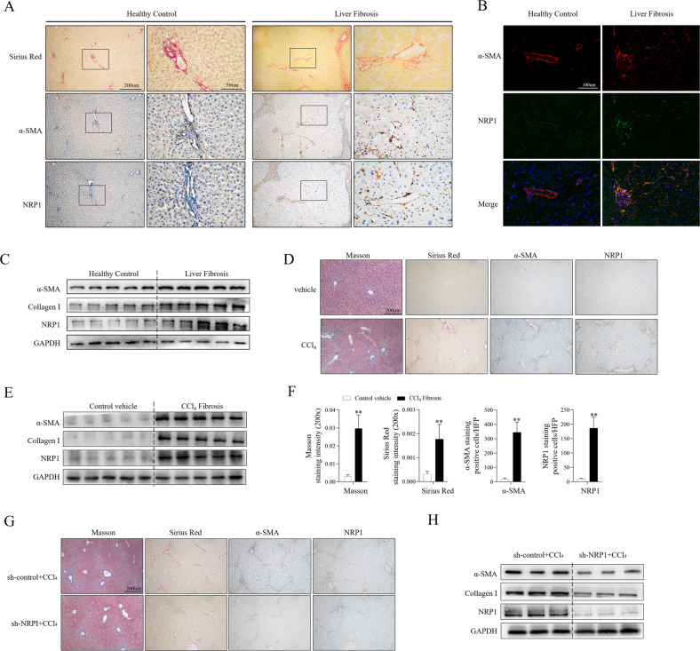
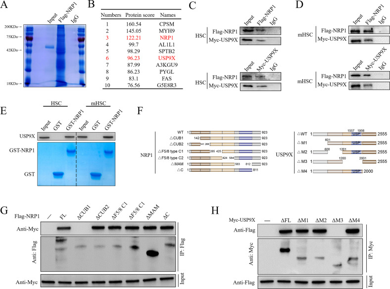
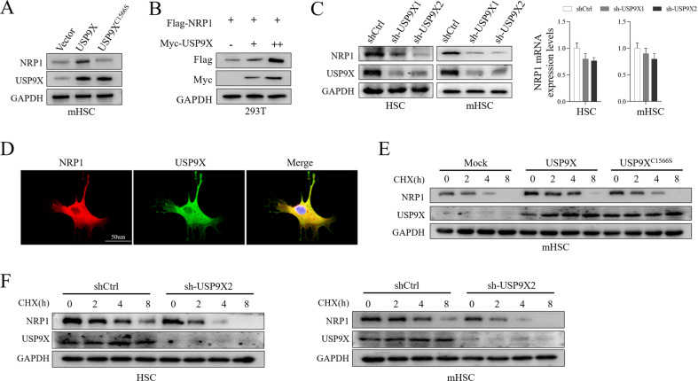
Liver fibrosis is a complex fibrotic process that develops early in the course of cirrhosis and is caused by chronic liver damage. The activation of hepatic stellate cells is primarily responsible for the fibrosis process. Studies show that NRP1 influences HSC motility and migration. However, whether NRP1 regulates HSC activation remains unknown. C57BL/6 male mice (6-8 weeks old) were intraperitoneally injected with 10% CCl4 in olive oil (5 μl/g body weight) every three days for four weeks to create an animal model of liver fibrosis. Control mice received olive oil (5 μl/g body weight). Different assays such as immunohistochemistry, immunostaining, Western blotting, qRT-PCR, immunoprecipitation, immunoprecipitation, and GST pull-down assays, and in vivo and in vitro ubiquitination assays were conducted. We found that NRP1 expression was significantly elevated both in mouse and human fibrotic livers, mainly in activated HSCs at the fibrotic foci. NRP1 promoted HSC activation via the cytokine TGF-β1, VEGFA, and PDGF-BB. Moreover, USP9X was found to be a critical deubiquitinating enzyme for the stability and high activity of NRP1 and NRP1 deubiquitination mediated by USP9X enhanced HSC activation and liver fibrosis. NRP1 deubiquitination mediated by USP9X enhances HSC activation, implying that targeting NRP1 or USP9X potentiates novel options in the treatment of liver fibrosis.


Conflict of interest statement

The authors declare no competing interests.

Figures

Fig. 1
Fig. 1. Elevated expression of NRP1 in fibrotic human and mouse liver tissues.
A Immunohistochemical staining of Sirus Red, α-SMA and NRP1 in liver sections of healthy control and fibrotic human liver. B Immunofluorescence staining of α-SMA (red) and NRP1 (green) in liver sections as in (A). C WB analysis of α-SMA, collagen I and NRP1 in fibrotic and healthy human liver tissues. DF Mice were treated with olive oil or carbon tetrachloride (CCl4) solution (5 μl/g body weight) via intraperitoneal injection three times a week for four weeks. Liver fibrosis was examined by Masson blue and Sirus Red staining (D). α-SMA, collagen I and NRP1 were evaluated by WB (E). Each group’s Masson positive and Sirius red staining region, and the number of NRP1+ and α-SMA+ cells in the given group was quantitatively evaluated (F). G, H CCl4 Mice were treated with adenovirus containing NRP1 interference sequence (sh-NRP1) or control sequence (sh-control) via vein injection. IHC (G) and WB (H) showed reduced liver fibrosis and decreased NRP1 and α-SMA expression in CCl4-treated mice after transfection with sh-NRP1. The graphs are represented as mean ± SD of at least three independent experiments. *P < 0.05; **P < 0.01.
Fig. 2
Fig. 2. NRP1 was required for the mouse primary HSCs’ activation.
A, B. HSCs isolated from mice were cultured in vitro for 24 h, 72 h, 120 h, or 168 h. A qRT-PCR analysis of α-SMA, collagen I and NRP1 in activated primary HSCs. B WB results of α-SMA, collagen I and NRP1 in activated primary HSCs. CG Primary HSCs were transfected with NRP1 shRNA (sh-NRP1) or control sequence (sh-control) for 72 h, following 48 h of in vitro culture. The expression of NRP1, α-SMA, and collagen I was evaluated by qRT-qPCR (C) and WB (D). E Apoptosis and the cycle of primary HSC cells were detected by flow cytometry, after NRP1 shRNA or control shRNA transfection. The percentages of cell numbers in G0/G1, S and G2/M stages were 32.82%, 48.91% and 18.26% (sh-control), 27.40%, 66.20% and 6.40% (sh-NRP1), respectively. F The co-expression of α-SMA (green) and NRP1 (red) in HSCs was observed by immunofluorescence. G Primary HSCs were treated with PDGF-BB (20 ng/mL), TGF-β1 (5 ng/mL) and VEGFA (5 ng/mL) to accelerate activation, while transfected with NRP1 shRNA or control shRNA. The expression levels of collagen I and α-SMA were assessed by WB. The obtained data have been represented as mean ± SD. *P < 0.05; **P < 0.01.
Fig. 3
Fig. 3. Direct interaction between USP9X and NRP1.
A Mouse primary HSCs were extracted and transfected with Flag-NRP1 for 24 h, and then CO-IP (with Flag beads) was used to evaluate cell lysates, followed by IB with antibodies against Flag. Post-staining with coomassie blue, proteins were collected, followed by identification through LC-MS/MS. B In mouse primary HSCs, LC-MS/MS analysis revealed NRP1 interacting proteins. Each peptide’s name and the number of peptides are mentioned. C IP was conducted to evaluate cell lysates from primary mouse HSCs using antibodies against USP9X and NRP1, followed by IB evaluation. As an isotype control, IgG was utilized. D IP was conducted to evaluate cell lysates from murine HSCs using antibodies against USP9X and NRP1, followed by IB evaluation. As an isotype control, IgG was utilized. E GST or GST-NRP1 coupled glutathione-Sepharose beads were used to incubate cell lysates from primary mouse HSCs or murine HSCs. Proteins that remained on Sepharose were then evaluated by IB with the appropriate antibodies. SDS-PAGE and Coomassie blue staining were used to examine recombinant GST-NRP1 isolated from bacteria. F NRP1, USP9X, and their several deletion mutants are depicted schematically. G The cotransfection of HEK293T cells was carried out with Myc-USP9X and Flag-NRP1 or its deletion mutants for 24 h, and then cell lysates were evaluated through IP with Flag beads followed by IB with antibodies against Flag and Myc. H The cotransfection of HEK293T cells was carried out with Flag-NRP1 and Myc-USP9X or its deletion mutants for 24 h, and then the evaluation of cell lysates was carried out through IP with His beads followed by IB with antibodies against Flag and Myc.
Fig. 4
Fig. 4. USP9X was primarily responsible for NRP1 stability.
A Murine HSCs were transfected with a plasmid (control, USP9X WT or USP9X C1566S mutant), and cell lysates were evaluated through IB with antibody against NRP1. B The transfection of HEK293T cells was carried out with a high level of Myc-USP9X for 24 h, followed by evaluation of cell lysates through IB with antibody against Flag. C Primary mouse HSCs and murine HSCs were transfected with 2 independent USP9X shRNA for 72 h, and then USP9X and NRP1 were analyzed using WB and qRT-PCR. D Immunofluorescence detection of USP9X and NRP1 cells expressing location. E Transfection of murine HSCs was carried out with adenovirus (control, USP9X WT or USP9X C1566S mutant), followed by exposure to CHX (100 μg/ml), and collection at the given times, and then IB evaluations were carried out with antibodies against NRP1 and USP9X. F Primary mouse HSCs or murine HSCs (stably expressing control shRNA or USP9X shRNA) were exposed to CHX (100 μg/ml), followed by collection at the given time, and then IB analysis was carried out with antibodies against NRP1 and USP9X.
Fig. 5
Fig. 5. USP9X deubiquitinates NRP1.
A Primary mouse HSCs and murine HSCs were co-transfected with Flag-NRP1HA-ubiquitin (HA-Ub), and Myc-USP9X WT or Myc-USP9X C1566S for 24 h, cells were then exposed to MG132 (10 μM) for 8 h. Ubiquitination level of NRP1 was evaluated IP with Flag beads and IB analysis antibodies against HA a (B). Primary mouse HSCs and murine HSCs were transfected with HA-Ub for 24 h, followed by USP9X knockdown with shRNA targeting USP9X for 72 h. Cells were then exposed to MG132 (10 μM) for 8 h. Ubiquitination level of NRP1 was evaluated IP with Flag beads and IB analysis antibodies against HA (C). GST-USP9X C1566S or GST-USP9X WT coupled to glutathione-Sepharose beads were treated with unubiquitylated or ubiquitylated Flag-NRP1, followed by Flag-NRP1 evaluation by IP with Flag beads and then IB evaluation was carried out with antibodies against HA and Flag. SDS-PAGE and Coomassie blue staining were used to examine recombinant GST-USP9X or GST-USP9X C1566S. D, E Murine HSCs (D) or primary mouse HSCs (E) were co-transfected with Flag-NRP1, Myc-USP9X, in the absence or presence of HA-Ub, HA-Ub K11-only, K48-only or K63-only plasmids, NRP1 ubiquitylation linkage was determined by IP with Flag and IB with HA.
Fig. 6
Fig. 6. Inactive USP9X attenuates mouse LF via destabilizing NRP1.
A Immunohistochemical staining of USP9X in liver sections of healthy control and fibrotic human liver. B WB analysis of USP9X in fibrotic and healthy human liver tissues. C Immunofluorescence staining of USP9X (red) and NRP1 (green) in liver sections as in (A). DG Mice were treated with olive oil or carbon tetrachloride (CCl4) solution (5 μl/g body weight) via intraperitoneal injection three times a week for four weeks. CCl4-induced LF mice were then infected with adenovirus containing USP9X interference sequence (sh-USP9X) or control sequence (sh-control) via vein injection. D Liver fibrosis was examined by Sirus Red staining. E The Sirius red staining region quantitative analysis and the number of NRP1+ and SMA+ cells in the underlined group in each group are shown. F WB analysis of USP9X, NRP1, α-SMA, and Collagen I expression in the liver of the CCl4-induced plus USP9X knockout group and the CCl4-induced group alone. G HSCs isolated from mice were cultured in vitro for 120 h. Immunofluorescence staining of USP9X (red) and NRP1 (green) in HSC. H WB results of USP9X, NRP1, α-SMA, and Collagen I expression of primary HSCs isolated from CCl4-induced plus USP9X knockdown group, compared to those from CCl4 induction group alone. The obtained data have been indicated as mean ± SD. *P < 0.05; **, P < 0.01.
Fig. 7
Fig. 7. The molecular mechanism underlying USP9X-mediated NRP1 deubiquitination enhances liver fibrosis by triggering HSCs.
NRP1 is stabilized and upregulated by the deubiquitinase USP9X. Increased NRP1 levels promote HSC activation and the secretion of elevated levels of ECM and inflammatory factors through cytokine TGF-β1, VEGF and PDGF-BB.

References

    1. Devaraj E, Perumal E, Subramaniyan R, Mustapha N. Liver fibrosis: extracellular vesicles mediated intercellular communication in perisinusoidal space. Hepatology. 2022;76:275–85. doi: 10.1002/hep.32239. - DOI - PubMed
    1. Kisseleva T, Brenner D. Molecular and cellular mechanisms of liver fibrosis and its regression. Nat Rev Gastroenterol Hepatol. 2021;18:151–66. doi: 10.1038/s41575-020-00372-7. - DOI - PubMed
    1. Gandhi CR. Hepatic stellate cell activation and pro-fibrogenic signals. J Hepatol. 2017;67:1104–5. doi: 10.1016/j.jhep.2017.06.001. - DOI - PMC - PubMed
    1. Tsuchida T, Friedman SL. Mechanisms of hepatic stellate cell activation. Nat Rev Gastroenterol Hepatol. 2017;14:397–411. doi: 10.1038/nrgastro.2017.38. - DOI - PubMed
    1. Friedman SL, Pinzani M. Hepatic fibrosis 2022: Unmet needs and a blueprint for the future. Hepatology. 2022;75:473–88. doi: 10.1002/hep.32285. - DOI - PMC - PubMed

Publication types