. 2023:2608:147-162.
doi: 10.1007/978-1-0716-2887-4_10.

Live-Imaging Analysis of Epithelial Zippering During Mouse Neural Tube Closure

Affiliations

Live-Imaging Analysis of Epithelial Zippering During Mouse Neural Tube Closure

Matteo A Molè et al. Methods Mol Biol. 2023.

Abstract

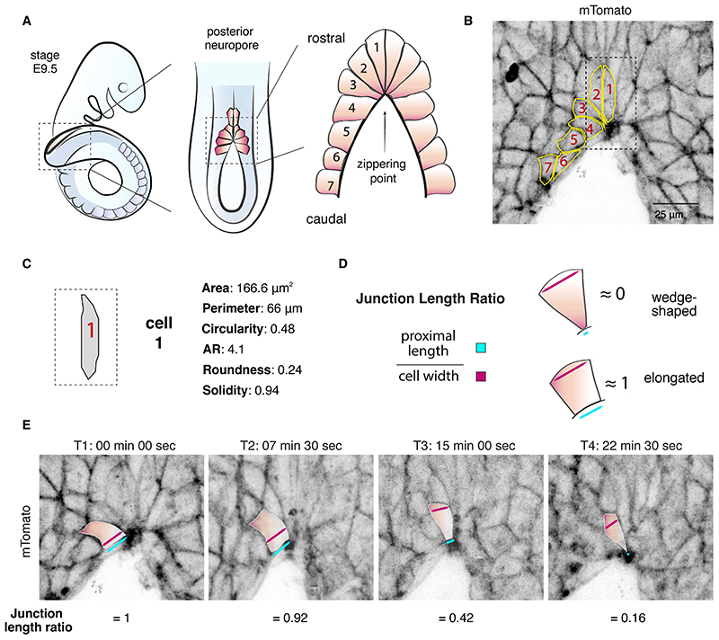
Zippering is a phenomenon of tissue morphogenesis whereby fusion between opposing epithelia progresses unidirectionally over significant distances, similar to the travel of a zip fastener, to ultimately ensure closure of an opening. A comparable process can be observed during Drosophila dorsal closure and mammalian wound healing, while zippering is employed by numerous organs such as the optic fissure, palatal shelves, tracheoesophageal foregut, and presumptive genitalia to mediate tissue sealing during normal embryonic development. Particularly striking is zippering propagation during neural tube morphogenesis, where the fusion point travels extensively along the embryonic axis to ensure closure of the neural tube. Advances in time-lapse microscopy and culture conditions have opened the opportunity for successful imaging of whole-mouse embryo development over time, providing insights into the precise cellular behavior underlying zippering propagation. Studies in mouse and the ascidian Ciona have revealed the fine-tuned cell shape changes and junction remodeling which occur at the site of zippering during neural tube morphogenesis. Here, we describe a step-by-step method for imaging at single-cell resolution the process of zippering and tissue remodeling which occurs during closure of the spinal neural tube in mouse. We also provide instructions and suggestions for quantitative morphometric analysis of cell behavior during zippering progression. This procedure can be further combined with genetic mutant models (e.g., knockouts), offering the possibility of studying the dynamics of tissue fusion and zippering propagation, which underlie a wide range of open neural tube defects.

Keywords: Cell shape; Epithelial closure; Live imaging; Neural tube; Neural tube defects; Tissue fusion.


Figures

Fig. 1
Fig. 1. Zippering during mouse neural tube closure.
(A) Schematic of zippering. Closure of the neural tube initiates at the boundary between the hindbrain and spinal cord (Closure 1). The wave of zippering then propagates bidirectionally. A second initiation site, Closure 2, occurs at the boundary between forebrain and hindbrain. This may be specific to mouse and probably does not occur in humans. Closure 3 forms at the very rostral end of the forebrain. Regions of open neural tube between sites of closure initiation are termed neuropores. In mouse, cranial closure completes by sealing of the anterior and, later, the hindbrain neuropore. Spinal closure progresses from Closure 1 along the caudal axis, until the entire open region of the posterior neuropore is eventually closed. Spinal zippering is estimated to cover a total length of approx. 5 mm to seal the caudal neural tube. (B) Representative brightfield image of mouse embryo at E9.5 with visible open posterior neuropore.
Fig. 2
Fig. 2. Post-acquisition processing steps.
(A) Images, after initial acquisition, tend to be noisy, limiting the ability to clearly identify the precise boundary of each cell. (B) Digital surface subtraction of the surface ectoderm from the underlying neural folds further improves signal and contrast. (C) Deconvolution processing significantly improves image resolution by increasing signal and correcting noise, allowing easier segmentation for subsequent analysis.
Fig. 3
Fig. 3. Segmentation and morphometric analysis of zippering.
(A) Schematics of cell arrangements at the site of zippering (dorsal view). Surface ectoderm cells 1-3 display a wedge-shaped morphology. Cells bordering the open PNP (cells 4–7) exhibit a more rectangular morphology. (B) Representative image of an un-recombined embryo expressing tdTomato fluorescence (Rosa26mTmG/mTmG) and manual segmentation of the cells at the site of zippering. (C) Morphometric analysis of cell 1. (D) Junction measurements and cell shape classification. Junction length ratio is calculated by dividing the length of the proximal junction by the cell maximum width. A junction length ratio ≈0 indicates a wedge-shaped morphology; a ratio ≈1 indicates an elongated morphology. (E) Stills from a live imaging movie showing cell shape changes during zippering. Cells shorten their proximal junctions over time to form a semi-rosette configuration at the zippering point.

References

    1. Begnaud S, Chen T, Delacour D, et al. Mechanics of epithelial tissues during gap closure. Curr Opin Cell Biol. 2016;42:52–62. - PMC - PubMed
    1. Martin P, Parkhurst SM. Parallels between tissue repair and embryo morphogenesis. Development. 2004;131:3021–34. - PubMed
    1. Jacinto A, Martinez-Arias A, Martin P. Mechanisms of epithelial fusion and repair. Nat Cell Biol. 2001;3:E117-23. - PubMed
    1. Nikolopoulou E, Galea GL, Rolo A, et al. Neural tube closure: cellular, molecular and biomechanical mechanisms. Development. 2017;144:552–566. - PMC - PubMed
    1. Gestri G, Bazin-Lopez N, Scholes C, Wilson SW. Cell Behaviors during Closure of the Choroid Fissure in the Developing Eye. Front Cell Neurosci. 2018;12:42. - PMC - PubMed

Publication types

LinkOut - more resources