. 2023 Feb 10;9(2):365-377.
doi: 10.1021/acsinfecdis.2c00566. Epub 2023 Jan 18.

Structural Basis for Cyclosporin Isoform-Specific Inhibition of Cyclophilins from Toxoplasma gondii

Affiliations

Structural Basis for Cyclosporin Isoform-Specific Inhibition of Cyclophilins from Toxoplasma gondii

Filippo Favretto et al. ACS Infect Dis. .

Abstract

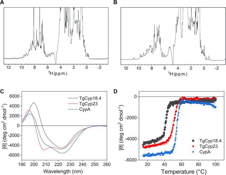
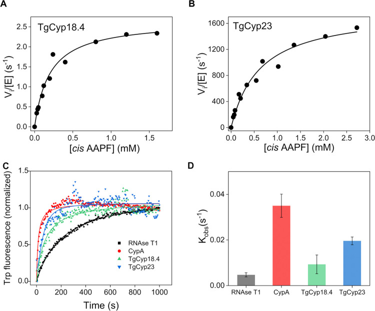
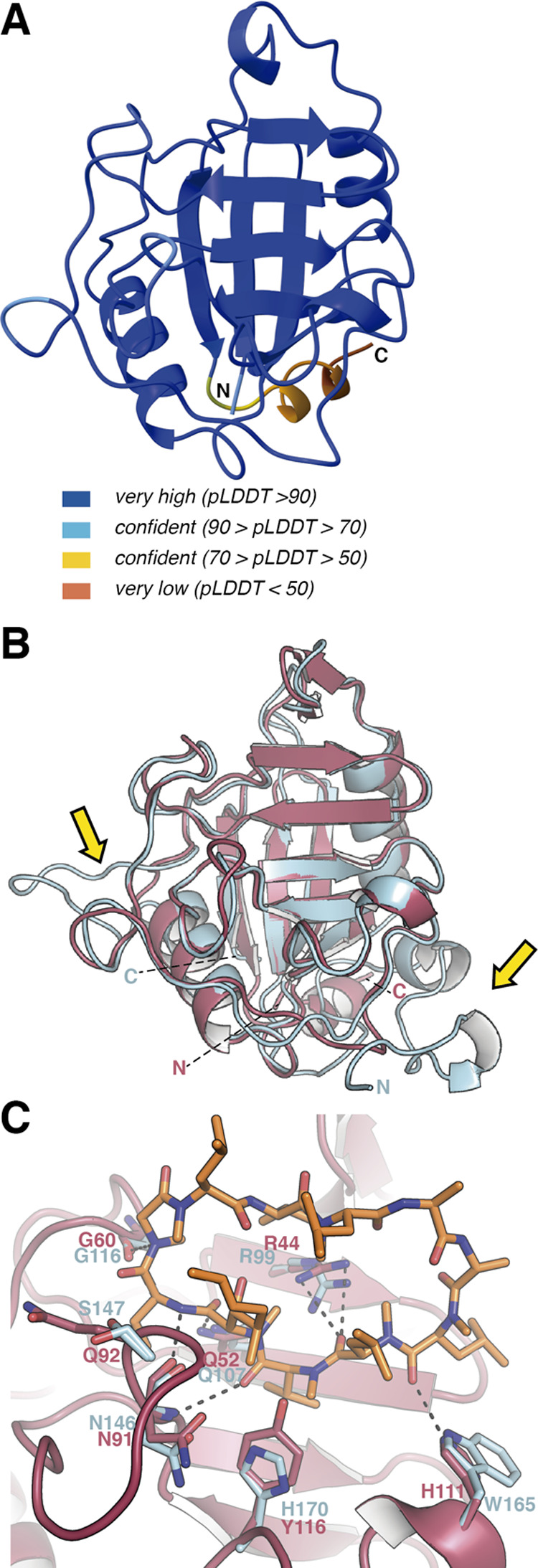
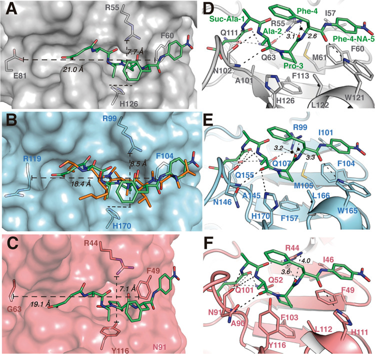
Cyclosporin (CsA) has antiparasite activity against the human pathogen Toxoplasma gondii. A possible mechanism of action involves CsA binding to T. gondii cyclophilins, although much remains to be understood. Herein, we characterize the functional and structural properties of a conserved (TgCyp23) and a more divergent (TgCyp18.4) cyclophilin isoform from T. gondii. While TgCyp23 is a highly active cis-trans-prolyl isomerase (PPIase) and binds CsA with nanomolar affinity, TgCyp18.4 shows low PPIase activity and is significantly less sensitive to CsA inhibition. The crystal structure of the TgCyp23:CsA complex was solved at the atomic resolution showing the molecular details of CsA recognition by the protein. Computational and structural studies revealed relevant differences at the CsA-binding site between TgCyp18.4 and TgCyp23, suggesting that the two cyclophilins might have distinct functions in the parasite. These studies highlight the extensive diversification of TgCyps and pave the way for antiparasite interventions based on selective targeting of cyclophilins.

Keywords: Toxoplasma gondii; chaperone-like activity; crystal structure; cyclophilin; cyclosporin; peptidyl-prolyl isomerization.


Conflict of interest statement

The authors declare no competing financial interest.

Figures

Figure 1
Figure 1
Sequence alignment of TgCyp18.4, TgCyp23, and human CypA. Amino acids critical for Cyp enzymatic activity and CsA binding are shown in bold. The sequences shown are TgCyp18.4 (TgCyp289250), TgCyp23 (TgCyp285760), and human CypA.
Figure 2
Figure 2
Structural characterization of TgCyp18.4 and TgCyp23. (A, B) 1H-1D NMR spectra of (A) 70 μM TgCyp18.4 (64 scans) and (B) 100 μM TgCyp23 (16 scans). (C) Far-UV CD spectra and (D) thermal denaturation profiles of 0.2 mg mL–1 TgCyp18.4 (black) and TgCyp23 (red). CypA was included as comparison (blue).
Figure 3
Figure 3
PPIase and chaperone-like activity of TgCyps. (A, B) Representative steady-state initial velocity kinetics for the PPIase activity of (A) TgCyp18.4 and (B) TgCyp23. (C) Catalytic effect of TgCyps on protein folding of RNase T1. The increase in fluorescence at 320 nm is shown as a function of the time of refolding in the presence of a fixed concentration of TgCyp18.4 (green), TgCyp23 (blue), and human CypA (red). The control experiment showing the spontaneous refolding of RNase T1 in the absence of Cyps is also displayed (black). (D) Histogram shows the mean value of the exponential folding rate constants kobs of RNase T1 in the absence and presence of Cyp variants. Color coding as in C. n = 3 independent experiments were performed. The error bars represent standard error from the mean value.
Figure 4
Figure 4
CsA inhibition of recombinant Cyps. (A) PPIase activity of 7 nM TgCyp23 (red) and 7 nM human CypA (black) measured at increasing concentrations of CsA, ranging from 0 to 30 nM. (B) PPIase activity of 3 μM TgCyp18.4 (black) and 3 μM TgCyp18.4 H111W (red) in the presence of increasing amounts of CsA, ranging from 0 to 6 μM. The concentration of AAPF used in the assays was fixed to 100 μM, corresponding to a cis substrate concentration of 40 μM, which is much smaller than the calculated Km. Therefore, no deviations from the expected first-order kinetics were observed in the presence of CsA.
Figure 5
Figure 5
Three-dimensional structure of TgCyp23 in complex with CsA. (A) Chemical structure of CsA. (B) Overall structure of the TgCyp23:CsA complex. TgCyp23 is displayed in blue cartoons, and CsA is depicted as orange sticks. N indicates the N-terminal region. (C) 2FoFc electron density map observed for CsA. Map contoured at 1σ. (D) Representation of the molecular surface of TgCyp23 (colored in blue) showing the CsA-binding pocket (CsA depicted as capped sticks). (E) CsA stabilization by TgCyp23. Relevant residues implicated in the interaction are labeled and shown as sticks. (F) Interaction network between nearby protein amino acids (distance in angstroms are indicated). Dashed lines indicate polar interactions.
Figure 6
Figure 6
AlphaFold-predicted model for TgCyp18.4. (A) Predicted structure for TgCyp18.4 with colors indicating the reliability of the model. (B) Structural superimposition of the predicted structure of TgCyp18.4 (salmon) onto the crystal structure of TgCyp23 (blue). C and N show C-terminus and N-terminus, respectively. Yellow arrows indicate regions showing larger differences between TgCyp23 and TgCyp18.4. (C) Comparison of CsA-binding pockets in TgCyp23 and TgCyp18.4. TgCyp23 amino acids are displayed in blue sticks, and TgCyp18.4 residues are displayed in red. Dashed lines indicate predicted polar interactions for the TgCyp18.4:CsA complex.
Figure 7
Figure 7
Structural comparison of substrate versus CsA recognition in CypA and Toxoplasma Cyps. (A) Detailed view of the binding site in the CypA:AAPF complex. All distances are indicated in dashed lines between the shown atoms in the extremes. (B) TgCyp23 cavity is displayed as the blue surface. AAPF in green sticks and superposed with half-CsA chains (residues 2–6) in orange sticks. (C) TgCyp18.4 surface in salmon and AAPF superposed is depicted as green sticks. (D) CypA:AAPF interaction network (polar contacts are indicated with dotted lines). (E) TgCyp23:AAPF predicted interaction network. (F) TgCyp18.4:AAPF predicted interaction network.

References

    1. Fischer G.; Wittmann-Liebold B.; Lang K.; Kiefhaber T.; Schmid F. X. Cyclophilin and peptidyl-prolyl cis-trans isomerase are probably identical proteins. Nature 1989, 337, 476–478. 10.1038/337476a0. - DOI - PubMed
    1. Göthel S. F.; Marahiel M. A. Peptidyl-prolyl cis-trans isomerases, a superfamily of ubiquitous folding catalysts. Cell. Mol. Life Sci. 1999, 55, 423–436. 10.1007/s000180050299. - DOI - PMC - PubMed
    1. Singh H.; Kaur K.; Singh M.; Kaur G.; Singh P. Plant Cyclophilins: Multifaceted Proteins With Versatile Roles. Front. Plant Sci. 2020, 11, 58521210.3389/fpls.2020.585212. - DOI - PMC - PubMed
    1. Radhakrishnan J.; Bazarek S.; Chandran B.; Gazmuri R. J. Cyclophilin-D: a resident regulator of mitochondrial gene expression. FASEB J. 2015, 29, 2734–2748. 10.1096/fj.14-263855. - DOI - PubMed
    1. Ünal C. M.; Steinert M. Microbial peptidyl-prolyl cis/trans isomerases (PPIases): virulence factors and potential alternative drug targets. Microbiol. Mol. Biol. Rev. 2014, 78, 544–571. 10.1128/MMBR.00015-14. - DOI - PMC - PubMed

Publication types