

Additive Fabrication of a Vascular 3D Phantom for Stereotactic Radiosurgery of Arteriovenous Malformations
- PMID: 36654837
- PMCID: PMC9828616
- DOI: 10.1089/3dp.2020.0305
Additive Fabrication of a Vascular 3D Phantom for Stereotactic Radiosurgery of Arteriovenous Malformations
Abstract
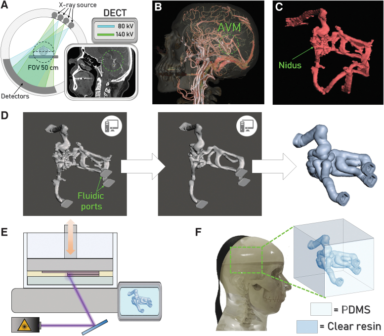
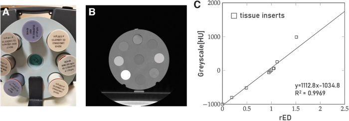
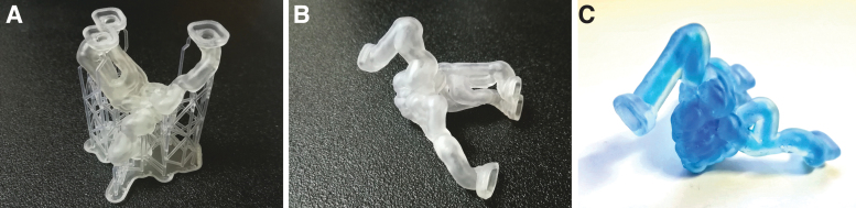
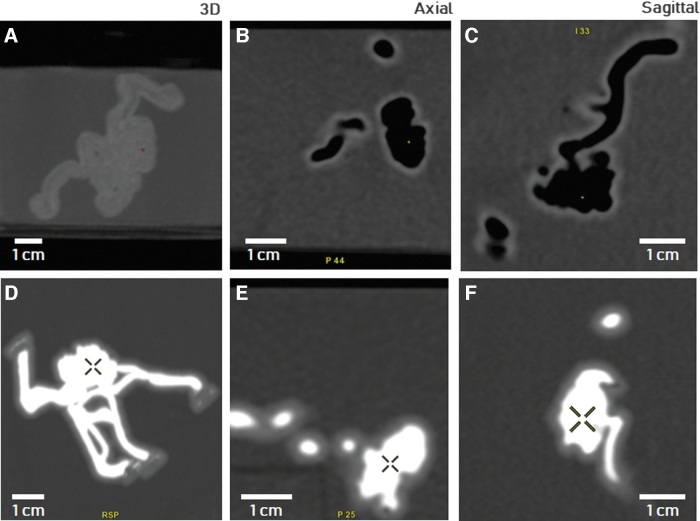
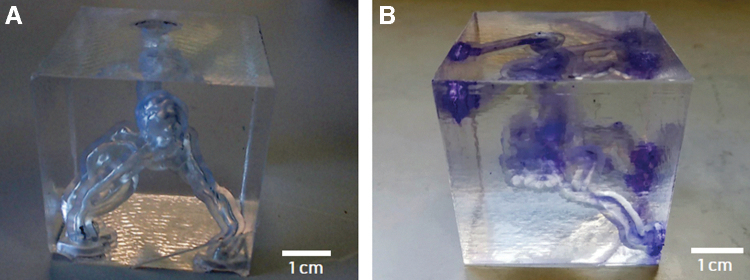
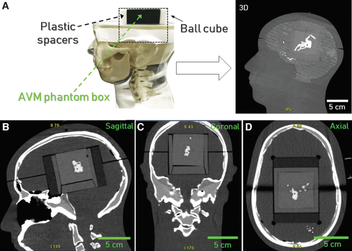
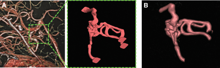
In this study, an efficient methodology for manufacturing a realistic three-dimensional (3D) cerebrovascular phantom resembling a brain arteriovenous malformation (AVM) for applications in stereotactic radiosurgery is presented. The AVM vascular structure was 3D reconstructed from brain computed tomography (CT) data acquired from a patient. For the phantom fabrication, stereolithography was used to produce the AVM model and combined with silicone casting to mimic the brain parenchyma surrounding the vascular structure. This model was made with tissues-equivalent materials for radiology. The hollow vascular system of the phantom was filled with a contrast agent usually employed on patients for CT scans. The radiological response of the phantom was tested and compared with the one of the clinical case. The constructed model demonstrated to be a very accurate physical representation of the AVM and its vasculature and good morphological consistency was observed between the model and the patient-specific source anatomy. These results suggest that the proposed method has potential to be used to fabricate patient-specific phantoms for neurovascular radiosurgery applications and medical research.
Keywords: additive manufacturing; anthropomorphic phantoms; stereolithography; surgical applications; tissue-equivalent materials.
Copyright 2021, Mary Ann Liebert, Inc., publishers.
Conflict of interest statement
No competing financial interests exist.
Figures
Similar articles
-
Three-dimensional assessment of the effects of high-density embolization material on the absorbed dose in the target for Gamma Knife radiosurgery of arteriovenous malformations.J Neurosurg. 2016 Dec;125(Suppl 1):123-128. doi: 10.3171/2016.7.GKS161545. J Neurosurg. 2016. PMID: 27903193
-
Application of Time-Resolved 3D Digital Subtraction Angiography to Plan Cerebral Arteriovenous Malformation Radiosurgery.AJNR Am J Neuroradiol. 2017 Apr;38(4):740-746. doi: 10.3174/ajnr.A5074. Epub 2017 Jan 26. AJNR Am J Neuroradiol. 2017. PMID: 28126751 Free PMC article.
-
Learning-based automatic segmentation of arteriovenous malformations on contrast CT images in brain stereotactic radiosurgery.Med Phys. 2019 Jul;46(7):3133-3141. doi: 10.1002/mp.13560. Epub 2019 May 21. Med Phys. 2019. PMID: 31050804 Free PMC article.
-
3D printing methods for radiological anthropomorphic phantoms.Phys Med Biol. 2022 Jul 27;67(15). doi: 10.1088/1361-6560/ac80e7. Phys Med Biol. 2022. PMID: 35830787 Review.
-
Stereotactic radiosurgery alone or combined with embolization for brain arteriovenous malformations: a systematic review and meta-analysis.J Neurosurg. 2018 May;128(5):1338-1348. doi: 10.3171/2016.11.JNS162382. Epub 2017 May 12. J Neurosurg. 2018. PMID: 28498057
Cited by
-
Development of a patient-specific chest computed tomography imaging phantom with realistic lung lesions using silicone casting and three-dimensional printing.Sci Rep. 2023 Mar 9;13(1):3941. doi: 10.1038/s41598-023-31142-5. Sci Rep. 2023. PMID: 36894618 Free PMC article.
-
Conductive Additive Manufactured Acrylonitrile Butadiene Styrene Filaments: Statistical Approach to Mechanical and Electrical Behaviors.3D Print Addit Manuf. 2023 Dec 1;10(6):1423-1438. doi: 10.1089/3dp.2022.0287. Epub 2023 Dec 11. 3D Print Addit Manuf. 2023. PMID: 38116220 Free PMC article.
-
Hybrid Additive Fabrication of a Transparent Liver with Biosimilar Haptic Response for Preoperative Planning.Diagnostics (Basel). 2021 Sep 21;11(9):1734. doi: 10.3390/diagnostics11091734. Diagnostics (Basel). 2021. PMID: 34574075 Free PMC article.
References
-
- Doi K. Diagnostic imaging over the last 50 years: Research and development in medical imaging science and technology. Phys Med Biol 2006;51:R5–R27. - PubMed
-
- Tam MDBS. Building virtual models by postprocessing radiology images: A guide for anatomy faculty. Anat Sci Educ 2010;3:261–266. - PubMed
-
- Knisely JPS, Apuzzo MLJ. Historical aspects of stereotactic radiosurgery: Concepts, people, and devices. World Neurosurg 2019;130:593–607. - PubMed
-
- Ding C, Saw CB, Timmerman RD. Cyberknife stereotactic radiosurgery and radiation therapy treatment planning system. Med Dosim 2018;43:129–140. - PubMed
-
- Zhang XQ, Shirato H, Aoyama H, et al. . Clinical significance of 3D reconstruction of arteriovenous malformation using digital subtraction angiography and its modification with CT information in stereotactic radiosurgery. Int J Radiat Oncol Biol Phys 2003;57:1392–1399. - PubMed
LinkOut - more resources
Full Text Sources