Case Reports
. 2023 Jan 10;18(3):1140-1143.
doi: 10.1016/j.radcr.2022.12.004. eCollection 2023 Mar.

Aberrant right gastric vein mimicking hepatic spread of prostate cancer on PSMA-PET/CT

Affiliations
Case Reports

Aberrant right gastric vein mimicking hepatic spread of prostate cancer on PSMA-PET/CT

Genevieve C Muñoz et al. Radiol Case Rep. .

Abstract

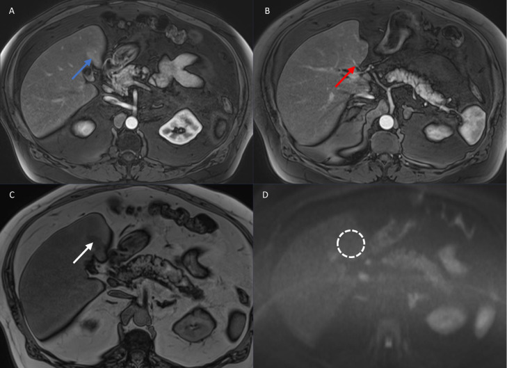
Hepatic vasculature can exhibit a wide variety of variants, some of which may resemble pathologic findings. In this case, a 53-year-old man presenting for staging of biochemically recurrent prostatic adenocarcinoma was found to have focally increased prostate-specific membrane antigen (PSMA) tracer uptake on positron emission tomography (PET) imaging in hepatic segment IV. This finding was initially concerning for hepatic metastasis of the patient's primary prostate adenocarcinoma. However, the area of radiotracer uptake was not associated with a discrete lesion on CT, and the geographic morphology of the uptake raised the possibility of a vascular etiology. Magnetic resonance imaging (MRI) of the liver showed no hepatic metastases and confirmed the presence of an aberrant right gastric vein directly perfusing the corresponding portion of hepatic segment IV. This case highlights PSMA uptake in the liver secondary to vascular variants as a potential mimic for metastatic disease on PSMA-PET/CT.

Keywords: Hepatic vasculature; PSMA-PET/CT; Prostate adenocarcinoma; Vascular variants.


Figures

Fig 1
Fig. 1
PSMA-PET/CT showed ill-defined moderate tracer uptake in posterior hepatic segment IV (red arrows, images A1-A3), corresponding to a vague area of hypodensity without a definite focal lesion (red arrow, image A1). There was also intense tracer uptake in the T4 vertebral body (not shown) and left fourth rib (blue arrows, images B1-B3), compatible with sites of osseous metastatic disease.
Fig 2
Fig. 2
MRI of the liver including T1-weighted arterial phase post-contrast images (images A and B), T1-weighted opposed-phase images (image C), and diffusion-weighted images (image D) was performed. In the area in question on the PSMA-PET/CT, there was vague arterial phase hyperenhancement (blue arrow) associated with focal fat deposition (white arrow). An anomalous vessel (red arrow) was noted to enter the liver directly through the capsule at this site, corresponding to an aberrant right gastric vein. There was no restricted diffusion (dashed circle) to indicate a metastasis at this location or elsewhere in the liver.

References

    1. Elsayes KM, Shaaban AM, Rothan SM, Javadi S, Madrazo BL, Castillo RP, et al. A comprehensive approach to hepatic vascular disease. Radiographics. 2017;37(3):822. doi: 10.1148/rg.2017160161. - DOI - PubMed
    1. Unal E, Ozmen MN, Akata D, Karcaaltincaba M. Imaging of aberrant left gastric vein and associated pseudolesions of segments II and III of the liver and mimickers. Diagn Interv Radiol. 2015;21(2):105–110. doi: 10.5152/dir.2014.14360. - DOI - PMC - PubMed
    1. Seong NJ, Chung JW, Kim HC, Park JH, Jae HJ, An SB, et al. Right gastric venous drainage: angiographic analysis in 100 patients. Korean J Radiol. 2012;13(1):53–60. doi: 10.3348/kjr.2012.13.1.53. - DOI - PMC - PubMed
    1. Deneve E, Caty L, Fontaine C, Guillem P. Simultaneous aberrant left and right gastric veins draining directly into the liver. Ann Anat. 2003;185(3):263–266. doi: 10.1016/S0940-9602(03)80037-7. - DOI - PubMed
    1. Takayasu K, Aoki K, Ichikawa T, Ohmura T, Sekiguchi R, Terauchi T, et al. Aberrant right gastric vein directly communicating with left portal vein system. Incidence and implications. Acta Radiol. 1990;31:575–577. - PubMed

Publication types

LinkOut - more resources