. 2023 Jan 13:11:e14319.
doi: 10.7717/peerj.14319. eCollection 2023.

Systematics and diversification of the Ichthyomyini (Cricetidae, Sigmodontinae) revisited: evidence from molecular, morphological, and combined approaches

Affiliations

Systematics and diversification of the Ichthyomyini (Cricetidae, Sigmodontinae) revisited: evidence from molecular, morphological, and combined approaches

Jorge Salazar-Bravo et al. PeerJ. .

Abstract

Ichthyomyini, a morphologically distinctive group of Neotropical cricetid rodents, lacks an integrative study of its systematics and biogeography. Since this tribe is a crucial element of the Sigmodontinae, the most speciose subfamily of the Cricetidae, we conducted a study that includes most of its recognized diversity (five genera and 19 species distributed from southern Mexico to northern Bolivia). For this report we analyzed a combined matrix composed of four molecular markers (RBP3, GHR, RAG1, Cytb) and 56 morphological traits, the latter including 15 external, 14 cranial, 19 dental, five soft-anatomical and three postcranial features. A variety of results were obtained, some of which are inconsistent with the currently accepted classification and understanding of the tribe. Ichthyomyini is retrieved as monophyletic, and it is divided into two main clades that are here recognized as subtribes: one to contain the genus Anotomys and the other composed by the remaining genera. Neusticomys (as currently recognized) was found to consist of two well supported clades, one of which corresponds to the original concept of Daptomys. Accordingly, we propose the resurrection of the latter as a valid genus to include several species from low to middle elevations and restrict Neusticomys to several highland forms. Numerous other revisions are necessary to reconcile the alpha taxonomy of ichthyomyines with our phylogenetic results, including placement of the Cajas Plateau water rat (formerly Chibchanomys orcesi) in the genus Neusticomys (sensu stricto), and the recognition of at least two new species (one in Neusticomys, one in Daptomys). Additional work is necessary to confirm other unanticipated results, such as the non-monophyletic nature of Rheomys and the presence of a possible new genus and species from Peru. Our results also suggest that ichthyomyines are one of the main Andean radiations of sigmodontine cricetids, with an evolutionary history dating to the Late Miocene and subsequent cladogenesis during the Pleistocene.

Keywords: Amazon; Andean; Chibchanomys orcesi; Daptomys; Ichthyomyini; Neotropics; Sigmodontalia; Subtribes; Water rats.


Conflict of interest statement

The authors declare that they have no competing interests.

Figures

Figure 1
Figure 1. Map of collecting localities of ichthyomyines specimens used in the present study.
Map of localities of specimens of the Ichthyomyini included in genetic and morphological analyses in the present study. Numbered localities correspond to those of Table S1.
Figure 2
Figure 2. Phylogenetic hypothesis (ML) for the Ichthyomyini based on a concatenated database of molecular characters.
Maximum likelihood phylogenetic inference for the Ichthyomyini, based on the combined analysis of one mitochondrial (Cytb) and three nuclear markers (RBP3, RAG1, and GHR) for 59 ichthyomyine specimens and multiple outgroup taxa in IQ-TREE. Using the same best-fit model selected by PartitionFinder, Log-likelihood of the tree: −6,3187.3930 (s.e. 1,387.0787). Nodal support from the ML bootstrap pseudo-replicates are indicated at each node along with Bayesian posterior probabilities (BI). White wedges indicate bootstrap values ≤ 75% and black indicates bootstrap frequencies ≥ 75%. For BI, white indicates PP < 0.95, whereas black indicates PP ≥ 0.95. Scale at the bottom represents substitutions per site. Terminals are named with original, museum-based identifications, followed by museum/identification voucher numbers and geographic information (two-letter country code and Department/State). Two-letter country codes as follows: EC, Ecuador; CO, Colombia; PE, Peru; BR, Brazil; GU, French Guiana; BO, Bolivia; CR, Costa Rica; ES, El Salvador. Colored boxes next to names indicate presumed species limits, recognized based on original identifications and/or monophyly.
Figure 3
Figure 3. Total evidence phylogenetic analysis for the Ichthyomyini.
Bayesian majority-rule consensus depicting the phylogenetic relationships among members of the Ichthyomyini based on combined DNA and morphological data (total evidence approach). A total of 56 morphological characters and four protein-coding genes (4,652 bp) were included in the analysis. Values adjacent to nodes are Bayesian posterior probabilities (PP).
Figure 4
Figure 4. Fit of ClaDS to the Ichthyomyini.
Inferred mean speciation rate (A) and number of lineages (B) through time, with individual MCMC iterations (thin blue line), the 95% credibility interval for each time point (thick blue line), and the mean for each time point (dotted green). Time runs from the past (starting at 0) to the present (crown age, here 5 Myrs). (C) Inferred lineage-specific speciation rates for the icthyomyine phylogeny. Marginal posteriors of the heterogeneity parameter σ (D), the trend parameter α (E), the turnover rate ε (F), and the mean change in rate m = αeσ2/2 (G). The orange lines indicate the value of the MAP estimate.
Figure 5
Figure 5. Divergence time estimates obtained from four analyses in BEAST, with ancestral range estimations based on DEC analysis.
Bayesian maximum clade credibility chronogram based on the combined analyses of four nuclear, one mitochondrial protein-coding genes, and 56 morphological characters for the Ichthyomyini and outgroup taxa. Hypothesized ancestral biogeographic ranges for Ichthyomyini were estimated based on the dispersal-extinction-cladogenesis (DEC) model as implemented in RASP 4.0. States at nodes represent the ancestral bioregion (or combination thereof) occupied immediately before diversification. Arrows indicate instances where the model suggested diversification via dispersal, whereas the stars indicate instances of diversification by ancestral range vicariance. Six-point stars indicate both, suggested speciation by dispersal and vicariance. Numbers above epochs indicate Ma. The blue box next to Pleistocene indicates the Holocene.
Figure 6
Figure 6. Ancestral ranges in the evolution of the Ichthyomyini estimated using rase.
Ancestral estimation according to rase at six time slices, from 4.96 Ma to the current distribution of members of the tribe Ichthyomyini. (A–E) Plot the 10%, 15%, and 25% highest posterior density for each extant branch (10% with highest color intensity). (F) plots current species distributions. The branches of the phylogenetic tree are color coded as in (A–F) to identify its position in the map. For further discussion, see main text.
Figure 7
Figure 7. Details of the head in two species of ichthyomyine rodents.
The figure illustrates the variation on the presence of pale spots on the auricular region in species of the Ichthyomyini (character 5). (A) Absent (Ichthyomys tweedii; MECN 5772); (B) present (Rheomys underwoodi; APG sn).
Figure 8
Figure 8. Details of the ventral tail coloration in the Ichthyomyini.
The figure illustrates the three types of ventral tail coloration in the Icthyomyini (character 6). (A) Unicolored (Rheomys raptor; MZUCR 5000); (B) entirely bicolored (Ichthyomys pinei; MZUA 234); (C) bicolored except distal inch (Ichthyomys orientalis; MECN 4914).
Figure 9
Figure 9. Plantar views of the left hind-feet in ichthyomyine rodents.
The figure illustrates variations in the width of the hindfeet (character 13) in ichthyomyine rodents. (A) Narrow (Neusticomys vossi; MECN 4332); (B) broad (Ichthyomys orientalis; MECN 4914).
Figure 10
Figure 10. Dorsal views of the tail.
The figure illustrates the general morphology of the tail (character 14) in ichthyomyine rodents. (A) Cylindrical or conical (Ichthyomys orientalis; MECN 4914); (B) compressed (Chibchanomys n. sp.; MUSA 13864).
Figure 11
Figure 11. Detail of the morphology of the rostrum in ichthyomyine rodents.
This figure illustrates variation in the general morphology of the rostrum (character 16), in ichthyomyine rodents. (A) Slender (Neusticomys vossi; MECN 4332); (B) broad (Ichthyomys orientalis; MECN 4914).
Figure 12
Figure 12. Differences in the development of the gnathic process in ichthyomyine rodents.
Gnathic process, appearance in lateral view, in representative taxa of ichthyomyine rodents (character 17). (A) Small (Ichthyomys orientalis; MECN 4914); and (B) large (Anotomys leander; AMNH 244605).
Figure 13
Figure 13. Dorsal views of adult crania.
The figure highlights differences in the overall morphology of the braincase in ichthyomyine rodents (character 19). (A) Non-inflated (Ichthyomys orientalis; MECN 4914); (B) rounded (Rheomys thomasi; ROM 101294); (C) globular (Anotomys leander; AMNH 244605).
Figure 14
Figure 14. Dorsal views of the posterior braincase region.
The figure illustrates variations in the position and development of the occipital condyles [occ] on the dorsal view of the braincase (character 20) in ichthyomyines. (A) Occ visible from above (Ichthyomys orientalis; MECN 4914); (B) occ not visible from above (Neusticomys vossi; MECN 4332).
Figure 15
Figure 15. Details of the interorbital region.
Shape and size of the interorbital region (appearance in dorsal view; lateral margins highlighted) in the Ichthyomyini (character 22). (A) Hourglass (Anotomys leander; AMNH 244605); (B) large (Neusticomys vossi; MECN 4332); (C) short (Rheomys thomasi; ROM 101294); and (D) very constrained (Ichthyomys orientalis; MECN 4914).
Figure 16
Figure 16. Ventral view of the diastemal palate.
Notice the differential development of the premaxillary process [eps] of the incisive foramina [if] (character 25). (A) eps short (Daptomys sp.; JBM 2500); (B) large (Rheomys thomasi; ROM 101294). Other abbreviations: I, lumen, maxillary septum.
Figure 17
Figure 17. Lateral views of the rostral and zygomatic regions.
Lateral views of the rostral and zygomatic regions Illustrating the variations in the position of the posterior margin of the zygomatic plate [izr] relative to the anterior face (marked with a dotted line) of the first upper molar [M1] (character 26). (A) Anterior (Ichthyomys orientalis; MECN 4914); (B) even with or posterior (Neusticomys vossi; MECN 4332).
Figure 18
Figure 18. Details of the auditory bullae.
Left otic capsule of ichthyomyine rodents illustrating variations in the morphology of the auditory bullae [ab] morphology (character 27). (A) Flask-shaped, not inflated (Ichthyomys orientalis; MECN 4914); (B) globular (Neusticomys vossi; MECN 4332).
Figure 19
Figure 19. Detail of the left otic capsule.
Ventral views of the left auditory bulla illustrating variations in the morphology of the petrotympanic (character 28). (A) Largely exposed (Ichthyomys orientalis; MECN 4914); (B) covered by the ectotympanic (Anotomys leander; AMNH 244605). Other abbreviations: ab, auditory bulla; cc, carotid canal.
Figure 20
Figure 20. Labial views of the left mandible (vertical ramus).
The figure shows the variation in the height of the coronoid process [cd] relative to the height of the condyle [cr] in the Ichthyomyini (character 29). (A) More or less at the same level (Chibchanomys n. sp.; MUSA 13864); and (B) cd clearly surpassing the cr (Ichthyomys orientalis; MECN 4914). Other abbreviation: ag, angular process.
Figure 21
Figure 21. Details of the cutting edge of the upper incisors.
Close up of the frontal view of upper incisors in ichthyomyine rodents highlighting the variation in the shape of the cutting edge (character 31). (A) Shallow inverted “v” (Neusticomys vossi; MECN 4332). (B) Deep inverted “v” (Ichthyomys orientalis; MECN 4914).
Figure 22
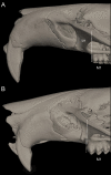
Figure 22. Lateral view of the rostral region.
Left lateral view of the rostrum in ichthyomyine rodents highlighting the variation in incisor procumbency (character 32). (A) Opisthodon (Anotomys leander; AMNH 244605); and (B) tending to orthodont (Ichthyomys orientalis; MECN 4914). Dotted lines conform to the incisive index of Thomas (1919).
Figure 23
Figure 23. Detail of the upper molar row (lateral view).
Figure highlights the variation in tubercular hypsodonty in the Ichthyomyini (character 33). (A) Moderate (Neusticomys vossi; MECN 4332); (B) marked (Anotomys leander; AMNH 244605).
Figure 24
Figure 24. Ventral view of adult crania.
Figure illustrates variations in the ratio of upper toothrow length to the length of the adult skull in ichthyomyine rodents (character 34). (A) Normal (Neusticomys vossi; MECN 4332); (B) “reduced” (Ichthyomys orientalis; MECN 4914); (C) “enlarged” (Anotomys leander; AMNH 244605). Please see Table 3 for details.
Figure 25
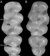
Figure 25. Details of the first two upper molars.
Occlusal view of right M1-M2 illustrating variations in the presence of the posteroloph [po] in the upper molars of ichthyomine rodents (character 35). (A) po present in M1-M2 (Anotomys leander; AMNH 244605); (B) po present in M1 (Neusticomys vossi; MECN 4332); (C) po absent in M1-M2 (Rheomys thomasi; ROM 101294).
Figure 26
Figure 26. Detail of the occlusal view of the right M1-M2.
Occlusal view of right M1-M2 illustrating variation in the presence of the paralophule [pa] in ichthyomine rodents (character 36). (A) Present (Neusticomys vossi ; MECN 5027); (B) absent (Daptomys oyapocki; AMNH 267597).
Figure 27
Figure 27. Details of the right first upper molar.
The figure (right M1 in occlusal view) highlights the variation in the presence of the anteromedian style [as] in M1 (character 37) in ichthyomyine rodents. (A) Present (Anotomys leander; AMNH 244605); (B) absent (Neusticomys vossi; MECN 4332).
Figure 28
Figure 28. Details of the M1.
The figure (left M1 in occlusal view) illustrates the variation in the presence of the anteromedian flexus [af] in M1 (character 38) in the Ichthyomyini: (A) present (Daptomys oyapocki; AMNH 267597) and (B) absent (Ichthyomys orientalis; MECN 4914).
Figure 29
Figure 29. Variability in the M1.
The figure (left M1 in occlusal view), illustrates the observed variation in the presence/absence of the enterostyle [es] in M1 of ichthyomyine rodents (character 39). (A) Present (Anotomys leander; AMNH 244605) and (B) absent (Ichthyomys orientalis; MECN 4914).
Figure 30
Figure 30. Ventral view of the alveoli of right upper molars of ichthyomyines.
Illustrates the variation in the number of roots in M1 (character 40). (A) 3 (Ichthyomys orientalis; MECN 4914); (B) 4 (Neusticomys monticolus; FMNH 71221).
Figure 31
Figure 31. Variability in the M2 in ichthyomyine rodents.
This figure (left M2 in occlusal view) illustrates the observed variation in the relative development (in size) between the posterior lobe [pl] and the anterior lobe [al] in the M2 (character 41) in ichthyomyine rodents. (A) pl smaller (Neusticomys vossi; MECN 4332); (B) pl subequal (Rheomys raptor; USNM 565826).
Figure 32
Figure 32. Occlusal view of the left upper molar toothrow in ichthyomyines.
The figure illustrates variation in the presence of the M3 (character 42). (A) Present (Rheomys raptor; USNM 565826); (B) barely absent (one side) or absent (both sides) (Daptomys oyapocki; AMNH 267597).
Figure 33
Figure 33. Detail of the morphology of M3.
This figure (left M3 in occlusal view) illustrates the variation in the general morphology of the M3 (character 43). (A) “complex” (Anotomys leander; AMNH 66202); (B) peg-like (Neusticomys vossi; MECN 4332).
Figure 34
Figure 34. Occlusal view of right lower molar rows of species of ichthyomyines.
Illustrates the observed variation in the presence of the posterolophid [pl] in the lower molars (character 44). (A) Present (Anotomys leander; AMNH 244605); (B) absent (Ichthyomys teweedii; MECN 5772). Other abbreviation: af, anteromedian flexid.
Figure 35
Figure 35. Detail of the first lower molar in ichthyomyines.
This figure (right m1 in occlusal view) illustrates the variation in the presence of the anteromedian flexid [af] in m1 (character 45). (A) Present (Anotomys leander; AMNH 244605); (B) absent (Ichthyomys orientalis; MECN 4914).
Figure 36
Figure 36. Occlusal views of the first lower molar in ichthyomyines.
The figure (left m1 in occlusal view) illustrates variations in the general morphology of m1 in ichthyomyines as they attain the bilobate condition with age (character 46). (A) Present (Ichthyomys orientalis; MECN 4914); (B) absent (Neusticomys orcesi; MEPN 12230). Acronyms: l1–l3, lobes.
Figure 37
Figure 37. Detail of the m2.
Occlusal views of the left m2 in ichthyomyine rodents showing variations in the presence of a entolophulid-like structure [en] in m2 (character 47). (A) Barely present (Neusticomys vossi; MECN 4332); (B) absent (Anotomys leander; AMNH 244605).
Figure 38
Figure 38. Detail of the m3 in ichthyomyines.
The figure (left m3 in occlusal view) shows the variation in the general morphology of the m3 (character 48) in ichthyomyines. (A) With two distinct lobes (Ichthyomys tweedii; MECN 5772); (B) with the posterior lobe small or absent (Neusticomys orcesi; MEPN 12230). Acronyms: l1–l2, lobes.
Figure 39
Figure 39. Details of the stomachs.
These ventral views (in midfrotal section) illustrate variations in the extent of gastric glandular epithelium (character 50) in ichthyomyine rodents. (A) Glandular epithelium present between the incisura angularis and the pyloric sphincter (A Daptomys sp.; JBM 2500 (same individual as MECN 6629)); (B) glandular epithelium restricted to the greater curvature (Ichthyomys orientalis; MEPN 12672). Abbreviations: b, bordering fold; d, duodenum; co, cornified epithelium; ge, glandular epithelium; i, incisura angularis.
Figure 40
Figure 40. Details of the soft palate.
The figure (soft palate in ventral view) shows variations in the number of interdental rugae (character 53) in ichthyomyine rodents. (A) Three (Daptomys sp.; JBM 2500 (same individual as MECN 6629)); (B) four (Ichthyomys tweedii; MECN 5772). Abbreviations: d1–d3, diastemal rugae; i1–i4, interdental rugae.

References

    1. Adams DC, Berns CM, Kozak KH, Wiens JJ. Are rates of species diversification correlated with rates of morphological evolution? Proceedings Royal Society B. 2009;276(1668):2729–2738. doi: 10.1098/rspb.2009.0543. - DOI - PMC - PubMed
    1. Alhajeri BH, Schenk JJ, Steppan S. Ecomorphological diversification following continental colonization in muroid rodents (Rodentia: Muroidea) Biological Journal of the Linnean Society. 2016;117(3):463–481. doi: 10.1111/bij.12695. - DOI
    1. Allen R, Ryan H, Davis BW, King C, Frantz L, Irving-Pease E, Barnett R, Linderholm A, Loog L, Haile J, Lebrasseur O, White M, Kitchener AC, Murphy WJ, Larson G. A mitochondrial genetic divergence proxy predicts the reproductive compatibility of mammalian hybrids. Proceedings of the Royal Society B: Biological Sciences. 2020;287(1928):20200690. doi: 10.1098/rspb.2020.0690. - DOI - PMC - PubMed
    1. Anderson S. Mammals of Bolivia, taxonomy and distribution. Bulletin of the American Museum of Natural History. 1997;231:1–652.
    1. Anthony HE. Preliminary report on Ecuadorean mammals No. 1. American Museum Novitates. 1921;20:6.

Publication types

LinkOut - more resources