. 2023 Jan 15;136(2):jcs260656.
doi: 10.1242/jcs.260656. Epub 2023 Jan 19.

Distinct role of ERp57 and ERdj5 as a disulfide isomerase and reductase during ER protein folding

Affiliations

Distinct role of ERp57 and ERdj5 as a disulfide isomerase and reductase during ER protein folding

Philip John Robinson et al. J Cell Sci. .

Abstract

Proteins entering the secretory pathway need to attain native disulfide pairings to fold correctly. For proteins with complex disulfides, this process requires the reduction and isomerisation of non-native disulfides. Two key members of the protein disulfide isomerase (PDI) family, ERp57 and ERdj5 (also known as PDIA3 and DNAJC10, respectively), are thought to be required for correct disulfide formation but it is unknown whether they act as a reductase, an isomerase or both. In addition, it is unclear how reducing equivalents are channelled through PDI family members to substrate proteins. Here, we show that neither enzyme is required for disulfide formation, but ERp57 is required for isomerisation of non-native disulfides within glycoproteins. In addition, alternative PDIs compensate for the absence of ERp57 to isomerise glycoprotein disulfides, but only in the presence of a robust reductive pathway. ERdj5 is required for this alternative pathway to function efficiently indicating its role as a reductase. Our results define the essential cellular functions of two PDIs, highlighting a distinction between formation, reduction and isomerisation of disulfide bonds.

Keywords: Disulfide formation; Endoplasmic reticulum; Protein disulfide isomerase; Protein folding; Protein secretion.


Conflict of interest statement

Competing interests The authors declare no competing or financial interests.

Figures
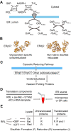
Fig. 1.
Fig. 1.
A translation system to assay the role of ERp57 and ERdj5 in the ER reducing pathway. (A) The cytosolic reducing pathway, involving glucose 6-phosphate dehydrogenase (G6PD) and thioredoxin reductase (TrxR1), is the source of reducing equivalents required to reduce ER localised oxidoreductases (OxR). OxRRed can either reduce other oxidoreductases (OxROx to OxRRed) or substrates (SubOx to SubRed). PGL, phosphoglucolactone; TrX, thioredoxin. (B) Ribbon structures of ERp57 (PDB: 3F8U) and ERdj5 (PDB: 3APO), two oxidoreductases that are involved in disulfide reduction or isomerisation during protein folding. (C) The possible routes by which reducing equivalents are transferred from the cytosol to nascent folding proteins in the ER. (D) Components of a typical translation reaction – rabbit reticulocyte lysate (RRL), amino acids minus methionine (aa-Met), S35-labelled methionine (S35 Met), potassium chloride (KCL) and RNA template. Microsomes or semi-permeabilised (SP) cells are added as an ER source. (E) The redox status of untreated rabbit reticulocyte lysate (RRL) is oxidised (ox) and is altered by adding G6P to form redox-balanced lysate (Bal) or DTT to form reducing lysate (Red). These supplements impact disulfide formation, reduction and isomerisation as shown in the table.
Fig. 2.
Fig. 2.
The 9EG7 epitope of β1-integrin forms before translocation is complete and requires ER exposure of downstream sequence. (A) Topology diagram of β1-integrin highlighting the location of the EGF domains (E1–E4) and the 9EG7 conformational epitope. The ribbon diagram (PDB: 7NXD) shows the structure and disulfide bonding of the EGF domains. (B) Diagram (left) of a stalled β1-integrin translation intermediate with a 5Met–V5 tag extension at the C-terminus. The diagram (right) shows the estimated N-terminal ER exposure of selected translation intermediates (621–817) with the position of the EGF domains (E1–E4) highlighted. SEC, the Sec61 translocon. (C) Non-reducing SDS-PAGE of radiolabelled translation intermediates (621–817 amino acids long) translated in either reducing (Red), redox balanced (Bal), or oxidising (Ox) lysates and immunoisolated with either V5 (left panel) or 9EG7 (right panel) antibodies. Annotations highlight immunoisolated translation product from reducing (*) redox balanced (<) and oxidising (+) lysates. (D) Non-reducing SDS-PAGE of translation intermediates (666–719 aa) translated in redox-balanced lysates and immunoisolated using the 9EG7 antibody (lanes 5–8) in comparison to protein G–Sepharose beads alone (lanes 1–4). The vertical bar highlights the gel position of the 9EG7 immunoisolated material in lanes 6–8. (E) A cartoon of a β1-integrin translation intermediate, at the minimal length required for formation of the 9EG7 conformational epitope. The diagram (bottom) shows the native disulfide connectivity in this exposed region. The location of the E2–E4 domains and the 9EG7 epitope is as indicated. All experiments shown in this figure have been repeated twice (n=3) from independent translation reactions and representative data are shown.
Fig. 3.
Fig. 3.
ERp57 knockout prevents the efficient folding of β1-integrin, whereas ERdj5 knockout has no impact on folding. (A) Experimental setup to assay disulfide isomerisation in translocated proteins. (B–D) Non-reducing SDS-PAGE of radiolabelled β1-integrin intermediate 817. (Bi) Translations were performed with reducing (Red), balanced (Bal), or oxidising (Ox) lysates. (Bii,C,D) Translations were performed with the indicated cell lines in oxidising lysate and then recovered with or without G6P. Castanospermine (Cas) was added where indicated. Samples were immunoisolated with a V5 or 9EG7 antibody. The bar charts to the right of each gel set show the fraction of β1-integrin folded as estimated by quantifying gel bands. For this purpose, each value was normalised to the wild-type (Wt) +G6P sample. The 9EG7 samples (folded protein) were then divided by the equivalent V5 samples (total protein) to calculate fraction folded [mean±s.d. for n=5 (B), n=3 (C) and n=4 (D)].
Fig. 4.
Fig. 4.
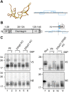
Disulfide rearrangements in the disintegrin domain of ADAM10 are independent of ERdj5 and ERp57. (A) Ribbon diagram of the ADAM10 disintegrin domain (left) with disulfide bonds in yellow, drawn using PDB file 6BE6. The diagram (right) shows the disulfide connectivity. (B) Topology diagram of the disintegrin 146 construct (left) showing the signal peptide (SP), neo-epitope tag (NE), the disintegrin domain sequence (Disintegrin), the V5 sequence and five methionine residues. The ribosome diagram (right) shows the expected ER exposure of the stalled disintegrin 146 intermediate in terms of the cysteine residues that make up the disulfide bonds. (C) Non-reducing SDS-PAGE of the radiolabelled disintegrin 146 intermediate. Translations were performed with the indicated cell lines in oxidising lysate and then recovered with or without G6P. Samples were immunoisolated using either a V5 or neoepitope (NE) antibody. A reduced control is also shown (Red). Wt, wild-type. Representative data is shown in this figure from at least three independent repeats. Annotations highlight immunoisolated reduced preprotein (*) or mature protein (○), and disulfide-bonded monomers (<) or dimers (▼). The vertical line highlights a smear of disulfide-bonded protein.
Fig. 5.
Fig. 5.
Knockout of ERdj5 and ERp57 together disrupts the folding of LDLr. (A) Topology diagram of wild-type LDLr, showing the location of the LA repeat domains (1–7), the EGF domains (E1, E2 and E3) and the β-propeller domain. The location of the C7 epitope is also indicated. The topology diagram of the truncated construct used in this study (LDLr-789-V5), consisting of LDLr residues 1–789 with the addition of five methionine residues and a V5 tag at the C-terminus. The green ribbon diagram shows the structure of the LA1 domain with disulfide bonds in yellow, drawn using PDB file 1LDL. (B) Non-reducing SDS-PAGE of radiolabelled LDLr-789-V5 produced as a stalled intermediate (diagram right) in translation reactions containing microsomes and either reduced (Red), balanced (Bal) or oxidised (Ox) lysates. (C–E) Non-reducing (non-Red) or reducing (Red) SDS-PAGE showing radiolabelled translation product of stalled LDLr-789-V5 translated with the indicated cell lines in oxidising lysate and recovered with or without G6P as indicated. Control samples translated in reducing lysate (+DTT) are also shown. Samples were immunoisolated using a V5 or C7 antibody. Annotations highlight differences between the mobility of translation products formed in the absence of G6P and the presence of Wt (*) or ERp57 KO (<) cells. In E, samples are EndoH treated as indicated. Wt, wild-type. All gels in this figure are representative of at least three independent experiments.
Fig. 6.
Fig. 6.
The cytosolic reducing pathway supplies reducing power to ERp57 and ERdj5 to enable efficient folding of LDLr, but both enzymes also influence folding when the pathway is inactive. Phosphoimages of SDS-PAGE gels run under reducing (Red) or non reducing (Non-red) conditions showing radiolabelled translation product of (A) LDLr-789-V5 and (B) LDLr-Wt translated with SP cells made from the indicated cell lines. Samples were initially translated in oxidising lysate before addition of G6P after 30 mins to +G6P samples as indicated. Samples were either immunoisolated using a V5 antibody or isolated using ConA beads. Annotations highlight the increased mobility of translation products formed in the presence of ERp57 KO (<) or ERp57/ERdj5 double KO (<<) cells. Wt, wild-type. All gels are representative of at least three independent experiments.
Fig. 7.
Fig. 7.
A model for the role of ERp57 and ERdj5 in the ER reducing pathway. (A) The correct folding of different substrates requires specific oxidoreductases. LDLr and β1-integrin are glycoproteins, whereas the disintegrin domain is not glycosylated. (B) The cytosolic reducing pathway is linked to isomerases such as ERp57 in the ER via reductases such as ERdj5 or ER selenoproteins. These reductases might well reduce folding proteins as well as other PDI family members. Other electron doners, such as ascorbate, might contribute to the reduction of ER proteins.

Similar articles

Cited by

References

    1. Addinsall, A. B., Wright, C. R., Andrikopoulos, S., Van Der Poel, C. and Stupka, N. (2018). Emerging roles of endoplasmic reticulum-resident selenoproteins in the regulation of cellular stress responses and the implications for metabolic disease. Biochem. J. 475, 1037-1057. 10.1042/BCJ20170920 - DOI - PubMed
    1. Askari, J. A., Tynan, C. J., Webb, S. E., Martin-Fernandez, M. L., Ballestrem, C. and Humphries, M. J. (2010). Focal adhesions are sites of integrin extension. J. Cell Biol. 188, 891-903. 10.1083/jcb.200907174 - DOI - PMC - PubMed
    1. Bazzoni, G., Shih, D. T., Buck, C. A. and Hemler, M. E. (1995). Monoclonal antibody 9EG7 defines a novel β1 integrin epitope induced by soluble ligand and manganese, but inhibited by calcium. J. Biol. Chem. 270, 25570-25577. 10.1074/jbc.270.43.25570 - DOI - PubMed
    1. Behnke, J., Mann, M. J., Scruggs, F. L., Feige, M. J. and Hendershot, L. M. (2016). Members of the Hsp70 family recognize distinct types of sequences to execute ER quality control. Mol. Cell 63, 739-752. 10.1016/j.molcel.2016.07.012 - DOI - PMC - PubMed
    1. Cao, X., Lilla, S., Cao, Z., Pringle, M. A., Oka, O. B. V., Robinson, P. J., Szmaja, T., Van Lith, M., Zanivan, S. and Bulleid, N. J. (2020). The mammalian cytosolic thioredoxin reductase pathway acts via a membrane protein to reduce ER-localised proteins. J. Cell Sci. 133, jcs241976. 10.1242/jcs.241976 - DOI - PMC - PubMed

Publication types

MeSH terms