. 2023 Feb 28;97(2):e0194722.
doi: 10.1128/jvi.01947-22. Epub 2023 Jan 19.

Chicken or Porcine Aminopeptidase N Mediates Cellular Entry of Pseudoviruses Carrying Spike Glycoprotein from the Avian Deltacoronaviruses HKU11, HKU13, and HKU17

Affiliations

Chicken or Porcine Aminopeptidase N Mediates Cellular Entry of Pseudoviruses Carrying Spike Glycoprotein from the Avian Deltacoronaviruses HKU11, HKU13, and HKU17

Qi-Zhang Liang et al. J Virol. .

Erratum in

Abstract

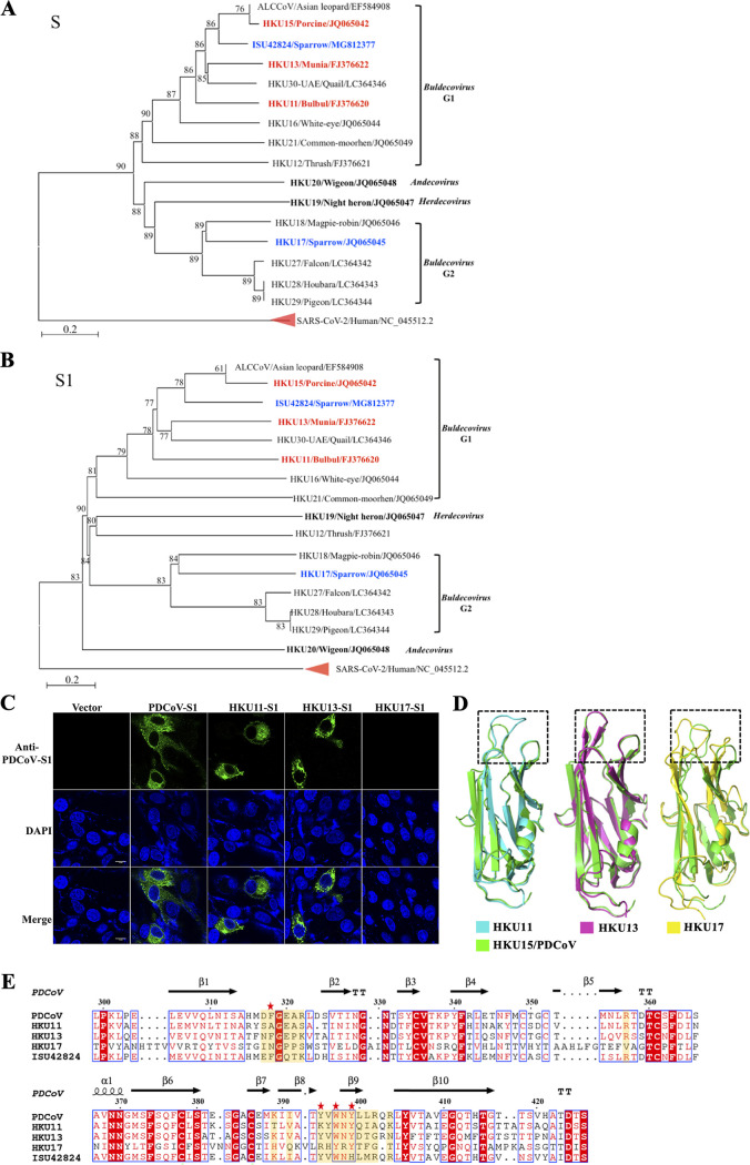
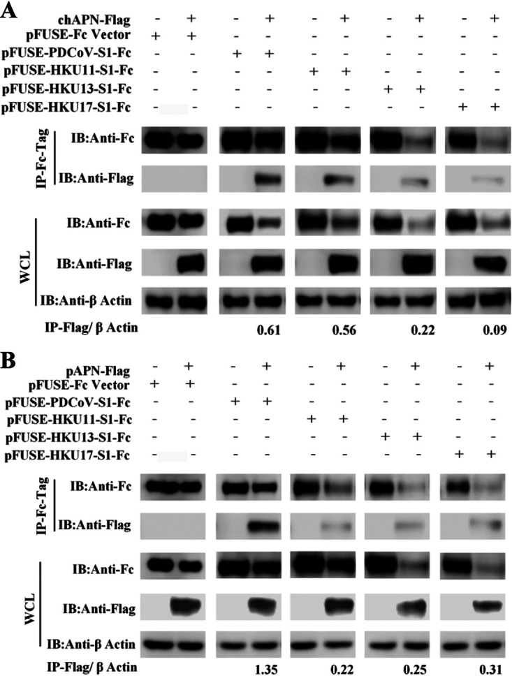
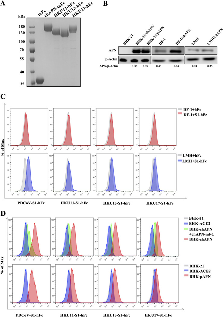
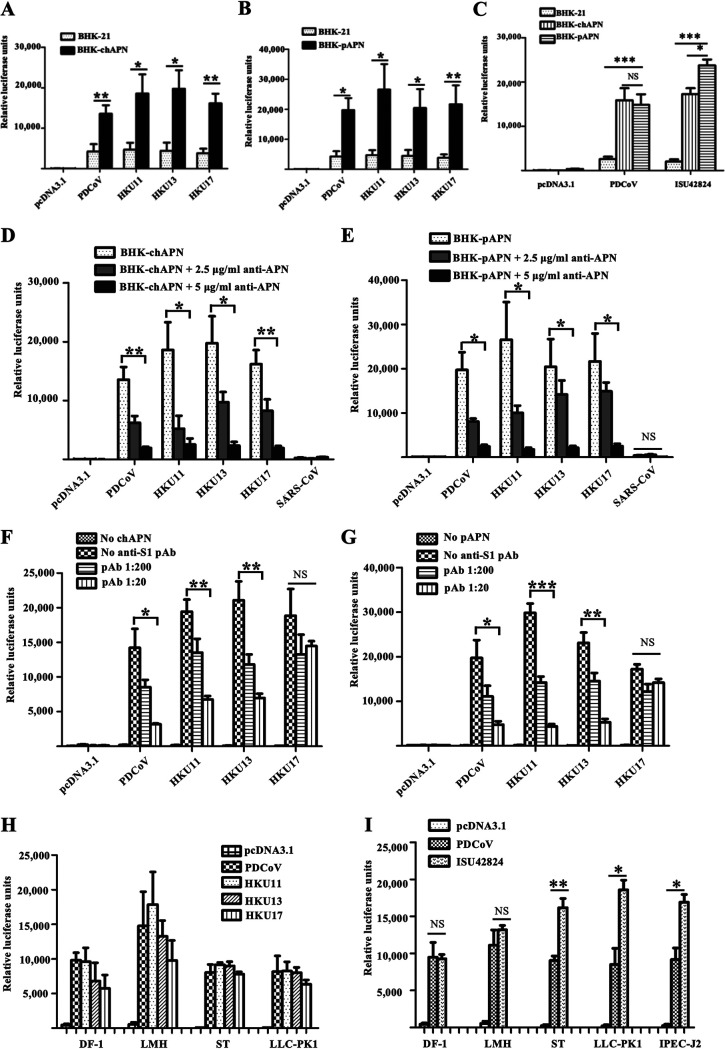
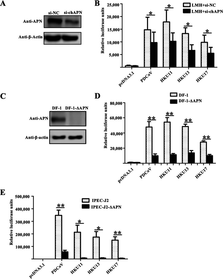
Members of deltacoronavirus (DCoV) have mostly been identified in diverse avian species as natural reservoirs, though the porcine DCoV (PDCoV) is a major swine enteropathogenic virus with global spread. The important role of aminopeptidase N (APN) orthologues from various mammalian and avian species in PDCoV cellular entry and interspecies transmission has been revealed recently. In this study, comparative analysis indicated that three avian DCoVs, bulbul DCoV HKU11, munia DCoV HKU13, and sparrow DCoV HKU17 (Chinese strain), and PDCoV in the subgenera Buldecovirus are grouped together at whole-genome levels; however, the spike (S) glycoprotein and its S1 subunit of HKU17 are more closely related to night heron DCoV HKU19 in Herdecovirus. Nevertheless, the S1 protein of HKU11, HKU13, or HKU17 bound to or interacted with chicken APN (chAPN) or porcine APN (pAPN) by flow cytometry analysis of cell surface expression of APN and by coimmunoprecipitation in APN-overexpressing cells. Expression of chAPN or pAPN allowed entry of pseudotyped lentiviruses with the S proteins from HKU11, HKU13 and HKU17 into nonsusceptible cells and natural avian and porcine cells, which could be inhibited by the antibody against APN or anti-PDCoV-S1. APN knockdown by siRNA or knockout by CRISPR/Cas9 in chicken or swine cell lines significantly or almost completely blocked infection of these pseudoviruses. Hence, we demonstrate that HKU11, HKU13, and HKU17 with divergent S genes likely engage chAPN or pAPN to enter the cells, suggesting a potential interspecies transmission from wild birds to poultry and from birds to mammals by certain avian DCoVs. IMPORTANCE The receptor usage of avian deltacoronaviruses (DCoVs) has not been investigated thus far, though porcine deltacoronavirus (PDCoV) has been shown to utilize aminopeptidase N (APN) as a cell receptor. We report here that chicken or porcine APN also mediates cellular entry by three avian DCoV (HKU11, HKU13, and HKU17) spike pseudoviruses, and the S1 subunit of three avian DCoVs binds to APN in vitro and in the surface of avian and porcine cells. The results fill the gaps in knowledge about the avian DCoV receptor and elucidate important insights for the monitoring and prevention of potential interspecies transmission of certain avian DCoVs. In view of the diversity of DCoVs, whether this coronavirus genus will cause novel virus to emerge in other mammals from birds, are worthy of further surveillance and investigation.

Keywords: aminopeptidase N (APN); avian deltacoronavirus; interspecies transmission; porcine deltacoronavirus (PDCoV); receptor.


Conflict of interest statement

The authors declare no conflict of interest.

Figures

FIG 1
FIG 1
Phylogenetic analysis, antigenic cross-reactivity, and structural comparison of the S and S1 proteins of selected deltacoronaviruses (DCoVs). (A) Phylogenetic tree of representative DCoVs generated based upon the S proteins. The tree was constructed by the neighbor-joining method (MEGA6 software) using SARS-CoV-2 as an outgroup. Bootstrap values are indicated for each node from 1,000 resamplings. The names of the CoV strains, hosts, and GenBank accession numbers are shown. (B) Phylogenetic tree of representative DCoVs generated based upon the S1 proteins. (C) Cross-reactivity of the anti-PDCoV-S1 polyclonal Ab with three avian DCoV S1 proteins. BHK-21 cells were transiently transfected with recombinant plasmids harboring the S1 genes from PDCoV or different avian DCoVs, and IFA was performed at 48 h posttransfection using polyclonal antibodies raised against PDCoV-S1. Fluorescent images were obtained using a confocal laser scanning microscope (Fluoviewver.3.1). (D) Homologous modeling of HKU11 RBD (cyan), HKU13 RBD (magentas), and HKU17 RBD (yellow) based on PDCoV/HKU15 spike glycoprotein (green; Protein Data Bank accession code 6BFU) were built by PyMOL software. Dashed boxes indicate two protruding tips supposedly mediating a receptor-RBD interaction. (E) Structural-based sequence alignment of RBD between PDCoV, HKU11, HKU13, HKU17, and US SpDCoV-ISU42824. The secondary structure elements determined in PDCoV (6BFU) are indicated above the sequences. Two protruding tips and surrounding sequences boxed in panel 4D are shaded in yellow. Four key aromatic residues (Phe-318, Tyr-394, Trp-396, and Tyr-398) at the tips of PDCoV RBD are marked with red stars.
FIG 2
FIG 2
Coimmunoprecipitation analysis of deltacoronavirus S1 proteins binding to chAPN or pAPN. 293T cells were cotransfected with plasmids carrying (A) chAPN or (B) pAPN with a FLAG tag and pFUSE plasmids including S1 genes from PDCoV or the avian DCoVs HKU11, HKU13, and HKU17 with hFc fusion tags. At 36 h posttransfection, the whole-cell lysate (WCL) was preadsorbed onto protein A-conjugated agarose beads prior to Western blot analysis (upper panels) or was used for Western blot directly (lower panels). Protein complexes were detected using an HRP-conjugated anti-Flag Ab or HRP-conjugated anti-Fc Ab as appropriate. β-actin was used as the loading control.
FIG 3
FIG 3
Soluble avian DCoV-S1 proteins bind to chicken permissive cells and nonsusceptible BHK-21 cells expressing chAPN or pAPN. (A) SDS-PAGE and Western blot analysis of purified mFc and hFc fusion proteins (mFc, chAPN-mFc, HKU11-S1-hFc, HKU13-S1-hFc and HKU17-S1-hFc) expressed in the supernatants of 293T cells. (B) Exogenous expressions of chAPN in BHK-21 cells and endogenous expression of chAPN orthologues in chicken DF-1 and LMH cells by Western blot analysis. β-actin was used as the loading control. (C) Flow cytometry analysis of three avian DCoV-S1-hFc proteins (20 μg/mL) and PDCoV-S1-hFc (10 μg/mL) binding to DF-1 or LMH cells. Equal amounts of hFc binding were used as the control (gray histogram). Cell surface binding was detected by a FITC-conjugated anti-human IgG Fc. (D) Flow cytometry analysis of three avian DCoV-S1-hFc proteins (20 μg/mL) and PDCoV-S1-hFc (10 μg/mL) binding to BHK-21 cells (gray histogram) and BHK-21 cells transfected with APN-expressing plasmids (BHK-chAPN or BHK-pAPN; red histograms). Soluble APN-mFc (10 μg/mL) preincubation was able to block avian DCoV-S1-hFc and PDCoV-S1-hFc binding to BHK-chAPN (green histogram). BHK-21 cells transfected with ACE2-expressing plasmid (BHK-ACE2) served as a negative control (blue histogram). Cell surface binding was detected by a FITC-conjugated antihuman IgG Fc.
FIG 4
FIG 4
APN-mediated pseudovirus entry into BHK-21 cells and avian and porcine natural cell lines. (A and B) BHK-21 cells were transfected or not with (A) chAPN or (B) pAPN and subsequently infected by lentiviruses pseudotyped with S proteins from PDCoV, HKU11, HKU13, or HKU17, with a mock pseudovirus (the empty expression vector pcDNA3.1 as packaging plasmid) as control. (C) BHK-21 cells were transfected or not with chAPN or pAPN and subsequently infected by the pseudoviruses (mock, PDCoV, or ISU42824). (D and E) BHK cells transfected with (D) chAPN or (E) pAPN were preincubated with anti-APN antibody at different concentrations to test for cell entry inhibition of the pseudoviruses (PDCoV, HKU11, HKU13, or HKU17) with mock pseudovirus (pcDNA3.1) and irrelevant pseudovirus with SARS-CoV S protein as controls. (F and G) BHK-21 cells transfected with empty plasmid (no APN), (F) chAPN, or (G) pAPN were infected by lentiviruses pseudotyped with PDCoV S, HKU11 S, HKU13 S, and HKU17 S proteins with mock pseudovirus (pcDNA3.1) as control. The cells were also preincubated with anti-PDCoV S1 neutralizing polyclonal antibody (pAb) to test cross neutralization reaction. (H) Cell lines with endogenous expression of chAPN (DF-1 and LMH) or pAPN (ST and LLC-PK1) were infected with the indicated pseudoviruses. (I) DF-1, LMH, ST, LLC-PK1, and IPEC-J2 cells were infected with the pseudoviruses (mock, PDCoV, or ISU42824). Cell entry efficiency was measured by luciferase activity at 48 h postinfection (hpi); error bars indicate SEM (two-tailed t test; *, P < 0.05; **, P < 0.01; ***, P < 0.001; n ≥ 3).
FIG 5
FIG 5
Effects of APN knockdown/knockout on pseudovirus entry into avian and porcine susceptible cells. (A) The efficacy of si-chAPN to knockdown expression of chAPN was determined by Western blot using an antibody specific for APN. β-actin was used as the loading control. (B) LMH cells were transfected with specific (si-chAPN) or nonspecific (si-NC) silencing RNA for 24 h, followed by infection with the indicated pseudoviruses. (C) Western blot analysis showing loss of chAPN expression in DF-1-ΔchAPN cells compared with the significant APN expression in DF-1 cells. β-actin was used as the loading control. (D) DF-1 and DF-1-ΔchAPN cells were infected with the indicated pseudoviruses. (E) IPEC-J2 and IPEC-J2-ΔpAPN cells were infected with the indicated pseudoviruses. Cell entry efficiencies were determined by luciferase activity measurement after 48 hpi; error bars indicate SEM (two-tailed t test; *, P < 0.05; **, P < 0.01; ***, P < 0.001; n ≥ 3).
FIG 6
FIG 6
Trypsin promotes but is not essential for PDCoV and avian DCoV S-mediated entry into porcine cells. (A) Pseudotyped lentiviruses displaying PDCoV, HKU11, HKU13, or HKU17 S proteins or empty vector pcDNA3.1 (mock) were incubated with trypsin (5 μg/mL) prior to entry assays in IPEC-J2 cells. Similar experiments were carried out using a gradient of different trypsin concentrations prior to infection in (B) ST cells or (C) LLC-PK1 cells. Cell entry efficiencies were assayed by luciferase activity measurement after 48 hpi; error bars indicate SEM (two-tailed t test; *, P < 0.05; **, P < 0.01; ***, P < 0.001; n ≥ 3).

Similar articles

Cited by

References

    1. de Groot RJ, Baker SC, Baric R, Enjuanes L, Gorbalenya AE, Holmes KV, Perlman S, Poon L, Rottier PJM, Talbot PJ, Woo PCY, Ziebuhr J. 2011. Coronaviridae, p 806–828. In King AMQ, Adams MJ, Carstens EB, Lefkowitz EJ (ed), Virus taxonomy: ninth report of the international committee on taxonomy of viruses. Elsevier Academic Press, London, England.
    1. Woo PCY, Lau SKP, Huang Y, Yuen K-Y. 2009. Coronavirus diversity, phylogeny and interspecies jumping. Exp Biol Med (Maywood) 234:1117–1127. 10.3181/0903-MR-94. - DOI - PubMed
    1. Woo PC, Lau SK, Lam CS, Lau CC, Tsang AK, Lau JH, Bai R, Teng JL, Tsang CC, Wang M, Zheng BJ, Chan KH, Yuen KY. 2012. Discovery of seven novel Mammalian and avian coronaviruses in the genus deltacoronavirus supports bat coronaviruses as the gene source of alphacoronavirus and betacoronavirus and avian coronaviruses as the gene source of gammacoronavirus and deltacoronavirus. J Virol 86:3995–4008. 10.1128/JVI.06540-11. - DOI - PMC - PubMed
    1. Woo PC, Lau SK, Lam CS, Lai KK, Huang Y, Lee P, Luk GS, Dyrting KC, Chan KH, Yuen KY. 2009. Comparative analysis of complete genome sequences of three avian coronaviruses reveals a novel group 3c coronavirus. J Virol 83:908–917. 10.1128/JVI.01977-08. - DOI - PMC - PubMed
    1. Lau SKP, Wong EYM, Tsang CC, Ahmed SS, Au-Yeung RKH, Yuen KY, Wernery U, Woo PCY. 2018. Discovery and sequence analysis of four deltacoronaviruses from birds in the middle east reveal interspecies jumping with recombination as a potential mechanism for avian-to-avian and avian-to-mammalian transmission. J Virol 92. 10.1128/JVI.00265-18. - DOI - PMC - PubMed

Publication types

Substances

LinkOut - more resources