. 2023 Feb 28;14(1):e0300122.
doi: 10.1128/mbio.03001-22. Epub 2023 Jan 19.

A Diverged Transcriptional Network for Usage of Two Fe-S Cluster Biogenesis Machineries in the Delta-Proteobacterium Myxococcus xanthus

Affiliations

A Diverged Transcriptional Network for Usage of Two Fe-S Cluster Biogenesis Machineries in the Delta-Proteobacterium Myxococcus xanthus

Mathieu Sourice et al. mBio. .

Abstract

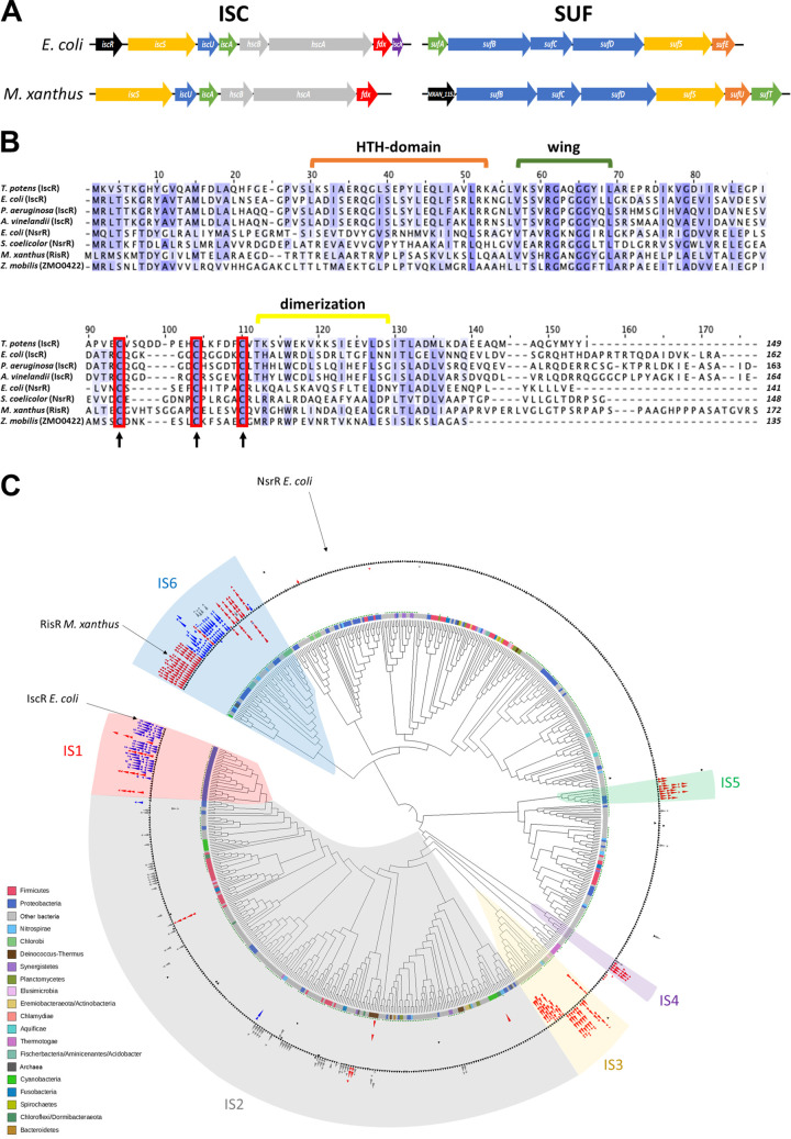
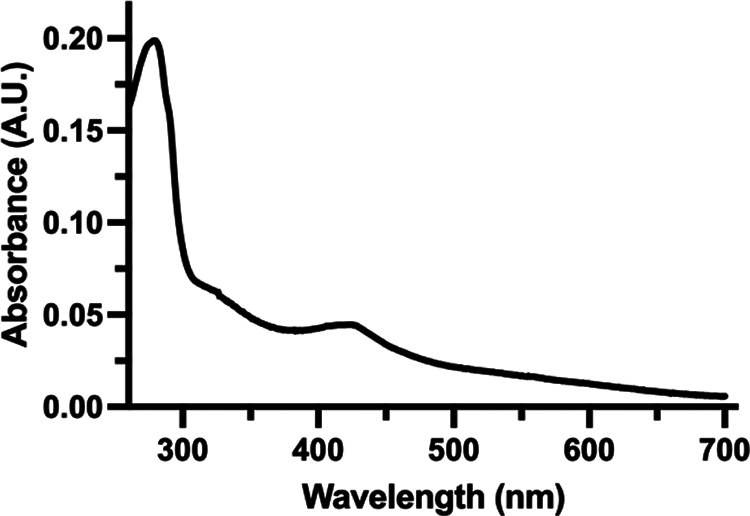
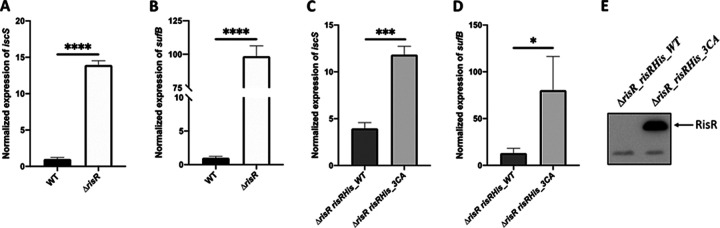
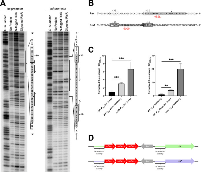
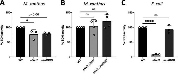
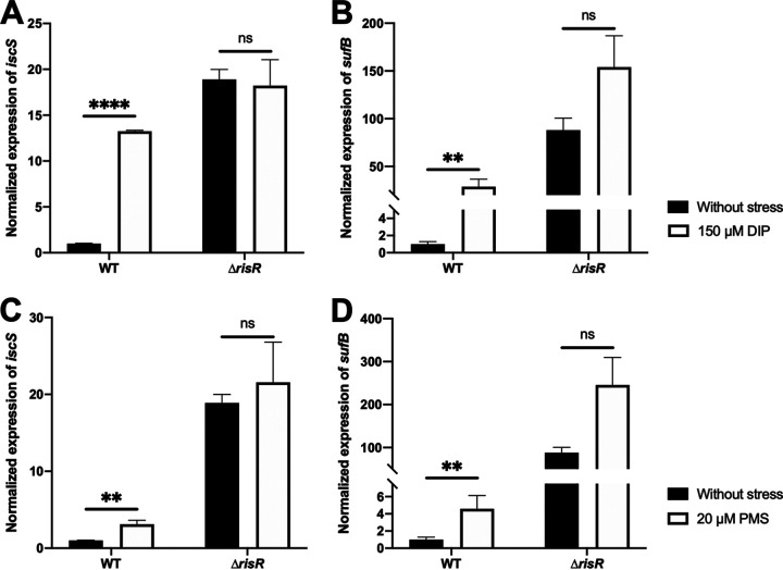
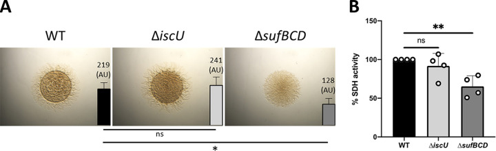
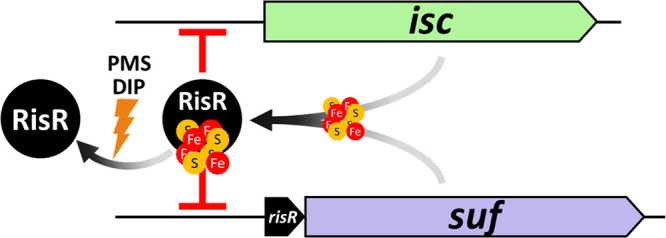
Myxococcus xanthus possesses two Fe-S cluster biogenesis machineries, ISC (iron-sulfur cluster) and SUF (sulfur mobilization). Here, we show that in comparison to the phylogenetically distant Enterobacteria, which also have both machineries, M. xanthus evolved an independent transcriptional scheme to coordinately regulate the expression of these machineries. This transcriptional response is directed by RisR, which we show to belong to a phylogenetically distant and biochemically distinct subgroup of the Rrf2 transcription factor family, in comparison to IscR that regulates the isc and suf operons in Enterobacteria. We report that RisR harbors an Fe-S cluster and that holo-RisR acts as a repressor of both the isc and suf operons, in contrast to Escherichia coli, where holo-IscR represses the isc operon whereas apo-IscR activates the suf operon. In addition, we establish that the nature of the cluster and the DNA binding sites of RisR, in the isc and suf operons, diverge from those of IscR. We further show that in M. xanthus, the two machineries appear to be fully interchangeable in maintaining housekeeping levels of Fe-S cluster biogenesis and in synthesizing the Fe-S cluster for their common regulator, RisR. We also demonstrate that in response to oxidative stress and iron limitation, transcriptional upregulation of the M. xanthus isc and suf operons was mediated solely by RisR and that the contribution of the SUF machinery was greater than the ISC machinery. Altogether, these findings shed light on the diversity of homeostatic mechanisms exploited by bacteria to coordinately use two Fe-S cluster biogenesis machineries. IMPORTANCE Fe-S proteins are ubiquitous and control a wide variety of key biological processes; therefore, maintaining Fe-S cluster homeostasis is an essential task for all organisms. Here, we provide the first example of how a bacterium from the Deltaproteobacteria branch coordinates expression of two Fe-S cluster biogenesis machineries. The results revealed a new model of coordination, highlighting the unique and common features that have independently emerged in phylogenetically distant bacteria to maintain Fe-S cluster homeostasis in response to environmental changes. Regulation is orchestrated by a previously uncharacterized transcriptional regulator, RisR, belonging to the Rrf2 superfamily, whose members are known to sense diverse environmental stresses frequently encountered by bacteria. Understanding how M. xanthus maintains Fe-S cluster homeostasis via RisR regulation revealed a strategy reflective of the aerobic lifestyle of this organsim. This new knowledge also paves the way to improve production of Fe-S-dependent secondary metabolites using M. xanthus as a chassis.

Keywords: Fe-S cluster biogenesis; Fe-S cluster homeostasis; Myxococcus xanthus; Rrf2-type regulator; iron starvation; oxidative stress; transcription regulation.


Conflict of interest statement

The authors declare no conflict of interest.

Figures

FIG 1
FIG 1
Identification of the Fe-S cluster biogenesis components in M. xanthus and phylogeny of Rrf2 family proteins. (A) Comparison of the genomic organization of the isc and suf gene clusters in E. coli and M. xanthus. Same color indicates gene function conservation, transcriptional regulator (black), cysteine desulfurase (yellow), sulfur acceptor (orange), scaffold (blue), Fe-S cluster carrier (green), chaperone/cochaperone, (gray), and electron donor (red). Purple indicates the iscX gene whose function in the Fe-S cluster biogenesis process remains to be characterized. (B) Multiple sequence alignment between M. xanthus RisR, IscR, and NrsR homologs from different bacterial species (Thermincola potens, E. coli, Pseudomonas aeruginosa, Azotobacter vinelandii, Streptomyces coelicolor, and Zymomonas mobilis). The protein sequences have been aligned using Clustal Omega, and BLOSUM 62 has been used for scoring alignments (threshold 25%). The red boxes represent the cysteine residues involved in the binding of the Fe-S cluster in E. coli IscR and conserved in M. xanthus RisR. Strictly conserved and partly conserved amino acids are highlighted in a blue color gradient from dark to light, respectively. Residues involved in IscR in wing and HTH-domain (green and orange brackets, respectively) and the dimerization helix (yellow bracket) are shown. (C) Cladogram of Rrf2 family proteins phylogeny. The circles at branches correspond to ultrafast bootstrap >95%. The strip around the tree indicates the corresponding major phyla at the bottom left side of the figure. The green dots indicate the presence of the three cysteines involved in the Fe-S cluster binding. Black triangles represent Rrf2 encoding genes, and the red, blue, and gray triangles indicate the SUF, ISC, and MIS machineries, respectively, at the rrf2 vicinity. Major groups in synteny with Fe-S cluster machinery genes are indicated (IS1-6). IS, iron sulfur.
FIG 2
FIG 2
The UV-visible spectrum suggests that RisR binds a Fe-S cluster. UV-visible spectrum of anaerobically isolated N-terminal Strep-tag RisR protein (6.2 μM). The spectrum shows two characteristic peaks with maxima at ~330 nm and ~420 nm that might indicate the presence of [4Fe-4S] clusters.
FIG 3
FIG 3
Holo-RisR downregulates the expression of both isc and suf operons. RT-qPCR analysis of the transcriptional levels of isc (A, C) and suf (B, D) operons in M. xanthus strains WT (panels A and B, black bars), ΔrisR (panels A and B, white bars), ΔrisR risR_WT (panels C and D, dark gray bars), and ΔrisR risR_3CA (panels C and D, light gray bars). The cultures used for RNA preparation were grown in CYE medium, and relative mRNA levels for each gene were normalized to 16S RNA levels. The amount of transcript in the WT strain is taken as reference. Error bars represent the standard deviation of three biological replicates. Statistical analysis was performed with Student’s test (panels A and B, ****, P < 0.0001; panel C, ***, P < 0.001; panel D, *, P < 0.05). Statistical analysis indicated that the difference between the expression levels of isc, or suf, in the ΔrisR risRHis_3CA mutant compared to ΔrisR was not significant. (E) Twenty-five micrograms of total protein extracts were analyzed for RisR protein levels by Western blot using antibodies raised against the histidine tag.
FIG 4
FIG 4
Holo-RisR binds to imperfect palindromic motifs in isc and suf promoters. (A) DNase I footprinting was used to identify the region where RisR binds within the suf and isc promoters from M. xanthus. RisR (1 μM) with or without the N-terminal StrepII tag was incubated with approximately 10 nM DNA prior to DNase I treatment. The vertical line indicates the protection area in each promoter. In the sequence, the gray box indicates the RisR protection region, and half arrows show bases corresponding to a palindromic motif present in both promoters. The predicted −10 and −35 hexamer RNA polymerase binding sites are boxed, and * shows a hypersensitive site evident after protein binding. (B) Sequence of RisR-binding motif identified in the suf and isc operon promoters from M. xanthus. The putative −10 and −35 hexamer RNA polymerase binding sites are boxed. The RisR-binding regions identified by DNase I footprinting are highlighted in gray, and in bold are shown bases corresponding to a palindromic motif present in both promoters. Mismatches in one of the half-sites are indicated in italic, and * shows hypersensitive sites. The nucleotides mutated by substitution are shown in red. (C) Relative fluorescence of the WT strain carrying the 3mCherry fused to the WT or mutated (4mut) promoters of suf or isc operons, and the ΔrisR strain with the WT promoters fused to 3mCherry. Results represent the normalization of the fluorescence over the OD600nm. These data were normalized to the WT strain carrying 3mCherry fused to the WT promoter of suf or isc operons. Each point represents the mean from 6 experiments; error bars represent standard deviations. Statistical analysis was performed with a Dunnet test; **, P < 0.01; ***, P < 0.001. (D) Schemes of the chromosomal reporter fusions used are shown.
FIG 5
FIG 5
SDH activity is not affected in strains lacking ISC or SUF machinery. (A) SDH activity was determined by measuring polarographically the O2 consumption using succinate as the substrate from membranes of the M. xanthus WT (black bar), ΔiscU (light gray bar), and ΔsufBCD (dark gray bar) strains. Bar graphs show the percentage of O2 consumption relative to the WT set to 100%. Error bars show the standard error of three biological replicates. Statistical analysis was performed using the Dunnet test (*, P < 0.05). (B) SDH activities were determined by measuring the O2 consumption from the M. xanthus WT (black), ΔrisR ΔiscU (light gray bar), and ΔrisR ΔsufBCD (dark gray bar) membranes using succinate as electron donor. Bar graphs show the percentage of O2 consumption relative to the WT set to 100%. Error bars show the standard error of four biological replicates. Statistical analysis was performed using the Dunnet test (ns, not significant). (C) SDH activities were determined by measuring the O2 consumption from the E. coli WT (black bar), ΔiscU (light gray bar), and ΔsufBCD (dark gray bar) membranes using succinate as electron donor. Bar graphs show the percentage of O2 consumption relative to the WT set to 100%. Error bars show the standard error of three biological replicates. Statistical analysis was performed using the Dunnet test (****, P < 0.0001; ns, not significant).
FIG 6
FIG 6
Activation of isc and suf operons during iron starvation and under oxidative stress depends only on RisR. Relative expression of (A) isc and (B) suf operons in M. xanthus WT and ΔrisR cells untreated (black bars) or treated with DIP (white bars) strains. The cultures used for RNA preparation were grown from OD600nm 0.2 to 0.5 on CYE containing or not 150 μM DIP. The relative mRNA levels for each gene were normalized to 16S RNA levels. The amount of transcript in the WT strain in the absence of stress is taken as reference. Error bars show the standard error of three biological replicates. Statistical analysis was performed with a Sidak test (**, P < 0.01; ****, P < 0.0001; ns, not significant). Relative expression of (C) isc and (D) suf operons in M. xanthus WT and ΔrisR cells untreated (black bars) or treated with PMS (white bars) strains. The cultures used for RNA preparation were grown until OD600nm 0.5 on CYE supplemented or not with 20 μM PMS. The amount of transcript in the WT strain grown in the absence of stress is taken as reference. Error bars show the standard error of three biological replicates. Statistical analysis was performed with a Sidak test (**, P < 0.01; ns, not significant).
FIG 7
FIG 7
The SUF machinery is required during iron starvation for M. xanthus growth and motility. (A) Colony morphology and motility of the WT, ΔiscU, and ΔsufBCD strains in the presence of 100 μM DIP. Pictures of the colonies of M. xanthus WT, ΔiscU, and ΔsufBCD strains were taken after 48 h of growth on CYE plates with 0.5% of agar and 100 μM DIP. The colony expansion of WT, ΔiscU, and ΔsufBCD strains at 48 h is shown as a bar (right side of each photograph). The colony expansion is calculated by subtracting the t0 spot diameter from the diameter of the colony at 48 h (tf) and dividing by 2 (scheme, Fig. S4B). The colony expansion is expressed in arbitrary units, and error bars show the standard error of three biological replicates. Statistical analysis was performed with a Dunnet test (*, P < 0.05; ns, not significant). (B) SDH activity was determined by measuring the O2 consumption from membranes of the M. xanthus WT (black bar), ΔiscU (light gray bar), and ΔsufBCD (dark gray bar) using succinate as the substrate. Membranes were prepared from cultures grown in CYE medium in the presence of 100 μM DIP at OD600nm between 0.4 and 0.6. Bar graphs show the percentage of O2 consumption relative to the WT set to 100%. Statistical analysis was performed with a Dunnet test (**, P < 0.01; ns, not significant).
FIG 8
FIG 8
Model of regulation of isc and suf operons by RisR in M. xanthus. Under optimal growth conditions, both ISC and SUF can mature RisR. The [4Fe-4S]-RisR (holo-form) acts as a repressor of both operons. This repression can be removed in stress conditions, such as iron starvation or oxidative stress. These stress conditions lead to a lack of or an oxidation of the iron atoms of the RisR Fe-S cluster, presumably converting RisR to its apo-form. Under its apo-form, RisR cannot bind to the promoters of isc and suf; thus, both operons are upregulated.

References

    1. Beinert H. 2000. Iron-sulfur proteins: ancient structures, still full of surprises. J Biol Inorg Chem 5:2–15. doi:10.1007/s007750050002. - DOI - PubMed
    1. Fontecave M. 2006. Iron-sulfur clusters: ever-expanding roles. Nat Chem Biol 2:171–174. doi:10.1038/nchembio0406-171. - DOI - PubMed
    1. Kiley PJ, Beinert H. 2003. The role of Fe-S proteins in sensing and regulation in bacteria. Curr Opin Microbiol 6:181–185. doi:10.1016/s1369-5274(03)00039-0. - DOI - PubMed
    1. Lénon M, Arias-Cartín R, Barras F. 2022. The Fe-S proteome of Escherichia coli: prediction, function, and fate. Met Integr Biometal Sci 14:mfac022. doi:10.1093/mtomcs/mfac022. - DOI - PubMed
    1. Ayala-Castro C, Saini A, Outten FW. 2008. Fe-S cluster assembly pathways in bacteria. Microbiol Mol Biol Rev 72:110–125. doi:10.1128/MMBR.00034-07. - DOI - PMC - PubMed

Publication types

MeSH terms

LinkOut - more resources