. 2023 Mar;61(1-2):e23507.
doi: 10.1002/dvg.23507. Epub 2023 Jan 19.

A knockout-first model of H3f3a gene targeting leads to developmental lethality

Affiliations

A knockout-first model of H3f3a gene targeting leads to developmental lethality

Kelly Bush et al. Genesis. 2023 Mar.

Abstract

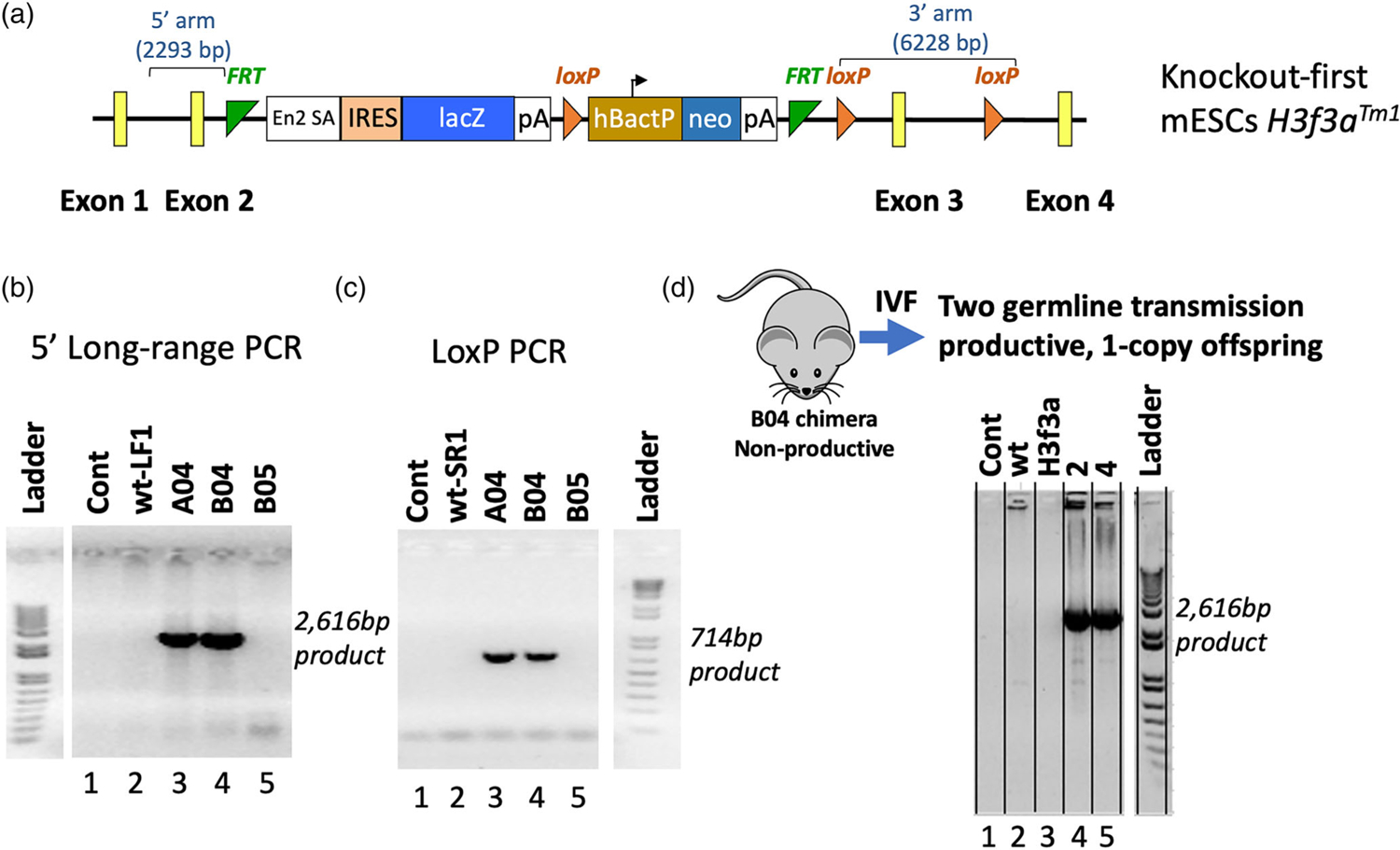
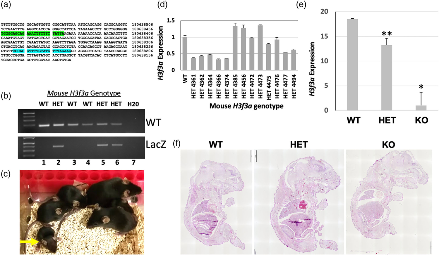
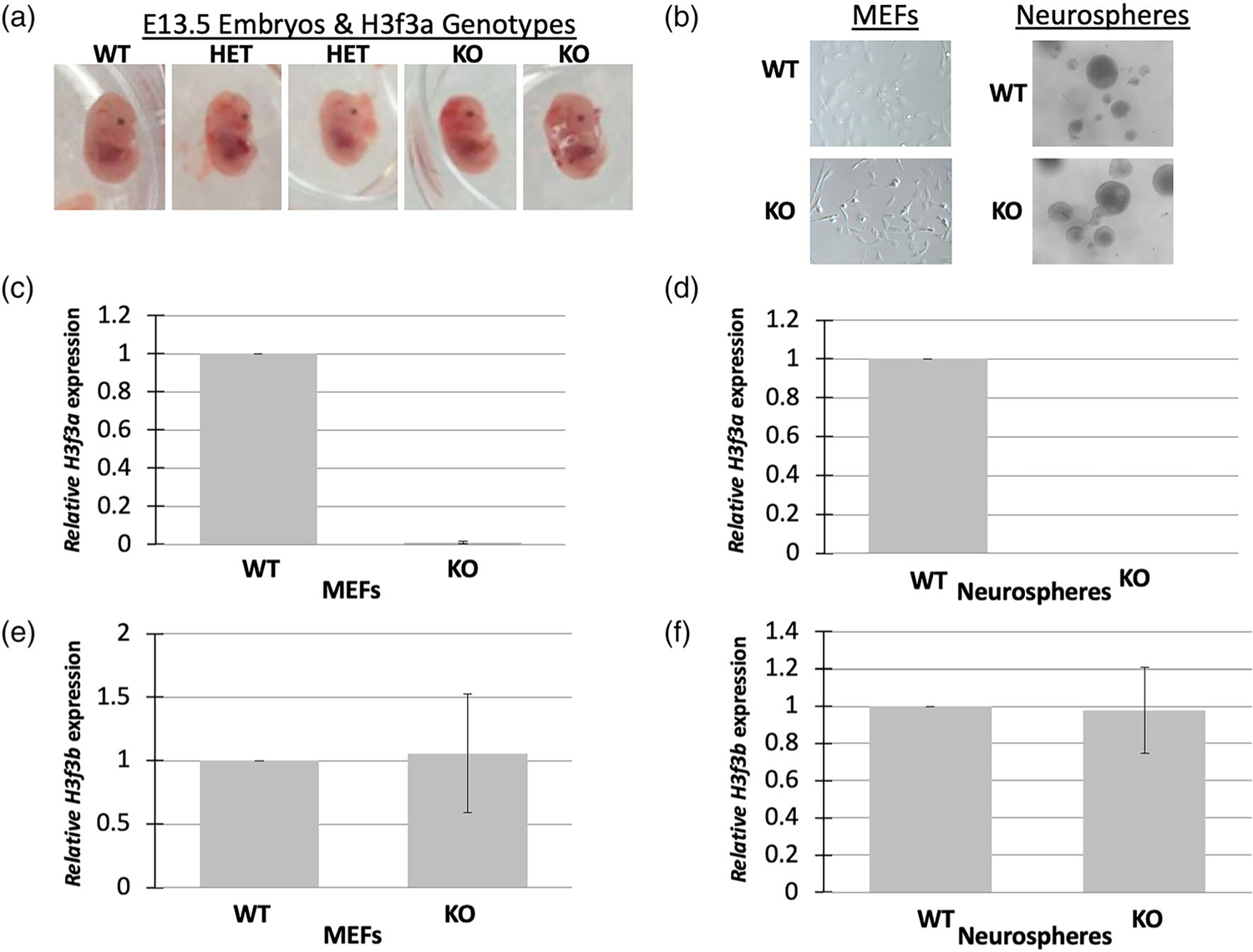
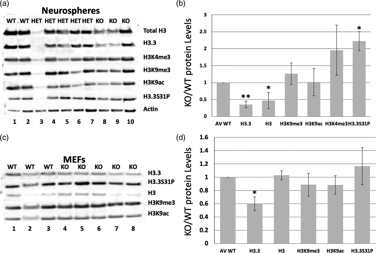
Histone variant H3.3 is encoded by two genes, H3f3a and H3f3b, which can be expressed differentially depending on tissue type. Previous work in our lab has shown that knockout of H3f3b causes some neonatal lethality and infertility in mice, and chromosomal defects in mouse embryonic fibroblasts (MEFs). Studies of H3f3a and H3f3b null mice by others have produced generally similar phenotypes to what we found in our H3f3b nulls, but the relative impacts of the loss of either H3f3a or H3f3b have varied depending on the approach and genetic background. Here we used a knockout-first approach to target the H3f3a gene for inactivation in C57BL6 mice. Homozygous H3f3a targeting produced a lethal phenotype at or before birth. E13.5 null embryos had some potential morphological differences from WT littermates including smaller size and reduced head size. An E18.5 null embryo was smaller than its control littermates with several potential defects including small head and brain size as well as small lungs, which would be consistent with a late gestation lethal phenotype. Despite a reduction in H3.3 and total H3 protein levels, the only histone H3 post-translational modification in the small panel assessed that was significantly altered was the unique H3.3 mark phospho-Serine31, which was consistently increased in null neurospheres. H3f3a null neurospheres also exhibited consistent gene expression changes including in protocadherins. Overall, our findings are consistent with the model that there are differential, cell-type-specific contributions of H3f3a and H3f3b to H3.3 functions in epigenetic and developmental processes.

Keywords: H3f3a; H3f3b; development; histone H3.3; mouse knockouts; neurospheres.


Figures

FIGURE 1
FIGURE 1
Production and validation of the H3f3a targeted mice. A knock-out first conditional-ready allele (H3f3atm1a) was generated in C57BL/6 N mice using targeted mouse embryonic stem cells (mESCs) from the KOMP Repository. (a) Map of the targeted allele. (b) and (c) Long-range and Lox-specific genomic PCR validation assays, respectively. Gene targeting of the H3f3atm1a allele in mESC by homologous recombination rather than by random insertion was validated via long-range genomic PCR using one primer in the H3f3a region but outside the homology arms of the targeting vector paired with an En2 SA primer inside the knockout-first cassette, producing the specific approximately 2.6 kb product. The retention and presence of the most distal LoxP site was confirmed by the LoxP PCR assay. (d) After IVF, the correct H3f3atm1a allele was detected in offspring. Data in panels b-d are from the UC Davis Mouse Biology Program as they validated the H3f3a targeted ES cells and mice. Because they run a variety of samples from different projects on the same gels, the data not relevant to this project were cropped out. We have included the ladder from the same gel in each case but only with the data relevant to the H3f3a project. Areas of cropping are indicated by white gaps between lanes. The Controls “Cont” in b-d contain no template negative controls, and in each case lane 2 is another negative control
FIGURE 2
FIGURE 2
Phenotypes of H3f3a targeted mice and evidence of embryonic lethality. (a) Sequence of the WT H3f3a allele with WT genotyping primer binding sites highlighted from UCSC Browser (mm39). (b) Example of WT and knockout-first allele (LacZ) amplicons detected via gene-specific genomic PCR. (c) A representative litter of pups generated from heterozygous crosses. All surviving pups were WT or heterozygous. A small heterozygous pup at the left is indicated by the yellow arrow. (d) H3f3a RNA levels by RT-qPCR in WT and heterozygous pups; heterozygous H3f3a RNA levels are heterogeneous. (e) H3f3a RNA levels in WT, heterozygous, and null animal ear tissue samples. (f) Representative H&E stained sections of E18.5 littermate embryos from heterozygous intercrosses are shown. Error bars are the standard deviation of triplicate samples in (d–e) and p values for WT versus heterozygous and null are = .0002 and .002, respectively for ** and *
FIGURE 3
FIGURE 3
H3f3a targeted MEFs and neurospheres. (a) Heterozygous and null mid-gestational embryos exhibit aberrant phenotypes including small heads. Timed matings were conducted and E13.5 embryos were isolated from heterozygous crosses. Representative embryos are shown. (b) MEFs and neurospheres were isolated from the embryos and cultures established. There were no obvious phenotypes of the cells of different genotypes. (c–d) H3f3a null MEFs and neurospheres exhibited nearly undetectable levels of H3f3a RNA, and (e–f) no significant changes in H3f3b RNA. MEFs and neurospheres were produced from one WT, two heterozygous, and two null embryos for RNA analysis
FIGURE 4
FIGURE 4
Loss of H3f3a strongly reduces both total H3 and H3.3 protein levels, but only impacts some histone H3 family marks. Histone extracts were isolated from H3f3a null MEFs and neurospheres, and analyzed by Western blot for total H3, H3.3, and a panel of histone marks. (a–b) Western blots for the indicated histone marks and controls of total H3, total H3.3, and b-Actin levels in neurospheres. (c–d) Western blots for the indicated histone marks and controls of total H3, total H3.3, and b-Actin levels in MEFs. Error bars are standard deviations. P values are <.05 and <.005 for * and ** marked samples. MEFs and neurospheres were derived from two WT, five heterozygous, and three null embryos here for histone mark protein level analysis

References

    1. Ahmad K, & Henikoff S (2002). The histone variant H3.3 marks active chromatin by replication-independent nucleosome assembly. Molecular Cell, 9(6), 1191–1200 http://www.ncbi.nlm.nih.gov/entrez/query.fcgi?cmd=Retrieve&db=PubMed&dop... - PubMed
    1. Bachu M, Tamura T, Chen C, Narain A, Nehru V, Sarai N, … Ozato K (2019). A versatile mouse model of epitope-tagged histone H3.3 to study epigenome dynamics. Journal of Biological Chemistry, 294(6), 1904–1914. 10.1074/jbc.RA118.005550 - DOI - PMC - PubMed
    1. Banaszynski LA, Wen D, Dewell S, Whitcomb SJ, Lin M, Diaz N, … Allis CD (2013). Hira-dependent histone H3.3 deposition facilitates PRC2 recruitment at developmental loci in ES cells. Cell, 155(1), 107–120. 10.1016/j.cell.2013.08.061 - DOI - PMC - PubMed
    1. Behjati S, Tarpey PS, Presneau N, Scheipl S, Pillay N, Van Loo P, … Flanagan AM (2013). Distinct H3F3A and H3F3B driver mutations define chondroblastoma and giant cell tumor of bone. Nature Genetics, 45(12), 1479–1482. 10.1038/ng.2814 - DOI - PMC - PubMed
    1. Bryant L, Li D, Cox SG, Marchione D, Joiner EF, Wilson K, … Bhoj EJ (2020). Histone H3.3 beyond cancer: Germline mutations in histone 3 family 3A and 3B cause a previously unidentified neurodegenerative disorder in 46 patients. Science Advances, 6(49), eabc9207. 10.1126/sciadv.abc9207 - DOI - PMC - PubMed

Publication types