Observational Study
. 2023 Mar;270(3):1195-1206.
doi: 10.1007/s00415-023-11561-0. Epub 2023 Jan 19.

MRI evidence of olfactory system alterations in patients with COVID-19 and neurological symptoms

Affiliations
Observational Study

MRI evidence of olfactory system alterations in patients with COVID-19 and neurological symptoms

Serena Capelli et al. J Neurol. 2023 Mar.

Abstract

Background and objective: Despite olfactory disorders being among the most common neurological complications of coronavirus disease 2019 (COVID-19), their pathogenesis has not been fully elucidated yet. Brain MR imaging is a consolidated method for evaluating olfactory system's morphological modification, but a few quantitative studies have been published so far. The aim of the study was to provide MRI evidence of olfactory system alterations in patients with COVID-19 and neurological symptoms, including olfactory dysfunction.

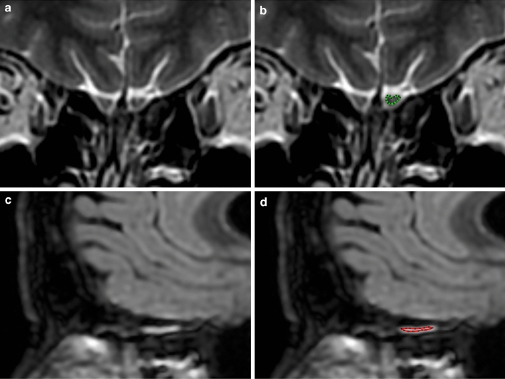
Methods: 196 COVID-19 patients (median age: 53 years, 56% females) and 39 controls (median age 55 years, 49% females) were included in this cross-sectional observational study; 78 of the patients reported olfactory loss as the only neurological symptom. MRI processing was performed by ad-hoc semi-automatic processing procedures. Olfactory bulb (OB) volume was measured on T2-weighted MRI based on manual tracing and normalized to the brain volume. Olfactory tract (OT) median signal intensity was quantified on fluid attenuated inversion recovery (FLAIR) sequences, after preliminary intensity normalization.

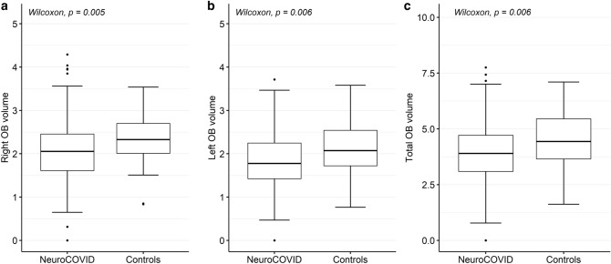
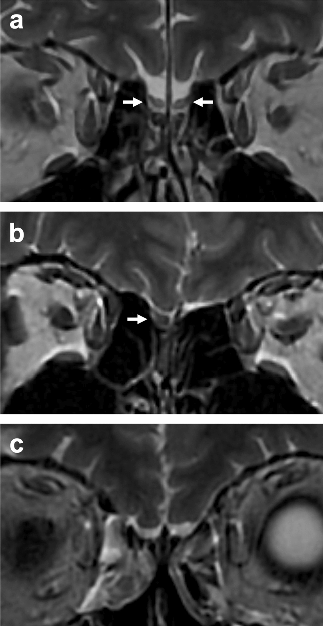
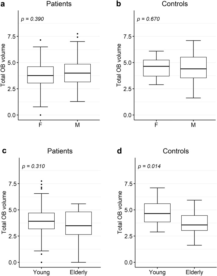
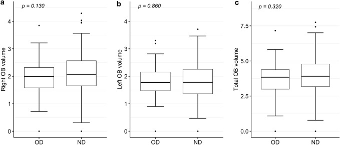
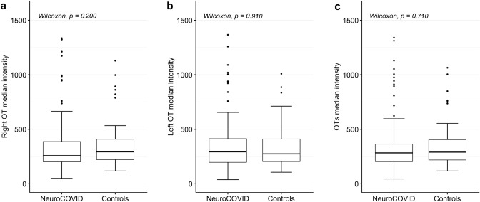
Results: COVID-19 patients showed significantly lower left, right and total OB volumes than controls (p < 0.05). Age-related OB atrophy was found in the control but not in the patient population. No significant difference was found between patients with olfactory disorders and other neurological symptoms. Several outliers with abnormally high OT FLAIR signal intensity were found in the patient group.

Conclusions: Brain MRI findings demonstrated OB damage in COVID-19 patients with neurological complications. Future longitudinal studies are needed to clarify the transient or permanent nature of OB atrophy in COVID-19 pathology.

Keywords: COVID-19; MRI; Olfactory bulbs; Olfactory dysfunction; Olfactory system.


Conflict of interest statement

There are no competing interests to declare.

Figures

Fig. 1
Fig. 1
Olfactory bulb and tract appearance on brain MRI scans. a Coronal T2-weighted MRI scan showing the location and appearance of the olfactory bulbs, surrounded by T2 hyperintense CSF. b Left olfactory bulb outlined on the T2-weighted MRI scan using ImageJ polygon selection tool. c Sagittal FLAIR MRI showing the location of the left olfactory tract, appearing hyperintense compared to normal. d Left olfactory tract outlined on the T2-weighted MRI scan using ImageJ polygon selection tool. Images were taken from a 55-year-old female patient with anosmia, attention and memory deficit, and neuropathy following COVID-19 infection
Fig. 2
Fig. 2
Olfactory bulb volume distribution in 196 COVID-19 patients and 39 controls. a Distribution of the right OB volume. b Distribution of the left OB volume. c Distribution of the total OB. OB volume was normalized to whole brain volume. Abbreviations: OB olfactory bulb
Fig. 3
Fig. 3
Representative MR images of complete atrophy of the olfactory bulbs in COVID-19 patients. Coronal T2-weighted MRI scan showing a normal-volume olfactory bulbs in a 63-year-old male control subject, b complete atrophy of the left OB in a 48-year-old female COVID-19 patient, and c bilateral complete OB atrophy in a 77-year-old female COVID-19 patient. Abbreviations: OB olfactory bulb
Fig. 4
Fig. 4
Olfactory bulb volume distribution in 196 COVID-19 patients and 39 controls, by gender and age. a Distribution of the total OB volume in the patient group, by gender (116 females and 80 males). b Distribution of the total OB volume in the control group, by gender (19 females and 20 males). c Distribution of the total OB volume in the patient group, by age (166 young patients < 65 years and 30 elderly patients). d Distribution of the total OB volume in the control group, by age (28 young controls and 11 elderly controls). OB volume was normalized to brain volume. Abbreviations: OB olfactory bulb
Fig. 5
Fig. 5
Olfactory bulb volume distribution in 196 COVID-19 patients, subgrouped by neurological complication. a Distribution of the right OB volume in patients complaining about olfactory dysfunction only (OD) and patients with other neurological disorders (ND). b Distribution of the left OB volume in OD and ND patients. d Distribution of the total OB volume in OD and ND patients. p-value was assessed by t-test. OB volume was normalized to whole brain volume. Abbreviations: OB olfactory bulb, OD = olfactory dysfunction only, ND  other neurological disorders
Fig. 6
Fig. 6
Median FLAIR signal intensity within the olfactory tract in 165 COVID-19 patients and 38 controls. a Distribution of the right OT intensity. b Distribution of the left OT intensity. c Distribution of the overall OT intensity. Abbreviations: OT  olfactory tract

Similar articles

Cited by

References

    1. WHO Coronavirus (COVID-19) Dashboard. https://covid19.who.int/. Accessed 28 Feb 2022
    1. Ellul MA, Benjamin L, Singh B, et al. Neurological associations of COVID-19. The Lancet Neurology. 2020;19:767–783. doi: 10.1016/S1474-4422(20)30221-0. - DOI - PMC - PubMed
    1. Veleri S. Neurotropism of SARS-CoV-2 and neurological diseases of the central nervous system in COVID-19 patients. Exp Brain Res. 2021 doi: 10.1007/s00221-021-06244-z. - DOI - PMC - PubMed
    1. Zubair AS, McAlpine LS, Gardin T, et al. Neuropathogenesis and Neurologic Manifestations of the Coronaviruses in the Age of Coronavirus Disease 2019: A Review. JAMA Neurol. 2020;77:1018. doi: 10.1001/jamaneurol.2020.2065. - DOI - PMC - PubMed
    1. Tsivgoulis G, Fragkou PC, Delides A, et al. Quantitative evaluation of olfactory dysfunction in hospitalized patients with Coronavirus [2] (COVID-19) J Neurol. 2020;267:2193–2195. doi: 10.1007/s00415-020-09935-9. - DOI - PMC - PubMed

Publication types