Randomized Controlled Trial
. 2023 Jan 3;6(1):e2251512.
doi: 10.1001/jamanetworkopen.2022.51512.

Assessment of Retinopathy of Prematurity Regression and Reactivation Using an Artificial Intelligence-Based Vascular Severity Score

Collaborators, Affiliations
Randomized Controlled Trial

Assessment of Retinopathy of Prematurity Regression and Reactivation Using an Artificial Intelligence-Based Vascular Severity Score

Sonja K Eilts et al. JAMA Netw Open. .

Abstract

Importance: One of the biggest challenges when using anti-vascular endothelial growth factor (VEGF) agents to treat retinopathy of prematurity (ROP) is the need to perform long-term follow-up examinations to identify eyes at risk of ROP reactivation requiring retreatment.

Objective: To evaluate whether an artificial intelligence (AI)-based vascular severity score (VSS) can be used to analyze ROP regression and reactivation after anti-VEGF treatment and potentially identify eyes at risk of ROP reactivation requiring retreatment.

Design, setting, and participants: This prognostic study was a secondary analysis of posterior pole fundus images collected during the multicenter, double-blind, investigator-initiated Comparing Alternative Ranibizumab Dosages for Safety and Efficacy in Retinopathy of Prematurity (CARE-ROP) randomized clinical trial, which compared 2 different doses of ranibizumab (0.12 mg vs 0.20 mg) for the treatment of ROP. The CARE-ROP trial screened and enrolled infants between September 5, 2014, and July 14, 2016. A total of 1046 wide-angle fundus images obtained from 19 infants at predefined study time points were analyzed. The analyses of VSS were performed between January 20, 2021, and November 18, 2022.

Interventions: An AI-based algorithm assigned a VSS between 1 (normal) and 9 (most severe) to fundus images.

Main outcomes and measures: Analysis of VSS in infants with ROP over time and VSS comparisons between the 2 treatment groups (0.12 mg vs 0.20 mg of ranibizumab) and between infants who did and did not receive retreatment for ROP reactivation.

Results: Among 19 infants with ROP in the CARE-ROP randomized clinical trial, the median (range) postmenstrual age at first treatment was 36.4 (34.7-39.7) weeks; 10 infants (52.6%) were male, and 18 (94.7%) were White. The mean (SD) VSS was 6.7 (1.9) at baseline and significantly decreased to 2.7 (1.9) at week 1 (P < .001) and 2.9 (1.3) at week 4 (P < .001). The mean (SD) VSS of infants with ROP reactivation requiring retreatment was 6.5 (1.9) at the time of retreatment, which was significantly higher than the VSS at week 4 (P < .001). No significant difference was found in VSS between the 2 treatment groups, but the change in VSS between baseline and week 1 was higher for infants who later required retreatment (mean [SD], 7.8 [1.3] at baseline vs 1.7 [0.7] at week 1) vs infants who did not (mean [SD], 6.4 [1.9] at baseline vs 3.0 [2.0] at week 1). In eyes requiring retreatment, higher baseline VSS was correlated with earlier time of retreatment (Pearson r = -0.9997; P < .001).

Conclusions and relevance: In this study, VSS decreased after ranibizumab treatment, consistent with clinical disease regression. In cases of ROP reactivation requiring retreatment, VSS increased again to values comparable with baseline values. In addition, a greater change in VSS during the first week after initial treatment was found to be associated with a higher risk of later ROP reactivation, and high baseline VSS was correlated with earlier retreatment. These findings may have implications for monitoring ROP regression and reactivation after anti-VEGF treatment.


Conflict of interest statement

Conflict of Interest Disclosures: Dr Pfeil reported receiving personal fees from Novartis Germany outside the submitted work. Dr Krohne reported receiving grants from Bayer and personal fees from Alimera Sciences, Allergan, Bayer, Novartis Germany, and Roche outside the submitted work. Dr Eter reported receiving grants from Bayer and personal fees from Alcon, Allergan, Apellis Pharmaceuticals, and Roche outside the submitted work. Dr Barth reported serving on the 2019 advisory board of Novartis Germany outside the submitted work. Dr Guthoff reported receiving grants from Bayer and personal fees from DORC outside the submitted work. Dr Busch reported receiving grants and personal fees from Bayer outside the submitted work. Dr Kalpathy-Cramer reported receiving grants from the National Institutes of Health during the conduct of the study and receiving grants from GE Healthcare and Genentech and serving as a consultant for Siloam Vision outside the submitted work. Dr Chiang reported receiving grants from Genentech and personal fees from Novartis Germany during the conduct of the study and owning equity in Inteleretina Professional Group outside the submitted work. Dr Chan reported receiving personal fees from Alcon and Genentech and being the owner of Siloam Vision outside the submitted work. Dr Coyner reported receiving personal fees from Boston AI Labs outside the submitted work. Dr Campbell reported receiving personal fees from Boston AI Labs and nonfinancial support from Siloam Vision during the conduct of the study and grants from Genentech outside the submitted work. Dr Stahl reported receiving personal fees from Novartis Germany and serving as a board member and speaker of the German Retina Society (Retinologische Gesellschaft) during the conduct of the study and receiving grants from Bayer and Novartis Germany and personal fees from Alcon, Apellis Pharmaceuticals, Bayer, Novartis Germany, and Roche outside the submitted work. No other disclosures were reported.

Figures

Figure 1.
Figure 1.. Comparison of Vascular Severity Scores Between Ranibizumab Dose Groups
Eyes that did and did not receive retreatment were included in this analysis; 2 eyes of 2 patients who required rescue treatment were excluded. Each marker represents the vascular severity score of 1 eye (using the median of all images available for an eye per visit). Black horizontal lines represent the mean values across the respective groups.
Figure 2.
Figure 2.. Development of the Vascular Severity Score During the Comparing Alternative Ranibizumab Dosages for Safety and Efficacy in Retinopathy of Prematurity (CARE-ROP) Trial
Baseline represents the severity of retinopathy of prematurity before the first injection of ranibizumab. Retreatment represents the time point immediately before a reinjection of ranibizumab was administered for reactivation of retinopathy of prematurity that required retreatment. A total of 36 eyes were included at baseline, 33 eyes at week 1, 29 eyes at week 4, 8 eyes at retreatment, and 27 eyes at week 24. Variations in the number of eyes included over the study period were due to missing data at specific time points. Dots and whiskers indicated mean and SD.
Figure 3.
Figure 3.. Vascular Severity Scores in Eyes Requiring Retreatment
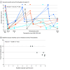
A, Each colored line represents the vascular severity score of the left or right eye of 1 infant requiring retreatment (8 eyes of 4 infants, with different colors representing different individual infants). The arrowheads represent the time points of reinjection with ranibizumab. One infant (dark blue lines) required 2 reinjections in both eyes. The x-axis is not to scale.
Figure 4.
Figure 4.. Comparison of Vascular Severity Scores Between Eyes With vs Without Retreatment
Two eyes of 2 patients who required rescue treatment were excluded. Each dot represents the vascular severity score of 1 eye (using the median of all images available for an eye per visit). Black horizontal lines represent the mean values across the respective groups.
Figure 5.
Figure 5.. Change in Vascular Severity Scores Between Baseline and Week 1
Each dot represents the vascular severity score of 1 eye (using the median of all images available for an eye per visit). Black horizontal lines represent the mean values across the respective groups.

References

    1. Terry TL. Extreme prematurity and fibroblastic overgrowth of persistent vascular sheath behind each crystalline lens. Am J Ophthalmol. 1942;25(2):203-204. doi:10.1016/S0002-9394(42)92088-9 - DOI - PubMed
    1. Stahl A, Krohne TU, Eter N, et al. ; Comparing Alternative Ranibizumab Dosages for Safety and Efficacy in Retinopathy of Prematurity (CARE-ROP) Study Group . Comparing alternative ranibizumab dosages for safety and efficacy in retinopathy of prematurity: a randomized clinical trial. JAMA Pediatr. 2018;172(3):278-286. doi:10.1001/jamapediatrics.2017.4838 - DOI - PMC - PubMed
    1. Stahl A, Lepore D, Fielder A, et al. . Ranibizumab versus laser therapy for the treatment of very low birthweight infants with retinopathy of prematurity (RAINBOW): an open-label randomised controlled trial. Lancet. 2019;394(10208):1551-1559. doi:10.1016/S0140-6736(19)31344-3 - DOI - PubMed
    1. Kennedy KA, Mintz-Hittner HA; BEAT-ROP Cooperative Group . Medical and developmental outcomes of bevacizumab versus laser for retinopathy of prematurity. J AAPOS. 2018;22(1):61-65. doi:10.1016/j.jaapos.2017.10.006 - DOI - PMC - PubMed
    1. Wallace DK, Kraker RT, Freedman SF, et al. ; Pediatric Eye Disease Investigator Group (PEDIG) . Assessment of lower doses of intravitreous bevacizumab for retinopathy of prematurity: a phase 1 dosing study. JAMA Ophthalmol. 2017;135(6):654-656. doi:10.1001/jamaophthalmol.2017.1055 - DOI - PMC - PubMed

Publication types

Substances