. 2023 Jan-Dec;15(1):2163838.
doi: 10.1080/19490976.2022.2163838.

An IBD-associated pathobiont synergises with NSAID to promote colitis which is blocked by NLRP3 inflammasome and Caspase-8 inhibitors

Affiliations

An IBD-associated pathobiont synergises with NSAID to promote colitis which is blocked by NLRP3 inflammasome and Caspase-8 inhibitors

Raminder Singh et al. Gut Microbes. 2023 Jan-Dec.

Abstract

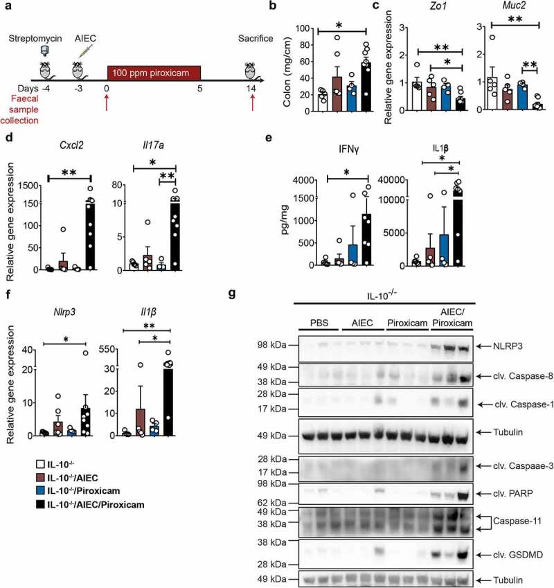
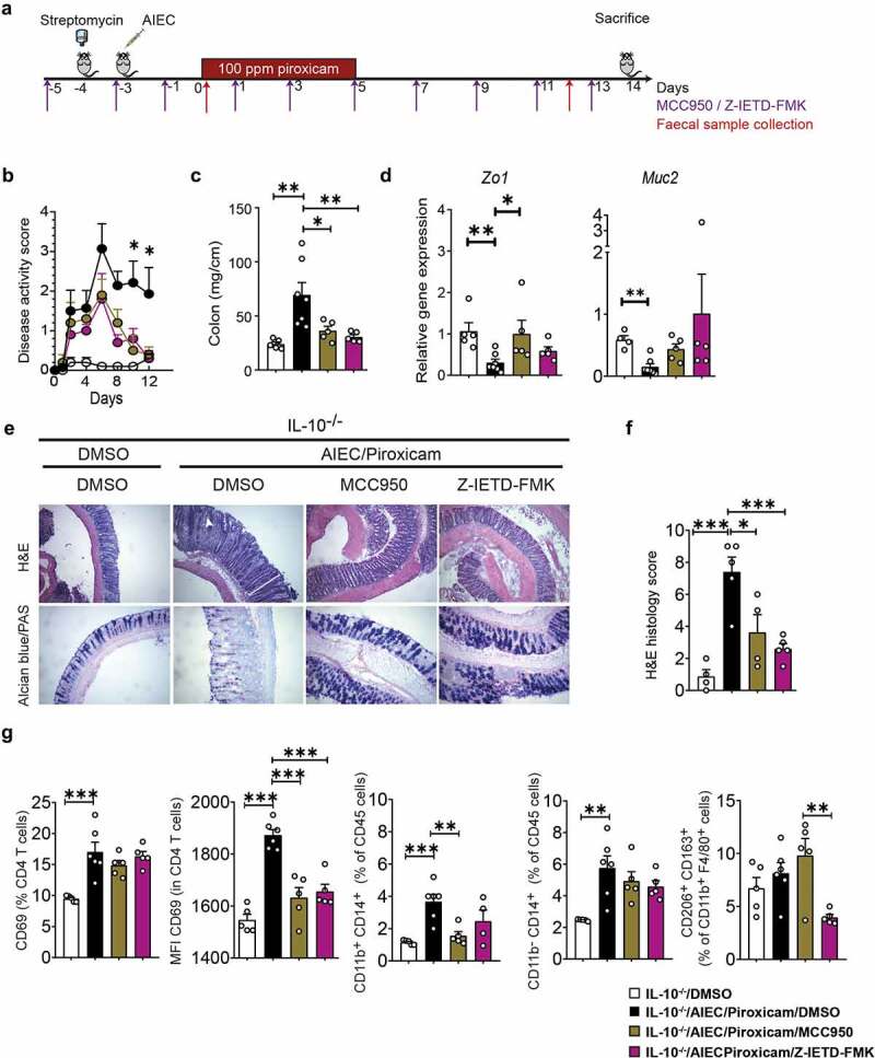
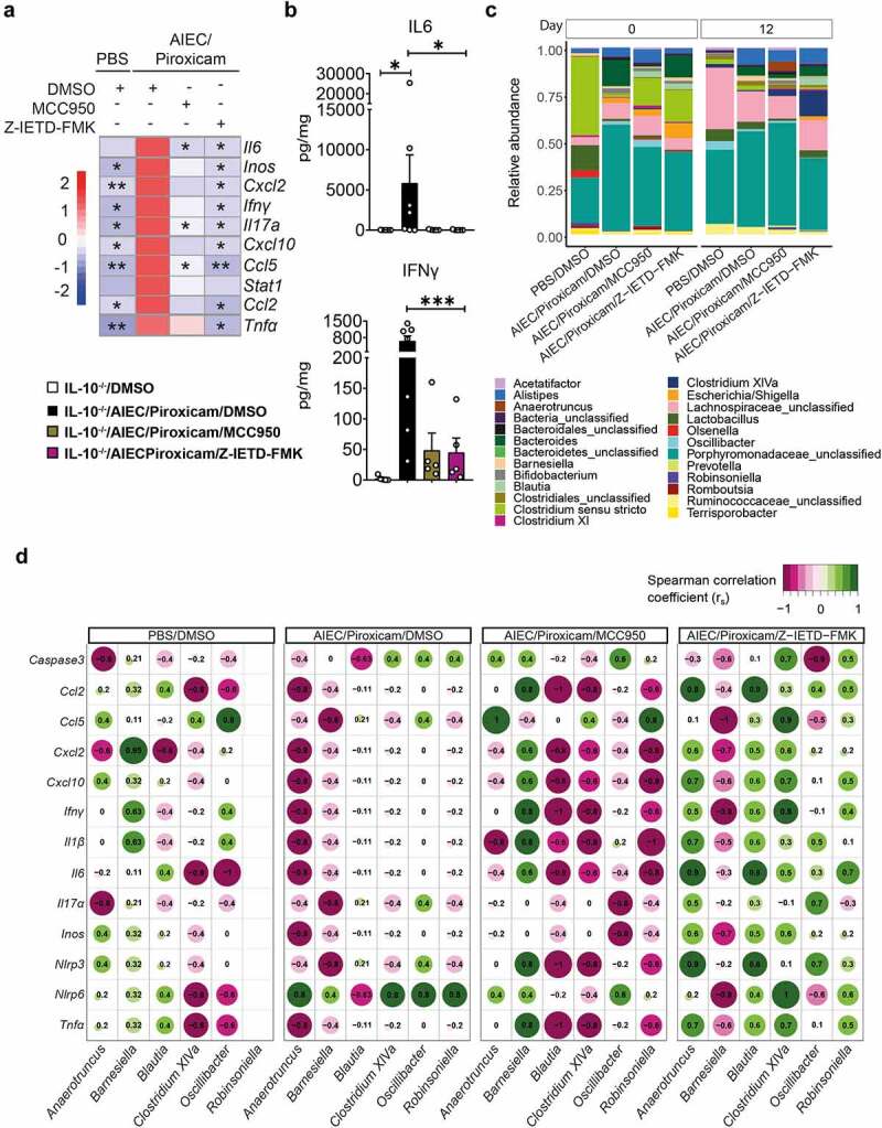
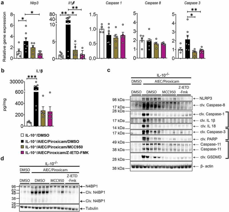
Conflicting evidence exists on the association between consumption of non-steroidal anti-inflammatory drugs (NSAIDs) and symptomatic worsening of inflammatory bowel disease (IBD). We hypothesized that the heterogeneous prevalence of pathobionts [e.g., adherent-invasive Escherichia coli (AIEC)], might explain this inconsistent NSAIDs/IBD correlation. Using IL10-/- mice, we found that NSAID aggravated colitis in AIEC-colonized animals. This was accompanied by activation of the NLRP3 inflammasome, Caspase-8, apoptosis, and pyroptosis, features not seen in mice exposed to AIEC or NSAID alone, revealing an AIEC/NSAID synergistic effect. Inhibition of NLRP3 or Caspase-8 activity ameliorated colitis, with reduction in NLRP3 inflammasome activation, cell death markers, activated T-cells and macrophages, improved histology, and increased abundance of Clostridium cluster XIVa species. Our findings provide new insights into how NSAIDs and an opportunistic gut-pathobiont can synergize to worsen IBD symptoms. Targeting the NLRP3 inflammasome or Caspase-8 could be a potential therapeutic strategy in IBD patients with gut inflammation, which is worsened by NSAIDs.

Keywords: AIEC; IL10−/− mice; cell death; inflammasome; inflammatory bowel disease; piroxicam.


Conflict of interest statement

No potential conflict of interest was reported by the authors.

Figures

Figure 1.
Figure 1.
IL10 −/− mice colonized with AIEC and fed piroxicam develop colitis associated with activation of N LRP 3 inflammasome and Caspase-8. (a) Study design. IL10 −/− mice were given streptomycin (5 g/L) in drinking water ad libitum for 24 hrs, followed by oral gavage with approx. 10 9 AIEC colony forming units (CFU), 100 ppm of piroxicam homogenized in normal chow for days followed by normal chow until day 14. Red arrows indicate timepoints for fecal collection. (b) Distal colon weight. (c) Real- time (RT)- qPCR for colonic epithelial and (d) inflammatory markers. (e) Protein levels of colonic inflammatory cytokines and chemokines. (f) RT- qPCR for colonic Nlrp3 and Il1b. (g) Western blot of markers of NLRP3 inflammasome, Caspase-8, apoptosis and pyroptosis markers. n = 3–8/group for RT- qPCR, protein, western blot, and necroscopy. Data are presented as mean ± SEM. Significance was determined using one- way ANOVA with Bonferroni or Kruskal–Wallis test with Dunn’s multiple comparison test, respectively. * p < .05; ** p < .01; *** p < .001.
Figure 2.
Figure 2.
Caspase-8 and NLRP3 inhibitors improve AIEC induced epithelial and immune alterations in IL10−/− mice fed with piroxicam. (a) Study design. Mice were injected intra-peritoneally with NLRP3 inhibitor (MCC950, 20 mg/kg) or Caspase-8 inhibitor (Z-IETD-FMK, 10 mg/kg) starting at day −5 and every second day as indicated with purple arrows, followed by euthanasia at day 14. Red arrows indicate timepoints for fecal collection. (b) Disease activity index. (c) Colon weight. (d) RT-qPCR of colonic epithelial genes. (e) Representative Hematoxylin and Eosin (H&E) and alcian blue (AB)/PAS staining of distal colon sections. In AB/PAS staining, Goblet cells are stained in dark purple color. White arrow indicates crypt abscess and white line indicate crypt hyperplasia in H&E stained sections. (f) Histology score. n = 4–7/group. (g) Isolated spleen T-cells (CD4 and CD69) and macrophages (CD11b, CD14, CD163, CD206) were immunophenotyped by Fluorescence-activated cell sorting (FACS) after gating on CD45 (Figure S2A-C) and expressed as MFI or percent of specific cell populations. n = 5–6/group. Significance was determined using one-way ANOVA with Bonferroni or Kruskal–Wallis test with Dunn’s multiple comparison test, respectively. * p < .05; ** p < .01; ***p < .001.
Figure 3.
Figure 3.
Caspase-8 and NLRP3 inhibitors have a differential effect on host–microbe interactions in AIEC-colonized IL10−/− mice treated with piroxicam. (a) Heat map of colonic gene inflammatory expression. AIEC-piroxicam/DMSO treated group was used as control for statistical analysis. (b) Protein level of colonic cytokines and chemokines n = 5–7/group. Data are presented as mean ± SEM. (c) Fecal samples were collected for 16S rRNA analysis at days 0 (upon piroxicam feeding) and 14 (end of trial). Data is presented as fecal bacteria relative-abundance at genus-level. (d) Spearman correlation between host inflammatory gene expression and specific microbial genera at day 14. n = 4–9/group. For (a) and (b), significance was determined using one-way ANOVA with Bonferroni or Kruskal–Wallis test with Dunn’s multiple comparison test, respectively. * p < .05; ** p < .01; ***p < .001.
Figure 4.
Figure 4.
Caspase-8 and NLRP3 inhibitors reduced NLRP3 inflammasome, apoptosis and pyroptosis in AIEC colonized IL10−/− mice after piroxicam feeding. (a) RT-qPCR of colonic NLRP3 inflammasome, and caspases. (b) IL-1β protein expression. (c) Western blot of Caspase-8, inflammasome and cell death markers. (d) Western blot of N4BP1. n = 3–7/group. Data are presented as mean ± SEM. Significance was determined using one-way ANOVA with Bonferroni or Kruskal–Wallis test with Dunn’s multiple comparison test, respectively. * p < .05; ** p < .01; *** p < .001.

References

    1. Round JL, Mazmanian SK.. The gut microbiota shapes intestinal immune responses during health and disease. Nat Rev Immunol. 2009;9(5):313–20. doi:10.1038/nri2515. - DOI - PMC - PubMed
    1. Bergsbaken T, Fink SL, Cookson BT. Pyroptosis: host cell death and inflammation. Nat Rev Microbiol. 2009;7(2):99–109. doi:10.1038/nrmicro2070. - DOI - PMC - PubMed
    1. Ananthakrishnan AN, Higuchi LM, Huang ES, Khalili H, Richter JM, Fuchs CS, Chan AT, Higuchi LM, Huang ES, Khalili H, et al. Aspirin, nonsteroidal anti-inflammatory drug use, and risk for Crohn disease and ulcerative colitis: a cohort study. Ann Intern Med. 2012;156(5):350–359. doi:10.7326/0003-4819-156-5-201203060-00007. - DOI - PMC - PubMed
    1. Klein A, Eliakim R. Non steroidal anti-inflammatory drugs and inflammatory bowel disease. Pharmaceuticals. 2010;3(4):1084–1092. doi:10.3390/ph3041084. - DOI - PMC - PubMed
    1. Moninuola OO, Milligan W, Lochhead P, Khalili H. Systematic review with meta-analysis: association between Acetaminophen and nonsteroidal anti-inflammatory drugs (NSAIDs) and risk of Crohn’s disease and ulcerative colitis exacerbation. Aliment Pharmacol Ther. 2018;47(11):1428–1439. doi:10.1111/apt.14606. - DOI - PMC - PubMed

Publication types

MeSH terms

LinkOut - more resources