

Cerebral perfusion using ASL in patients with COVID-19 and neurological manifestations: A retrospective multicenter observational study
- PMID: 36657613
- PMCID: PMC9842391
- DOI: 10.1016/j.neurad.2023.01.005
Cerebral perfusion using ASL in patients with COVID-19 and neurological manifestations: A retrospective multicenter observational study
Abstract
Background and purpose: Cerebral hypoperfusion has been reported in patients with COVID-19 and neurological manifestations in small cohorts. We aimed to systematically assess changes in cerebral perfusion in a cohort of 59 of these patients, with or without abnormalities on morphological MRI sequences.
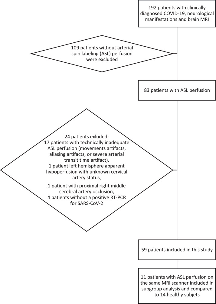
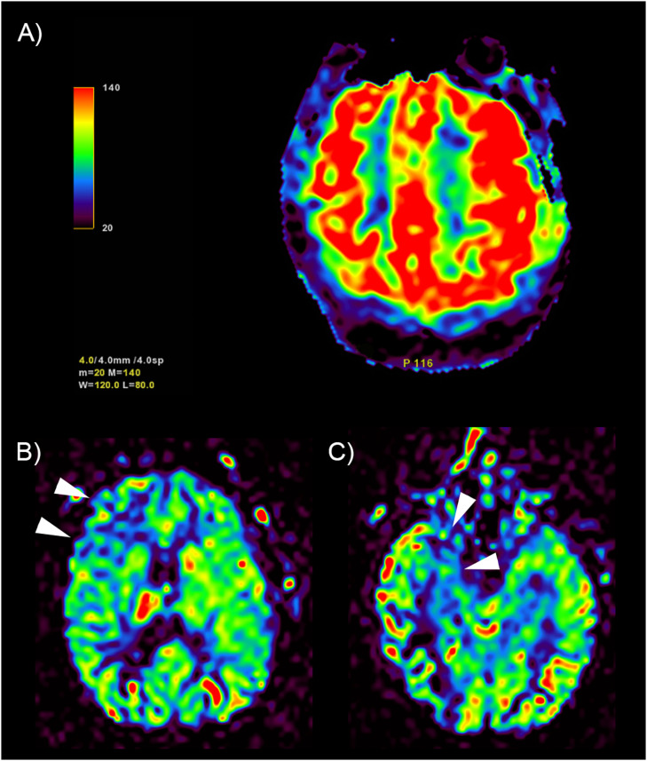
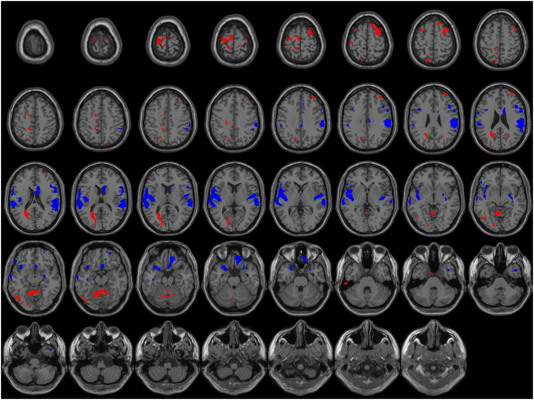
Methods: Patients with biologically-confirmed COVID-19 and neurological manifestations undergoing a brain MRI with technically adequate arterial spin labeling (ASL) perfusion were included in this retrospective multicenter study. ASL maps were jointly reviewed by two readers blinded to clinical data. They assessed abnormal perfusion in four regions of interest in each brain hemisphere: frontal lobe, parietal lobe, posterior temporal lobe, and temporal pole extended to the amygdalo-hippocampal complex.
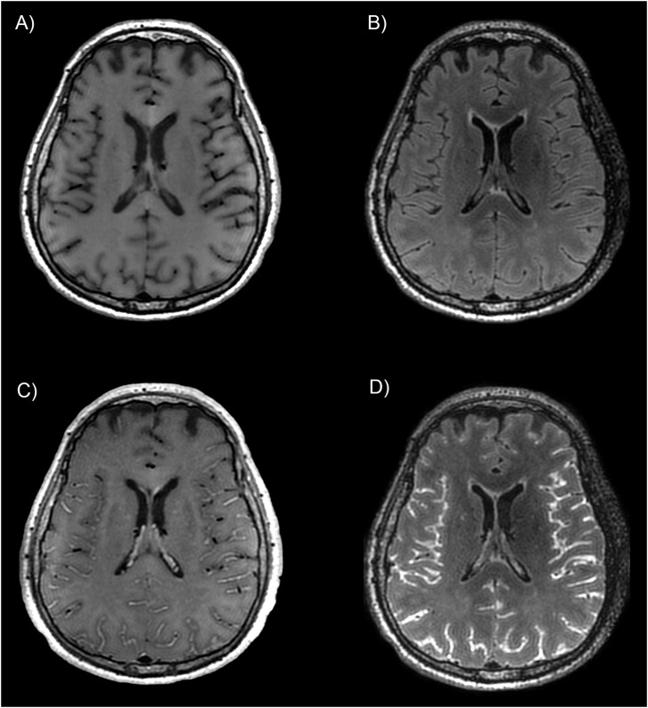
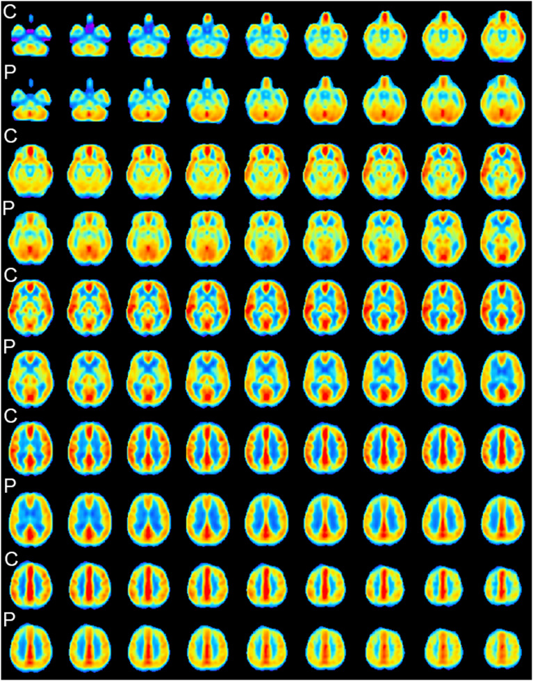
Results: Fifty-nine patients (44 men (75%), mean age 61.2 years) were included. Most patients had a severe COVID-19, 57 (97%) needed oxygen therapy and 43 (73%) were hospitalized in intensive care unit at the time of MRI. Morphological brain MRI was abnormal in 44 (75%) patients. ASL perfusion was abnormal in 53 (90%) patients, and particularly in all patients with normal morphological MRI. Hypoperfusion occurred in 48 (81%) patients, mostly in temporal poles (52 (44%)) and frontal lobes (40 (34%)). Hyperperfusion occurred in 9 (15%) patients and was closely associated with post-contrast FLAIR leptomeningeal enhancement (100% [66.4%-100%] of hyperperfusion with enhancement versus 28.6% [16.6%-43.2%] without, p = 0.002). Studied clinical parameters (especially sedation) and other morphological MRI anomalies had no significant impact on perfusion anomalies.
Conclusion: Brain ASL perfusion showed hypoperfusion in more than 80% of patients with severe COVID-19, with or without visible lesion on conventional MRI abnormalities.
Keywords: COVID-19; Cerebrovascular circulation; Magnetic resonance imaging; Multicenter study; Neuroimaging.
Copyright © 2023 Elsevier Masson SAS. All rights reserved.
Conflict of interest statement
Declaration of Competing Interest The authors declare that they have no known competing financial interests or personal relationships that could have appeared to influence the work reported in this paper.
Figures
References
Publication types
MeSH terms
Substances
LinkOut - more resources
Full Text Sources
Medical
