. 2023 Feb;16(1):17-26.
doi: 10.1016/j.mucimm.2022.11.004. Epub 2023 Jan 16.

Tissue-resident memory T cells trigger rapid exudation and local antibody accumulation

Affiliations

Tissue-resident memory T cells trigger rapid exudation and local antibody accumulation

Pamela C Rosato et al. Mucosal Immunol. 2023 Feb.

Abstract

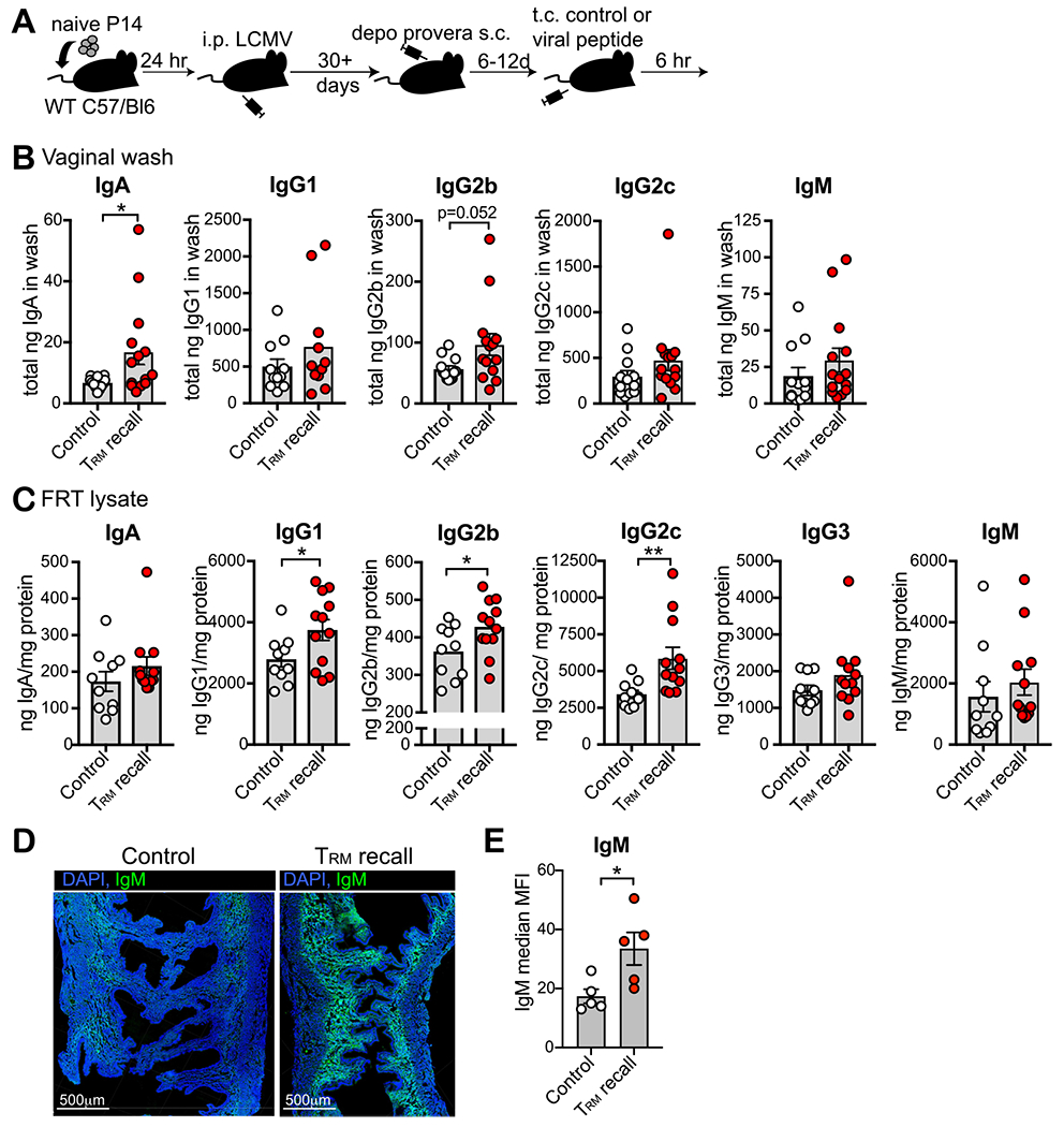
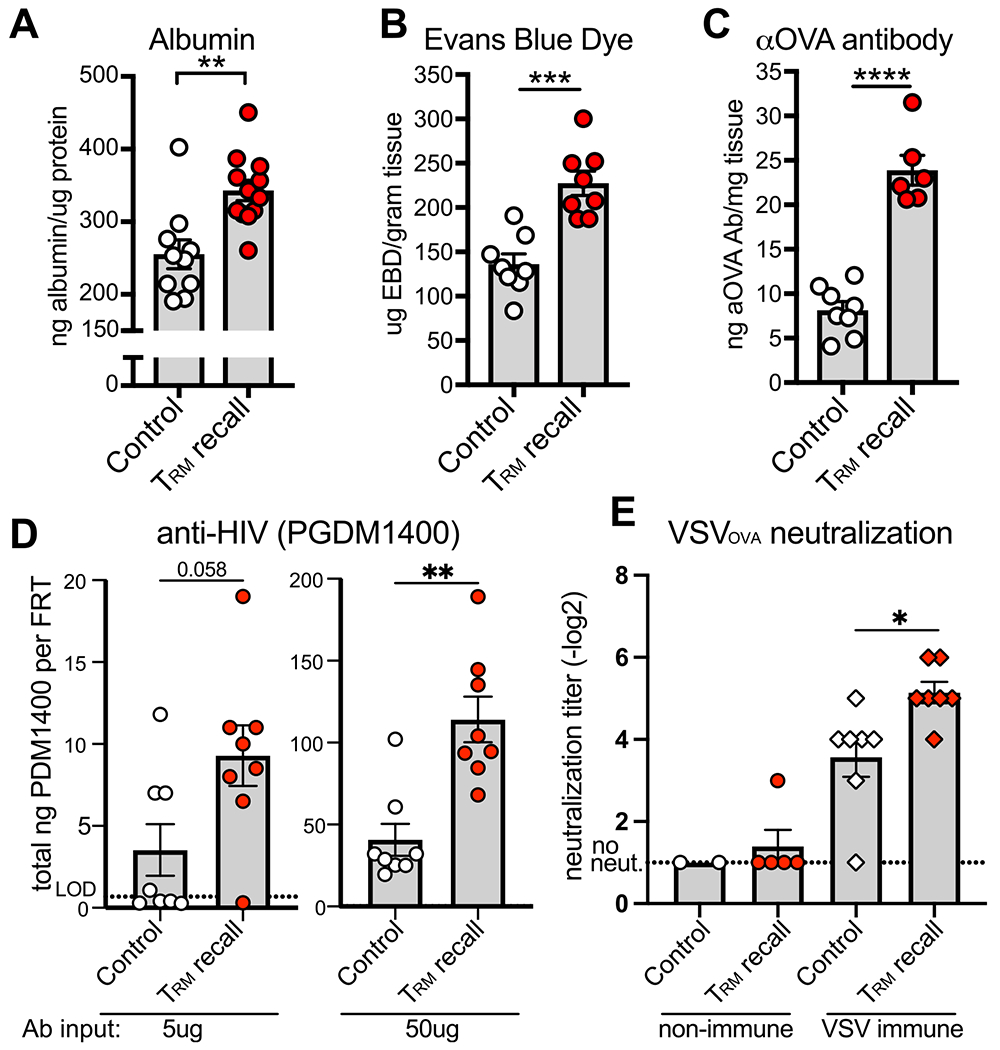
Adaptive immunity is didactically partitioned into humoral and cell-mediated effector mechanisms, which may imply that each arm is separate and does not function together. Here, we report that the activation of CD8+ resident memory T cells (TRM) in nonlymphoid tissues triggers vascular permeability, which facilitates rapid distribution of serum antibodies into local tissues. TRM reactivation was associated with transcriptional upregulation of antiviral signaling pathways as well as Fc receptors and components of the complement cascade. Effects were local, but evidence is presented that TRM in brain and reproductive mucosa are both competent to induce rapid antibody exudation. TRM reactivation in the mouse female genital tract increased local concentrations of virus-specific neutralizing antibodies, including anti-vesicular stomatitis virus, and passively transferred anti-HIV antibodies. We showed that this response was sufficient to increase the efficacy of ex vivo vesicular stomatitis virus neutralization. These results indicate that CD8+ TRM antigen recognition can enhance local humoral immunity.


Conflict of interest statement

DECLARATION OF COMPETING INTEREST

The authors have no competing interests to declare.

Figures

Fig. 1
Fig. 1
TRM reactivation induces immune-activating transcriptional changes. (A) Schematic of experimental set-up. P14 immune chimeras 120 days post-infection were challenged transcervically with gp33 or control SIINFEKL peptide and microarray was performed 9 hours later; (B) Volcano plot of differentially expressed genes in FRT where TRM were recalled versus control; (C) Top 100 differentially expressed genes categorized by function; (D) Fc receptor and complement gene expression in FRT tissue where TRM were recalled versus control. Data from n = 3 mice in each group; experiment performed once; (E) Representative PIGR staining in the uterine horns of Depo-Provera treated P14 immune chimera mice 35 days post-infection transcervically challenged with gp33 or control SIINFEKL peptide. Tissue taken 6 hours after peptide challenge. PIGR in magenta, DAPI in blue. < scale bar = 500 μm>; (F) quantification of E (n = 5 mice for TRM recall and n = 6 for control). Data are pooled from two independent experiments. Statistical significance was determined by unpaired two-tailed Mann–Whitney test where *p < 0.05. Error bars indicate standard error of mean. DAPI = 4′,6-diamidino-2-phenylindole dihydrochloride; LCMV = lymphocytic choriomeningitis virus; PIGR = polymeric immunoglobulin receptor; TRM = Tissue-resident memory T cells; WT = wild-type.
Fig. 2
Fig. 2
TRM reactivation induces changes in IV antibody staining in non-lymphoid tissue. (A) P14 immune chimeras were challenged transcervically with gp33 peptide (n = 6 mice) or control peptide (SIINFEKL; n = 6 mice) then 6 hours later biotinylated anti-CD8α antibody was transferred intravenously and mice were euthanized after 3 minutes. Representative flow cytometry plots from FRT tissue. Cells gated on CD8β +. Quantification of mean fluorescent intensity of IV-negative stained cells (gate indicated in histogram); (B) Levels of IgG2c, IgA, and IgM in the vaginal lumen of P14 immune chimera mice, stratified by estrus cycle (Di; n = 34 mice), (Pro; n = 13 mice), (Est; n = 16 mice), (Met; n = 6 mice); (C) Experiment was designed as in (A) except CD8α antibody was injected intravenously 12 hours after intracranial delivered peptides. Representative flow cytometry plots from brain tissue. Cells gated on CD8β +. Quantification of mean fluorescent intensity of IV-negative stained cells (gate indicated in histogram) with n = 4 for control and n = 4 for TRM recall. All data are pooled from at least two independent experiments. Statistical significance was determined by unpaired two-tailed t test in all instances except the IgM panel in (B) where an unpaired two-tailed Mann–Whitney was performed (due to non-normal distribution of the data) where * p < 0.05, ** p < 0.01, *** p < 0.001, **** p < 0.0001. Error bars indicate standard error of mean. Di = diestrus; Est = estrus; FRT = female reproductive tract; LCMV = lymphocytic choriomeningitis virus; Ig = immunoglobulin; i.p. = intraperitoneal; IV = intravenous; Met = metestrus; Pro = proestrus; TRM = Tissue-resident memory T cells; WT = wild-type.
Fig. 3
Fig. 3
TRM reactivation triggers rapid increase in antibody in the FRT. (A) Depo-Provera treated P14 chimeras were challenged with gp33 or control peptide transcervically and then 6 hours later, vaginal washes and FRT tissues were assayed for antibody by enzyme-linked immunosorbent assay; (B) Levels of antibody in vaginal washes 6 hours following TRM reactivation; n = 12 mice for control and n = 14 mice for TRM recall; (C) levels of antibody in FRT tissue 6 hours following TRM reactivation; n = 10 mice for control and n = 12 mice for TRM recall; (D) Representative IgM staining of FRT tissue where green = IgM and blue = DAPI stained nuclei. Scale bar = 500 μm; E: Quantification of D; n = 5 mice. All data are pooled from at least two independent experiments. Statistical significance was determined by unpaired two-tailed Mann–Whitney in (B) and (C) panel IgG2c, or by unpaired two-tailed t test in (E) and (C) panel IgG1 and IgG2b, where * p < 0.05, ** p < 0.01. Error bars indicate standard error of mean. DAPI = 4′,6-diamidino-2-phenylindole dihydrochloride; FRT = female reproductive tract; LCMV = lymphocytic choriomeningitis virus; Ig = immunoglobulin; TRM = Tissue-resident memory T cells.
Fig. 4
Fig. 4
TRM reactivation triggers vascular permeability and neutralizing ab infiltration into the FRT. (A-C) Levels of albumin (n = 10 mice for control and n = 12 for TRM recall) (A), intravenous transferred EBD (n = 8 mice) (B), and IV-transferred anti-OVA ab (n = 8 mice for control and n = 6 for TRM recall) (C) in FRT tissue 6 hours following transcervical delivery of gp33 or control peptide to Depo-Provera treated P14 immune chimera mice. (D) Levels of PGDM1400 HIV neutralizing ab in the FRT after transferring 5 μm (left; n = 8 mice) or 50 μm (right; n = 8 mice) ab intravenous 2.5–3 hours prior to transcervical delivery of gp33 or control peptide; (E) Plaque reduction neutralization assay against VSVOVA using FRT lysate from Depo-Provera treated P14 immune chimeras that were (right) or were not (left) previously infected with VSVOVA. Data are 6 hours following transcervical delivery of gp33 or control peptide. Non-immune; n = 2 mice for control, experiment performed once, and n = 5 for TRM recall. VSV immune; n = 7 mice for both groups. All data are pooled from at least two independent experiments unless noted. Statistical significance was determined by unpaired two-tailed t test in (A–C), unpaired two-tailed Mann–Whitney in (D), and a two-way analysis of variance with Sidak’s multiple comparisons test (E) where * p < 0.05, ** p < 0.01, *** p < 0.001, **** p < 0.0001. Error bars indicate standard error of mean. ab = antibody; aOVA = anti-chicken ovalbumin; EBD = evans blue dye; FRT = female reproductive tract; TRM = Tissue-resident memory T cells; VSV = virus expressing ova.

References

    1. Naluyima P et al. Terminal effector CD8 T cells defined by an IKZF2+IL-7R–transcriptional signature express FcγRIIIA, expand in HIV infection, and mediate potent HIV-specific antibody-dependent cellular cytotoxicity. J. Immunol 203, 2210–2221 (2019). - PMC - PubMed
    1. Arunachalam PS et al. T cell-inducing vaccine durably prevents mucosal SHIV infection even with lower neutralizing antibody titers. Nat. Med 26, 932–940 (2020). - PMC - PubMed
    1. Petitdemange C et al. Vaccine induction of antibodies and tissue-resident CD8 + T cells enhances protection against mucosal SHIV-infection in young macaques. JCI Insight 4, e126047 (2019). - PMC - PubMed
    1. Masopust D & Soerens AG Tissue-resident T cells and other resident leukocytes. Annu. Rev. Immunol 37, 521–546 (2019). - PMC - PubMed
    1. Szabo PA, Miron M & Farber DL Location, location, location: tissue-resident memory T cells in mice and humans. Sci. Immunol 4, eaas9673 (2019). - PMC - PubMed

Publication types

Substances