. 2023 Feb;44(2):199-204.
doi: 10.3174/ajnr.A7757. Epub 2023 Jan 19.

Early Fetal Corpus Callosum: Demonstrating Normal Growth and Detecting Pathologies in Early Pregnancy

Affiliations

Early Fetal Corpus Callosum: Demonstrating Normal Growth and Detecting Pathologies in Early Pregnancy

T Weissbach et al. AJNR Am J Neuroradiol. 2023 Feb.

Abstract

Background and purpose: A malformed corpus callosum carries a risk for abnormal neurodevelopment. The advent of high-frequency transducers offers the opportunity to assess corpus callosum development in early pregnancy. The aim of the study was to construct a reference chart of the fetal corpus callosum length on ultrasound between 13 and 19 weeks of gestation and to prospectively examine growth patterns in pathologic cases.

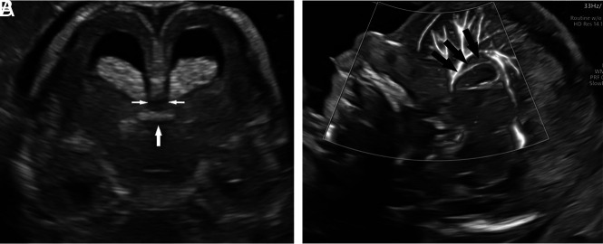
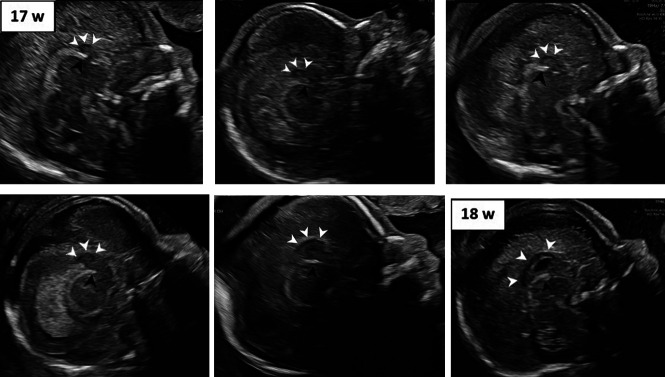
Materials and methods: We performed a prospective cross-sectional study between 2020 and 2022 in well-dated, low-risk, singleton pregnancies between 13 and 19 weeks of gestation. A standardized image was obtained in the midsagittal plane. Imaging criteria were used as a confirmation of the early corpus callosum. Measurements were taken by 4 trained sonographers. Intra- and interobserver variability was assessed. Corpus callosum length in centiles were calculated for each gestational week.

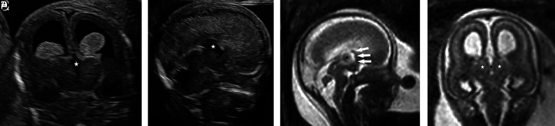
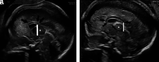
Results: One hundred eighty-seven fetuses were included in the study. All cases met inclusion criteria. At 13 weeks of gestation, the margins of the early corpus callosum were sufficiently clear to be measured in 80% (20/25) of fetuses. A cubic polynomial regression model best described the correlation between corpus length and gestational age. The correlation coefficient (r 2) was 0.929 (P < .001). Intra- and interobserver variability had high interclass correlation coefficients (>0.99). Presented is the earliest published case of agenesis of corpus callosum and a case of dysgenetic corpus callosum in Rubinstein-Taybi syndrome.

Conclusions: Provided is a nomogram of the early fetal corpus callosum. Applying imaging criteria helped to identify a case of complete agenesis of the corpus callosum as early as 14 weeks.


Figures

FIG 1.
FIG 1.
Imaging criteria for early CC identification. A, A hypoechogenic structure (double arrows) crossing the midline of the brain above the echogenic tela choroidea (single arrow), located beneath the pericallosal artery (black arrows, B).
FIG 2.
FIG 2.
The growing early CC between 13 and 18 weeks. Marked in each image are the tela choroidea (black arrowhead) and CC (white arrowheads). W indicates weeks.
FIG 3.
FIG 3.
Absent early CC. A, In the coronal plane of the third ventricle and thalami, structures crossing the midline are not apparent, resulting in an unroofed third ventricle (star). B, In the midsagittal plane, a prominent unroofed third ventricle (star) is observed in the designated location of the absent CC. C, Fetal MR imaging at 16 + 6 gestational weeks in the sagittal plane demonstrating the absence of the CC, resulting in an unroofed third ventricle (star) with the partial volume effect of the interthalamic adhesion and the anterior portion of the postcommissural fornix (white arrows). D, MR imaging in the coronal plane shows the unroofed third ventricle (black star) and the adjacent thalami (white stars).
FIG 4.
FIG 4.
A, Short and dysgenetic CC in Rubinstein-Taybi syndrome at 19.3 weeks. The rostrum is absent, the genu is underdeveloped (single arrow), and the splenium is short (double arrow), terminating prematurely at the anterior aspect (vertical line) of the massa intermedia (star). B, A normal CC at 19 weeks for comparison. The rostrum and genu (double arrow) are apparent and splenium (single arrow) is observed to extend beyond the posterior border (vertical line) of massa intermedia (star).

Similar articles

Cited by

References

    1. Blaauw J, Meiners LC. The splenium of the corpus callosum: embryology, anatomy, function and imaging with pathophysiological hypothesis. Neuroradiology 2020;62:563–85 10.1007/s00234-019-02357-z - DOI - PMC - PubMed
    1. Kier EL, Truwit CL. The normal and abnormal genu of the corpus callosum: an evolutionary, embryologic, anatomic, and MR analysis. AJNR Am J Neuroradiol 1996;17:1631–41 - PMC - PubMed
    1. Hewitt W. The development of the human corpus callosum. J Anat 1962;96(Pt 3):355–58 - PMC - PubMed
    1. Bernardes da Cunha S, Carneiro MC, Miguel Sa M, et al. . Neurodevelopmental outcomes following prenatal diagnosis of isolated corpus callosum agenesis: a systematic review. Fetal Diagn Ther 2021;48:88–95 10.1159/000512534 - DOI - PubMed
    1. Bartholmot C, Cabet S, Massoud M, et al. . Prenatal imaging features and postnatal outcome of short corpus callosum: a series of 42 cases. Fetal Diagn Ther 2021;48:217–26 10.1159/000512953 - DOI - PubMed

LinkOut - more resources