. 2023 Jan 19;10(2):e200080.
doi: 10.1212/NXI.0000000000200080. Print 2023 Mar.

CAR-T Cell-Mediated B-Cell Depletion in Central Nervous System Autoimmunity

Affiliations

CAR-T Cell-Mediated B-Cell Depletion in Central Nervous System Autoimmunity

Sasha Gupta et al. Neurol Neuroimmunol Neuroinflamm. .

Abstract

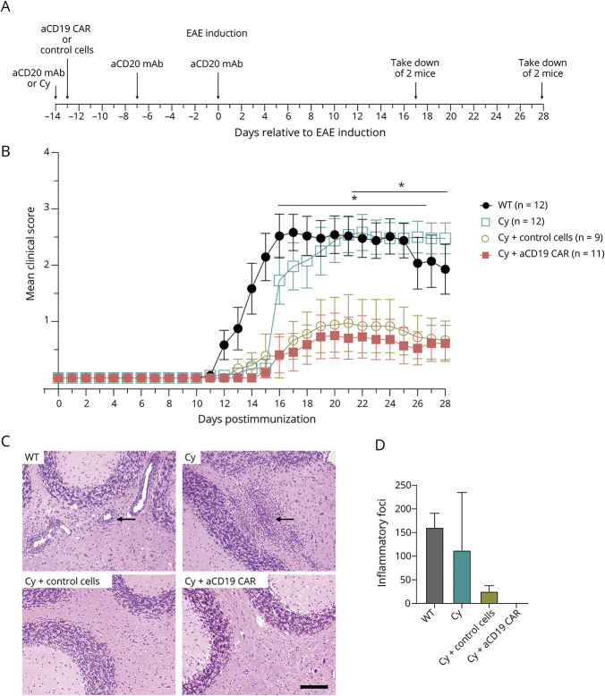
Background and objectives: Anti-CD20 monoclonal antibody (mAb) B-cell depletion is a remarkably successful multiple sclerosis (MS) treatment. Chimeric antigen receptor (CAR)-T cells, which target antigens in a non-major histocompatibility complex (MHC)-restricted manner, can penetrate tissues more thoroughly than mAbs. However, a previous study indicated that anti-CD19 CAR-T cells can paradoxically exacerbate experimental autoimmune encephalomyelitis (EAE) disease. We tested anti-CD19 CAR-T cells in a B-cell-dependent EAE model that is responsive to anti-CD20 B-cell depletion similar to the clinical benefit of anti-CD20 mAb treatment in MS.

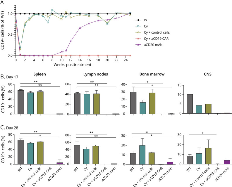
Methods: Anti-CD19 CAR-T cells or control cells that overexpressed green fluorescent protein were transferred into C57BL/6 mice pretreated with cyclophosphamide (Cy). Mice were immunized with recombinant human (rh) myelin oligodendrocyte protein (MOG), which causes EAE in a B-cell-dependent manner. Mice were evaluated for B-cell depletion, clinical and histologic signs of EAE, and immune modulation.

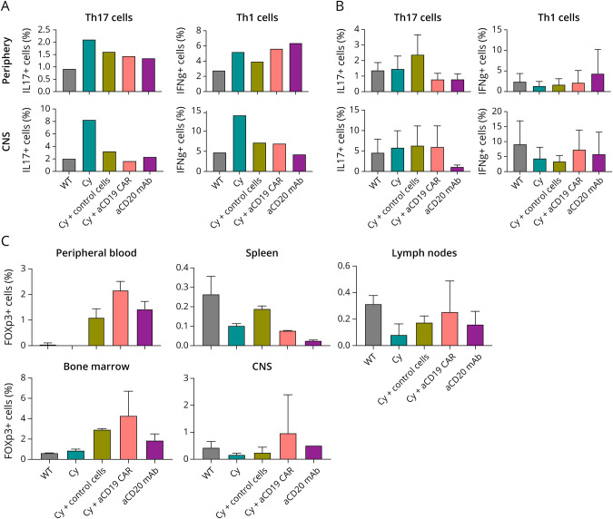
Results: Clinical scores and lymphocyte infiltration were reduced in mice treated with either anti-CD19 CAR-T cells with Cy or control cells with Cy, but not with Cy alone. B-cell depletion was observed in peripheral lymphoid tissue and in the CNS of mice treated with anti-CD19 CAR-T cells with Cy pretreatment. Th1 or Th17 populations did not differ in anti-CD19 CAR-T cell, control cell-treated animals, or Cy alone.

Discussion: In contrast to previous data showing that anti-CD19 CAR-T cell treatment exacerbated EAE, we observed that anti-CD19 CAR-T cells ameliorated EAE. In addition, anti-CD19 CAR-T cells thoroughly depleted B cells in peripheral tissues and in the CNS. However, the clinical benefit occurred independently of antigen specificity or B-cell depletion.


Figures

Figure 1
Figure 1. EAE Disease on CAR-T Cell Therapy
(A) Experimental timeline. (B) Average EAE clinical score after immunization with rhMOG. Multiple t tests revealed p < 0.05 from days 15–26 between WT vs cyclophosphamide (Cy) + anti-CD19 (aCD19) CAR-T cells and from days 21–28 from between Cy alone vs Cy + aCD19 CAR-T cells. Shown is average ± SD. (C) Cerebellar histology 28 days after EAE induction. WT and cyclophosphamide treated mice show typical perivascular mononuclear inflammatory cell foci in the white matter (indicated by arrows), whereas the Cy treated with aCD19 CAR-T cells or control cells are normal. Bar = 100 µm and applies to all. (D) Representative total numbers of meningeal and parenchymal foci within spinal cord and brain per sacrificed mouse shown, 2 mice per group. aCD20 as anti-CD20, Cy as cyclophosphamide, and aCD19 CAR as anti-CD19 CAR-T cell. CAR = chimeric antigen receptor; Cy = cyclophosphamide; EAE = experimental autoimmune encephalomyelitis; MOG = myelin oligodendrocyte protein; WT = wild type.
Figure 2
Figure 2. B Cell Depletion Based on Treatment
(A) Frequency of CD45+CD11b-CD19+ cells, average of 5 mice peripheral blood from 5 µL of tail vein blood as ratio against WT. (B) Frequency of CD45+CD11b-CD19+ cells, average of 2 mouse per disease cohort per compartment which included spleen, inguinal lymph node, and femoral bone marrow, and 1 mouse per CNS (brain and spinal cord) on day 17 postimmunization. Shown is average ± SD. (C) Frequency of CD45+CD11b-CD19+ cells, average of 2 mouse per disease cohort per compartment which included spleen, inguinal lymph node, and femoral bone marrow, and CNS (brain and spinal cord) on day 28 postimmunization. Paired t test, p value <0.05 considered significant (*), if p < 0.009 (**). WT = wild type.
Figure 3
Figure 3. Endogenous T Cell Response Based on Treatment
(A) Frequency of CD45+CD4+IL17+ (Th17) or CD45+CD4+IFNg+ (Th1) cells of 1 mouse per disease cohort from periphery (spleen) or CNS (brain and spinal cord) on day 17 postimmunization. (B) Frequency of CD45+CD4+IL17+ (Th17) or CD45+CD4+IFNg+ (Th1) cells, average of 3 mice per disease cohort from periphery (spleen) or CNS (brain and spinal cord) on day 28 postimmunization (C) Frequency of CD4+CD25+FOXp3+ cells average of 2 mice per disease cohort per compartment on day 28 postimmunization. Shown is average ± SD.

Similar articles

Cited by

References

    1. Wallin MT, Culpepper WJ, Campbell JD, et al. . The prevalence of MS in the United States: a population-based estimate using health claims data [published correction appears in Neurology. 2019,93(15):688]. Neurology. 2019,92(10):e1029-e1040. doi:10.1212/WNL.0000000000007035 - DOI - PMC - PubMed
    1. Hauser SL, Bar-Or A, Comi G, et al. . Ocrelizumab versus interferon beta-1a in relapsing multiple sclerosis. N Engl J Med. 2017,376(3):221-234. doi:10.1056/NEJMoa1601277 - DOI - PubMed
    1. Kappos L, Li D, Calabresi PA, et al. . Ocrelizumab in relapsing-remitting multiple sclerosis: a phase 2, randomised, placebo-controlled, multicentre trial. Lancet. 2011,378(9805):1779-1787. doi:10.1016/S0140-6736(11)61649-8 - DOI - PubMed
    1. Hawker K, O'Connor P, Freedman MS, et al. . Rituximab in patients with primary progressive multiple sclerosis: results of a randomized double-blind placebo-controlled multicenter trial. Ann Neurol. 2009,66(4):460-471. doi:10.1002/ana.21867 - DOI - PubMed
    1. Montalban X, Hauser SL, Kappos L, et al. . Ocrelizumab versus placebo in primary progressive multiple sclerosis. N Engl J Med. 2017,376(3):209-220. doi:10.1056/NEJMoa1606468 - DOI - PubMed

Publication types

Substances