. 2023 Jan 19;14(1):311.
doi: 10.1038/s41467-023-35949-8.

Structural basis for a conserved neutralization epitope on the receptor-binding domain of SARS-CoV-2

Affiliations

Structural basis for a conserved neutralization epitope on the receptor-binding domain of SARS-CoV-2

Kuan-Ying A Huang et al. Nat Commun. .

Abstract

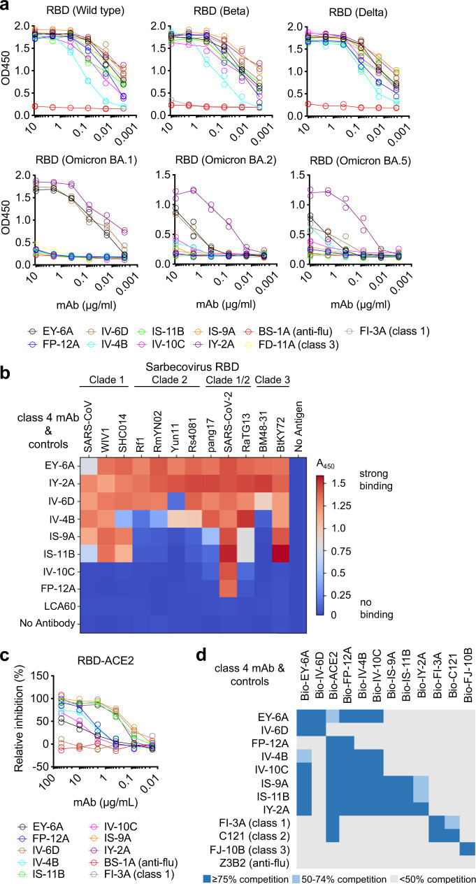
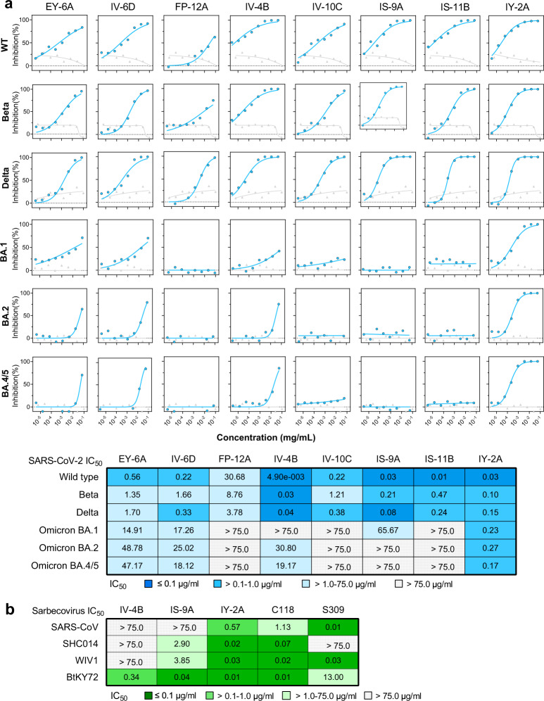
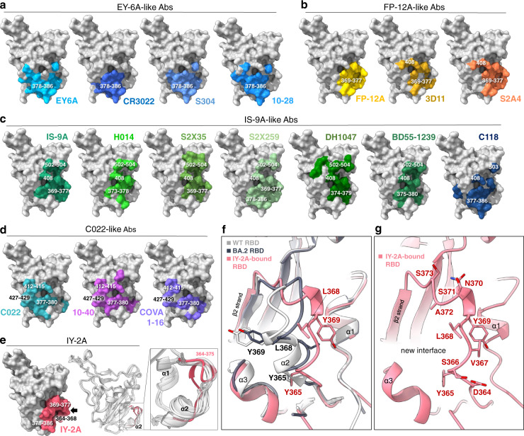
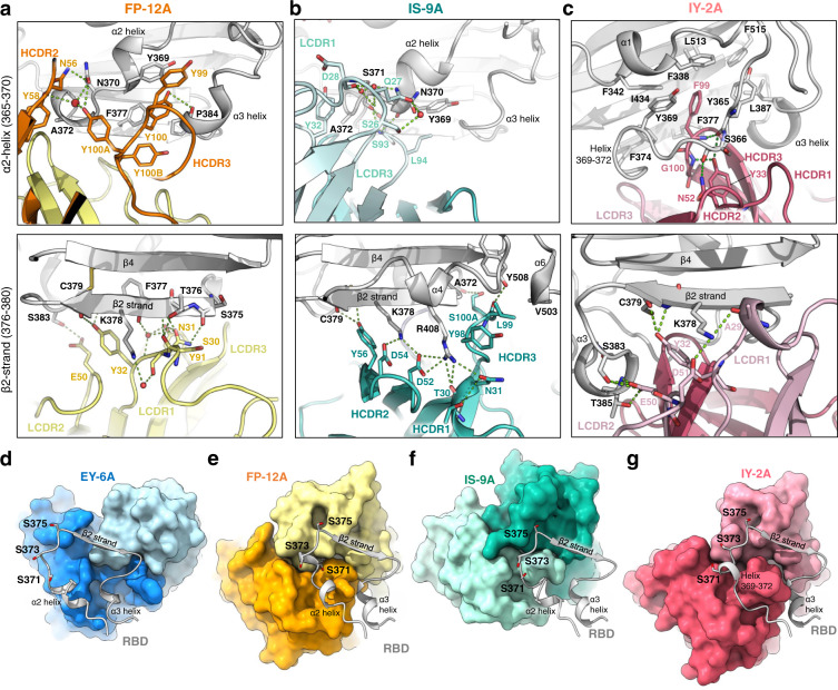
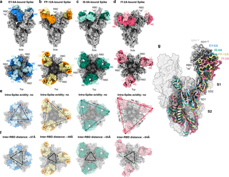
Antibody-mediated immunity plays a crucial role in protection against SARS-CoV-2 infection. We isolated a panel of neutralizing anti-receptor-binding domain (RBD) antibodies elicited upon natural infection and vaccination and showed that they recognize an immunogenic patch on the internal surface of the core RBD, which faces inwards and is hidden in the "down" state. These antibodies broadly neutralize wild type (Wuhan-Hu-1) SARS-CoV-2, Beta and Delta variants and some are effective against other sarbecoviruses. We observed a continuum of partially overlapping antibody epitopes from lower to upper part of the inner face of the RBD and some antibodies extend towards the receptor-binding motif. The majority of antibodies are substantially compromised by three mutational hotspots (S371L/F, S373P and S375F) in the lower part of the Omicron BA.1, BA.2 and BA.4/5 RBD. By contrast, antibody IY-2A induces a partial unfolding of this variable region and interacts with a conserved conformational epitope to tolerate all antigenic variations and neutralize diverse sarbecoviruses as well. This finding establishes that antibody recognition is not limited to the normal surface structures on the RBD. In conclusion, the delineation of functionally and structurally conserved RBD epitopes highlights potential vaccine and therapeutic candidates for COVID-19.


Conflict of interest statement

The authors declare no competing interests.

Figures

Fig. 1
Fig. 1. Specificity and epitope grouping of class 4 antibodies.
a Binding of antibody with RBD of wild type, Beta, Delta, Omicron BA.1, BA.2 and BA.5 variants measured by ELISA. Anti-RBD mAb FD-11 A (class 3) and mAb FI-3A (class 1) were included as controls. Anti-influenza H3 mAb BS-1A was included as a control. OD450, optical density at 450 nm. Each antibody was run with two technical replicates (n = 2) for each RBD antigen. b Heat-map showing binding of each antibody (50 nM) to the indicated sarbecovirus RBD, measured by ELISA as OD450 value. Anti-MERS RBD antibody LCA60 was included as a control. c The ability of antibody to inhibit binding of the RBD to MDCK-ACE2. Anti-influenza H3 mAb BS-1A was included as a control. Each antibody was run with two technical replicates (n = 2) in the experiment. d Cross-competition for RBD binding by class 4 antibodies and ACE2. Anti-RBD mAb FI-3A (class 1), mAb C121 (class 2), FJ-10B (class 3), and anti-influenza stem mAb Z3B2 were included as controls. Each antibody was run with four technical replicates (n = 4) in the experiment and values are presented as mean. Source data are provided as a Source Data file.
Fig. 2
Fig. 2. Neutralization breath of class 4 antibodies.
a Neutralization potency of each antibody against SARS-CoV-2 pseudotyped virus (wild type, Beta, Delta, and Omicron BA.1, BA.2 and BA.4/5 variants). Gray curves are anti-influenza H3 mAb BS-1A as control. Data are mean of technical duplicates (n = 2), and curves are fit by nonlinear regression for half-maximal inhibitory concentrations (IC50 values), as summarized in the table below. Each box of the table is colored accordingly: the higher the potency, the darker the color. b Neutralization potencies of class 4 antibodies using a pseudovirus-based assay of SARS-CoV and sarbecoviruses. C118 (class 4) and S309 (class 3) are anti-RBD mAbs and are included as controls. The IC50 values are summarized for each antibody, with each box colored accordingly. Source data are provided as a Source Data file.
Fig. 3
Fig. 3. Structural footprints of class 4 antibodies on the RBD and an induced fit of RBD by IY-2A.
Footprints of each class 4 antibody on the internal surface of RBD are shown. The footprint includes all the residues that are directly involved in hydrogen-bond (2.5-3.5 Å), salt-bridge (<4 Å), or hydrophobic (3.3-4.0 Å) interaction in the structure. a EY-6A, and similar antibodies share the binding site around the residues 378-386 region. PDB code 6ZER for EY-6A, 6W41 for CR3022, 7R6X for S304 and 7SI2 for 10–28. b FP-12A and similar antibodies share the binding site around the residues 369-377 region, with some reaching residue 408. PDB code 7M7B for 3D11 and 7JVA for S2A4. c IS-9A and similar antibodies extend their footprints upwards and contact residue 408 and the residues 502-504 region. PDB code 7CAH for H014, 7R6W for S2X35, 7M7W for S2X259, 7LD1 for DH1047, 7WRL for BD55-1239 and 7RKS for C118. d C022 and similar antibodies extend their footprints towards the left side, including residues 412–415 and 427–429. PDB code 7RKU for C022, 7SD5 for 10–40 and 7JMW for COVA1-16. e IY-2A recognizes a region (the 365-369 helix, black arrow), which is originally buried but is now exposed after a conformational change (enlarged view, the backbone shift highlighted in red). Structures (PDB code 6ZER, 7M7W, 7R6X, 7R6W, 6W41, light gray) are used for superimposition. f Superimposition of the ACE2-bound wild-type RBD (PDB 6M0J, white). The ACE2-bound Omicron BA.2 RBD (PDB 7ZF7, gray) with IY-2A-bound RBD (red) reveals a rotated view of the conformational change, highlighting residues Y365, L368, and Y369. g Detailed structure of the shifted 364–373 region in IY-2A-bound RBD, with all affected residues drawn in sticks and labeled.
Fig. 4
Fig. 4. Detailed structural interface between class 4 antibodies and RBD.
a The interface between FP-12A (heavy chain, orange; light chain, yellow) and two key binding regions on RBD (gray, upper, 365–370 α2 and 384-388 α3 helices; lower, 376–380 β2 strand). Heavy and light chain CDRs (HCDR and LCDR) and key interacting residues are labeled. Hydrogen bonds are shown in green dash lines, and water molecules in red spheres. b The same views of interface as in (a) between IS-9A (heavy chain, dark teal; light chain, light teal) and RBD (gray). c The same views of interface as in (a) between IY-2A (heavy chain, dark red; light chain, light red) and RBD (gray). dg The binding mode of class 4 antibodies (surface) with the epitope region including α2 helix, β2 strand and α3 helix of RBD (ribbon, in light gray) for EY-6A, (d in dark and light blue), FP-12A (e in orange and yellow), IS-9A (f in dark and light teal) and IY-2A (g in dark and light red). Residues of Variants of Concern (S371, S373, and S375) are shown in sticks and labeled. These panels (dg) are in exactly the same view.
Fig. 5
Fig. 5. Cryo-EM structures of class-4-mAb-bound Spike protein.
ad Surface representation of cryo-EM structures of the Spike-Fab complex for EY-6A, (a), heavy and light chains in dark and light blue, PDB code 6ZDH), FP-12A (b), in orange and yellow), IS-9A (c), in dark and light teal) and IY-2A (d), in dark and light red). Spike is shown in gray. All bound RBDs are in the “up” conformation. The constant region of Fab is modelled based on EM density at low-threshold rendering. e The potential for intra-spike avidity effects (simultaneous binding of both Fabs of a single IgG to adjacent RBDs on a Spike trimer) was evaluated by measuring the distance between the C-terminal (C224) of each bound Fab. Distances are shown in black dashed lines. f Inter-RBD distances measured between D428 of RBD for each complex structure. g The conformation of RBD of the class-4-mAb-bound Spike compared to that of the apo 2-RBD-up Spike of Delta variant (PDB code 7V7U and 7V7T as representative). Only one subunit of Spike is shown in licorice, with S2 subunit being superimposed. NTDs are omitted for clarity. Both RBD and SD1/2 show visible shifts to further widen the inter-subunit space. Each structure is colored accordingly as in (ad). The other two subunits are from IS-9A-bound Spike shown in transparent surface.

Similar articles

Cited by

References

    1. Khoury DS, et al. Neutralizing antibody levels are highly predictive of immune protection from symptomatic SARS-CoV-2 infection. Nat. Med. 2021;27:1205–1211. doi: 10.1038/s41591-021-01377-8. - DOI - PubMed
    1. Earle KA, et al. Evidence for antibody as a protective correlate for COVID-19 vaccines. Vaccine. 2021;39:4423–4428. doi: 10.1016/j.vaccine.2021.05.063. - DOI - PMC - PubMed
    1. Lan J, et al. Structure of the SARS-CoV-2 spike receptor-binding domain bound to the ACE2 receptor. Nature. 2020;581:215–220. doi: 10.1038/s41586-020-2180-5. - DOI - PubMed
    1. Liu L, et al. Potent neutralizing antibodies against multiple epitopes on SARS-CoV-2 spike. Nature. 2020;584:450–456. doi: 10.1038/s41586-020-2571-7. - DOI - PubMed
    1. Barnes CO, et al. SARS-CoV-2 neutralizing antibody structures inform therapeutic strategies. Nature. 2020;588:682–687. doi: 10.1038/s41586-020-2852-1. - DOI - PMC - PubMed

Publication types

Supplementary concepts