. 2023 Jul;44(7):1442-1454.
doi: 10.1038/s41401-023-01051-4. Epub 2023 Jan 19.

Kaempferide ameliorates cisplatin-induced nephrotoxicity via inhibiting oxidative stress and inducing autophagy

Affiliations

Kaempferide ameliorates cisplatin-induced nephrotoxicity via inhibiting oxidative stress and inducing autophagy

Yan-Fei Shao et al. Acta Pharmacol Sin. 2023 Jul.

Abstract

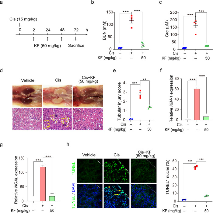
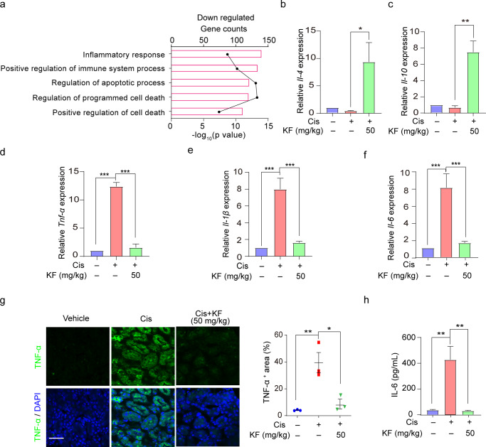
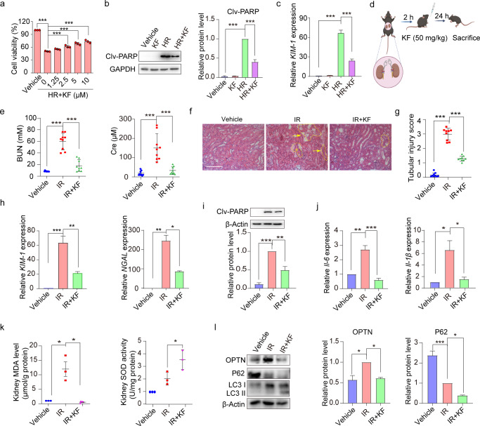
Acute kidney injury (AKI) caused by anti-tumor drugs, such as cisplatin, is a severe complication with no effective treatment currently, leading to the reduction or discontinuation of chemotherapy. Natural products or herbal medicines are gradually considered as promising agents against cisplatin-induced AKI with the advantages of multi-targeting, multi-effects, and less resistance. In this study, we investigated the effects of kaempferide, a natural flavonoid extracted from the rhizome of Kaempferia galanga, in experimental AKI models in vitro and in vivo. We first conducted pharmacokinetic study in mice and found a relative stable state of kaempferide with a small amount of conversion into kaempferol. We showed that both kaempferide (10 μM) and kaempferol (10 μM) significantly inhibited cisplatin-caused injuries in immortalized proximal tubule epithelial cell line HK-2. In AKI mice induced by injection of a single dose of cisplatin (15 mg/kg), oral administration of kaempferide (50 mg/kg) either before or after cisplatin injection markedly improved renal function, and ameliorated renal tissue damage. We demonstrated that kaempferide inhibited oxidative stress and induced autophagy in cisplatin-treated mice and HK-2 cells, thus increasing tubular cell viability and decreasing immune responses to attenuate the disease progression. In addition, treatment with kaempferide significantly ameliorated ischemia-reperfusion-induced renal injury in vitro and in vivo. We conclude that kaempferide is a promising natural product for treating various AKI. This study has great implications for promotion of its use in healthcare products, and help to break through the limited use of cisplatin in the clinic.

Keywords: acute kidney injury; autophagy; cisplatin; kaempferide; nephrotoxicity; oxidative stress.


Conflict of interest statement

The authors declare no competing interests.

Figures

Fig. 1
Fig. 1. Pharmacokinetic study of kaempferide and its protection in HK-2 cell toxicity.
a Chemical structure of kaempferide (KF) and kaempferol (KFR). b, c Concentration-time curve in plasma (b) and kidney (c), n = 3 animals/group. d, e The Cmax of kaempferide and kaempferol in plasma (d) and kidney (e). f, g The AUC of kaempferide and kaempferol content in plasma (f) and kidney (g). h Relative mRNA expression of KIM-1 in HK-2 treated with vehicle, cisplatin (5 μM), cisplatin plus kaempferide (10 μM), cisplatin plus kaempferol (10 μM), respectively. i Western blot analysis of the Clv-PARP expression in HK-2 treated with vehicle, cisplatin, cisplatin plus kaempferide, cisplatin plus kaempferol. β-Actin served as a loading control. j Relative mRNA expression of Bax and Bcl2 in HK-2 treated with vehicle, cisplatin, cisplatin plus kaempferide, cisplatin plus kaempferol, respectively. Data are presented as means ± SEM; *P < 0.05, **P < 0.01, ***P < 0.001; one-way ANOVA with multiple comparisons test.
Fig. 2
Fig. 2. Kaempferide prevents the nephrotoxicity caused by cisplatin.
a Schematic illustration of kaempferide (KF) treatment schedule for AKI prevention induced by cisplatin. b, c BUN and serum Cre levels of mice treated with vehicle, cisplatin, cisplatin plus kaempferide, respectively. n = 6 animals/group. d, e Representative H&E staining and the morphology of kidneys from each group. Arrows indicated tubules with necrosis, anoikis, or brush border loss, asterisks indicated cast formation. Scale bar: 100 μm. The tubular injury degree was scored in each group. n = 6 animals/group. f, g Relative mRNA expression of KIM-1 and NGAL in the renal cortex from mice treated with vehicle, cisplatin, cisplatin plus kaempferide, respectively. h Western blot analysis of the Clv-PARP expression after cisplatin and kaempferide treatment. GAPDH served as a loading control. Data are presented as means ± SEM; *P < 0.05, **P < 0.01, ***P < 0.001; one-way ANOVA with multiple comparisons test.
Fig. 3
Fig. 3. Kaempferide treatment attenuates cisplatin-induced AKI.
a Schematic illustration of kaempferide (KF) treatment schedule for AKI therapy induced by cisplatin. b, c BUN and serum Cre levels of mice treated with vehicle, cisplatin, cisplatin plus kaempferide, respectively. n = 4 animals/group. d, e Representative H&E staining and the morphology of kidneys from each group. The dashed area indicated the tubular damage, arrows indicated tubules with necrosis, anoikis, or brush border loss, and asterisks indicated cast formation. Scale bar: 200 μm. The tubular injury degree was scored in each group. n = 3 animals/group. f, g Relative mRNA expression of KIM-1 and NGAL in the renal cortex from mice treated with vehicle, cisplatin, cisplatin plus kaempferide, respectively. h Representative TUNEL staining images in kidneys from mice treated with vehicle, cisplatin, cisplatin plus kaempferide, respectively. Scale bar: 50 μm. Quantification of TUNEL positive nuclei in kidneys of each group. n = 3 animals/group. Data are presented as means ± SEM; **P < 0.01, ***P < 0.001; one-way ANOVA with multiple comparisons test.
Fig. 4
Fig. 4. The immune response is reduced in kaempferide-treated cis-AKI mice.
a GO analysis of the downregulated genes in the renal tissues from kaempferide treated mice. bf Relative mRNA expressions of Il-4, Il-10, Tnf-α, Il-1β, and Il-6 in the renal cortex from each group. g Representative images of immunofluorescence staining for TNF-α antibody in kidneys. Scale bar: 50 μm. n = 3 animals/group. h ELISA to detect the secretion level of IL-6 in kidney tissues from each group. Data are presented as means ± SEM; *P < 0.05, **P < 0.01, ***P < 0.001; one-way ANOVA with multiple comparisons test.
Fig. 5
Fig. 5. Cisplatin-induced oxidative stress is inhibited after kaempferide treatment.
a GO analysis of the upregulated genes in the renal tissues from kaempferide treated mices. b Representative ROS images in HK-2 cells detected by flow cytometry after cisplatin and kaempferide incubation. c Statistical results of ROS level in HK-2 cells with cisplatin and kaempferide treatment. d, e The MDA level and SOD activity from HK-2 cells exposed to cisplatin and kaempferide. f, g Detection of MDA level and SOD activity in renal cortex from mice treated with vehicle, cisplatin, cisplatin plus kaempferide, respectively. n = 3 animals/group. Data are presented as means ± SEM; **P < 0.01, ***P < 0.001; one-way ANOVA with multiple comparisons test.
Fig. 6
Fig. 6. Autophagy is induced after kaempferide treatment in HK-2 cells.
a Western blot analysis of the p-AKT, AKT, p-AMPK and AMPK expression in HK-2 cells treated with vehicle and kaempferide. GAPDH served as a loading control. b Relative mRNA expression of Beclin-1 in HK-2 cells treated with vehicle, kaempferide, respectively. c Western blot analysis of the OPTN, P62 and LC3 II expression in HK-2 cells treated with vehicle, kaempferide. GAPDH served as a loading control. d HK-2 cells transfected with mCherry-GFP-LC3 plasmid were treated with kaempferide for 12 h and the GFP, mCherry signal was detected by immunofluorescence. Scale bars: 10 μm. e Western blot analysis of the OPTN, P62 and LC3 II expression in the renal cortex treated with vehicle, cisplatin, cisplatin plus kaempferide. GAPDH served as a loading control. f HK-2 cells transfected with mCherry-GFP-LC3 plasmid were treated with cisplatin, cisplatin plus kaempferide, cisplatin and kaempferide plus 3-MA for 12 h and the GFP and mCherry signals were detected by immunofluorescence. Scale bars: 10 μm. g The survival rate of HK-2 cells upon treatment with cisplatin (5 μM), kaempferide (10 μM) and 3-MA (10 μM) for 24 h. Data are presented as means ± SEM; n.s. not significant, *P < 0.05, **P < 0.01, ***P < 0.001; Student’s t test or one-way ANOVA with multiple comparisons test.
Fig. 7
Fig. 7. Autophagy induction is required in the renal protection of kaempferide.
a Western blot analysis of the OPTN, P62 and LC3II expression in the renal cortex treated with vehicle, cisplatin, cisplatin plus kaempferide. GAPDH served as a loading control. b Schematic diagram of the model of AKI mice treated with cisplatin, kaempferide and 3-MA. c, d BUN and serum Cre levels of mice treated with vehicle, cisplatin, cisplatin plus kaempferide, cisplatin plus 3-MA, cisplatin plus kaempferide and 3-MA. n = 6 animals/group. e Western blot analysis of Clv-PARP expression in the renal cortex from each group. GAPDH served as a loading control. f, g Representative H&E sections of the kidney in each group. Arrows indicate the location of tubular damage, including tubular necrosis and brush border loss, and asterisks indicate protein cast deposition. Scale bar: 200 μm. The tubular injury degree was scored in each group. n = 6 animals/group. Data are presented as means ± SEM; n.s. not significant, *P < 0.05, **P < 0.01, ***P < 0.001; one-way ANOVA with multiple comparisons test.
Fig. 8
Fig. 8. Kaempferide treatment attenuates ischemia-reperfusion-induced AKI.
a Cell viability of HK-2 cells with HR treated with kaempferide for 24 h. b Western blot analysis of the Clv-PARP expression in HK-2 cells after HR and kaempferide (10 μM) treatment. GAPDH served as a loading control. c Relative mRNA expression of KIM-1 in HK-2 cells from each group. d Schematic diagram of kaempferide treatment schedule for IR-AKI. e BUN and serum Cre levels of IR-AKI mice treated with or without kaempferide. n = 9 animals/group. f Representative H&E staining of kidneys from each group. The dashed area indicated the tubular injury. The arrows indicated the loss of tubular structure and epithelial necrosis, asterisks indicated cast formation. Scale bar: 200 μm. n = 9 animals/group. g The tubular injury degree was scored in each group. h Relative mRNA expression of KIM-1 and NGAL in the renal cortex from each group. i Western blot analysis of the Clv-PARP expression from the renal tissues of each group. β-Actin served as a loading control. j Relative mRNA expression of IL-6 and IL-1β in the renal cortex from each group. k Detection of MDA level and SOD activity in the renal cortex from each group. n = 3 animals/group. l Western blot analysis of the OPTN, P62 and LC3II expression from the renal tissues of each group. β-Actin served as a loading control. Data are presented as means ± SEM; *P < 0.05, **P < 0.01, ***P < 0.001; one-way ANOVA with multiple comparisons test.
Fig. 9
Fig. 9. Schematic diagram of kaempferide’s protection against acute kidney injury.
Kaempferide can inhibit oxidative stress and induce autophagy in tubular cells, leading to reduced cell death and immune response in kidneys, thus protecting against both cisplatin-induced and ischemia-reperfusion-simulated acute kidney injury.

References

    1. Dasari S, Tchounwou PB. Cisplatin in cancer therapy: molecular mechanisms of action. Eur J Pharmacol. 2014;740:364–78. doi: 10.1016/j.ejphar.2014.07.025. - DOI - PMC - PubMed
    1. Stehlik J, Armstrong B, Baran DA, Bridges ND, Chandraker A, Gordon R, et al. Early immune biomarkers and intermediate-term outcomes after heart transplantation: Results of Clinical Trials in Organ Transplantation-18. Am J Transpl. 2019;19:1518–28. doi: 10.1111/ajt.15218. - DOI - PMC - PubMed
    1. Pierson Marchandise M, Gras V, Moragny J, Micallef J, Gaboriau L, Picard S, et al. The drugs that mostly frequently induce acute kidney injury: a case - noncase study of a pharmacovigilance database. Br J Clin Pharmacol. 2017;83:1341–9. doi: 10.1111/bcp.13216. - DOI - PMC - PubMed
    1. Beyer J, Rick O, Weinknecht S, Kingreen D, Lenz K, Siegert W. Nephrotoxicity after high-dose carboplatin, etoposide and ifosfamide in germ-cell tumors: incidence and implications for hematologic recovery and clinical outcome. Bone Marrow Transpl. 1997;20:813–9. doi: 10.1038/sj.bmt.1700980. - DOI - PubMed
    1. Winston JA, Safirstein R. Reduced renal blood flow in early cisplatin-induced acute renal failure in the rat. Am J Physiol. 1985;249:F490–6. - PubMed