

Identification of immune associated potential molecular targets in proliferative diabetic retinopathy
- PMID: 36658547
- PMCID: PMC9854219
- DOI: 10.1186/s12886-023-02774-y
Identification of immune associated potential molecular targets in proliferative diabetic retinopathy
Abstract
Background: Diabetic retinopathy (DR) is one of the most common microvascular complications of diabetes and causes of blindness in developed countries. Our study was designed to identify immune-related genes involved in the progression of proliferative diabetic retinopathy (PDR).
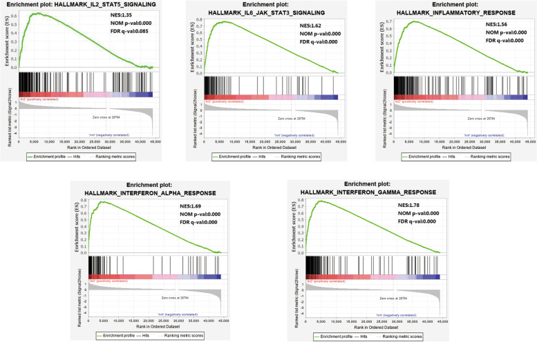
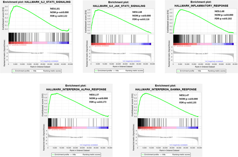
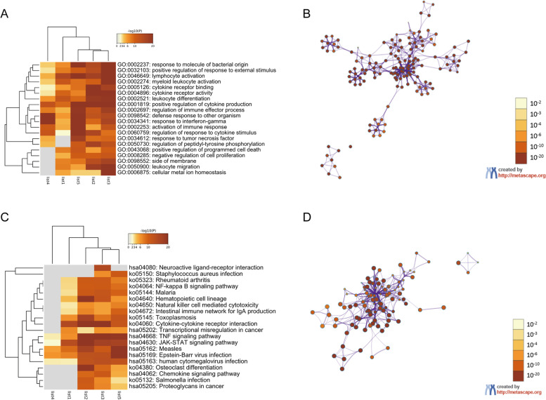
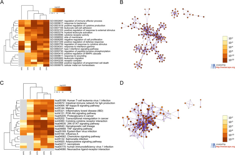
Methods: The "GSE102485" dataset of neovascular membrane samples (NVMs) from type 1 and 2 diabetes mellitus patients was downloaded from the Gene Expression Omnibus database. Functional enrichment analyses, protein-protein interaction network (PPI) construction, and module analysis of immune pathways in NVMs and controls were conducted via Gene Set Enrichment Analysis and Metascape.
Results: The significantly upregulated hallmark gene sets in DR2 and DR1 groups were involved in five immune pathways. Only CCR4, CXCR6, C3AR1, LPAR1, C5AR1, and P2RY14 were not previously reported in the context of PDR molecular pathophysiology. Except for P2RY14, all of the above were upregulated in retinal samples from experimental diabetes mouse models and human retina microvascular endothelial cells (HRMECs) treated with high glucose (HG) by quantitative Real Time Polymerase Chain Reaction (qRT-PCR).
Conclusion: The genes identified herein provide insight into immune-related differential gene expression during DR progression.
Keywords: Biomarkers; Genes; Immune; Inflammation; Neovascular membrane; Proliferative diabetic retinopathy.
© 2023. The Author(s).
Conflict of interest statement
The authors declare that they have no competing interests.
Figures


Similar articles
-
CD44 Drives M1 Macrophage Polarization in Diabetic Retinopathy.Curr Eye Res. 2023 Aug;48(8):770-780. doi: 10.1080/02713683.2023.2210273. Epub 2023 May 16. Curr Eye Res. 2023. PMID: 37191152
-
Molecular investigation of candidate genes for pyroptosis-induced inflammation in diabetic retinopathy.Front Endocrinol (Lausanne). 2022 Jul 25;13:918605. doi: 10.3389/fendo.2022.918605. eCollection 2022. Front Endocrinol (Lausanne). 2022. PMID: 35957838 Free PMC article.
-
Identification of RUNX1 as a Mediator of Aberrant Retinal Angiogenesis.Diabetes. 2017 Jul;66(7):1950-1956. doi: 10.2337/db16-1035. Epub 2017 Apr 11. Diabetes. 2017. PMID: 28400392 Free PMC article.
-
Identification of immune-related endoplasmic reticulum stress genes in proliferative diabetic retinopathy using bioinformatics analysis.Front Endocrinol (Lausanne). 2024 Aug 30;15:1341206. doi: 10.3389/fendo.2024.1341206. eCollection 2024. Front Endocrinol (Lausanne). 2024. PMID: 39280014 Free PMC article.
-
Molecular Surgery: Proteomics of a Rare Genetic Disease Gives Insight into Common Causes of Blindness.iScience. 2020 Oct 13;23(11):101667. doi: 10.1016/j.isci.2020.101667. eCollection 2020 Nov 20. iScience. 2020. PMID: 33134897 Free PMC article. Review.
Cited by
-
hsa_circ_0099682 exacerbates the development of diabetic retinopathy by promoting pathological neoangiogenesis via the miR-125b-5p/HNRNPU axis.J Mol Histol. 2025 Aug 9;56(4):264. doi: 10.1007/s10735-025-10535-y. J Mol Histol. 2025. PMID: 40782251
References
MeSH terms
Grants and funding
LinkOut - more resources
Full Text Sources
Medical
Miscellaneous
