. 2023 Mar;32(3):e4569.
doi: 10.1002/pro.4569.

The B domain of protein A retains residual structures in 6 M guanidinium chloride as revealed by hydrogen/deuterium-exchange NMR spectroscopy

Affiliations

The B domain of protein A retains residual structures in 6 M guanidinium chloride as revealed by hydrogen/deuterium-exchange NMR spectroscopy

Saeko Yanaka et al. Protein Sci. 2023 Mar.

Erratum in

  • CORRECTION.
    [No authors listed] [No authors listed] Protein Sci. 2023 Jul;32(7):e4696. doi: 10.1002/pro.4696. Protein Sci. 2023. PMID: 37382354 Free PMC article. No abstract available.

Abstract

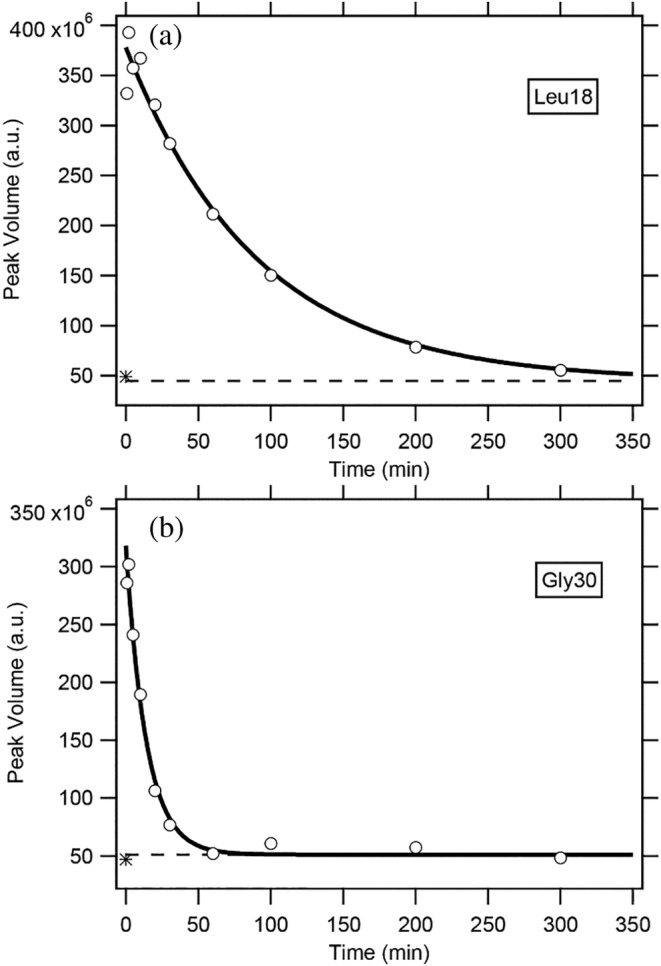
The characterization of residual structures persistent in unfolded proteins is an important issue in studies of protein folding, because the residual structures present, if any, may form a folding initiation site and guide the subsequent folding reactions. Here, we studied the residual structures of the isolated B domain (BDPA) of staphylococcal protein A in 6 M guanidinium chloride. BDPA is a small three-helix-bundle protein, and until recently its folding/unfolding reaction has been treated as a simple two-state process between the native and the fully unfolded states. We employed a dimethylsulfoxide (DMSO)-quenched hydrogen/deuterium (H/D)-exchange 2D NMR techniques with the use of spin desalting columns, which allowed us to investigate the H/D-exchange behavior of individually identified peptide amide (NH) protons. We obtained H/D-exchange protection factors of the 21 NH protons that form an α-helical hydrogen bond in the native structure, and the majority of these NH protons were significantly protected with a protection factor of 2.0-5.2 in 6 M guanidinium chloride, strongly suggesting that these weakly protected NH protons form much stronger hydrogen bonds under native folding conditions. The results can be used to deduce the structure of an early folding intermediate, when such an intermediate is shown by other methods. Among three native helical regions, the third helix in the C-terminal side was highly protected and stabilized by side-chain salt bridges, probably acting as the folding initiation site of BDPA. The present results are discussed in relation to previous experimental and computational findings on the folding mechanisms of BDPA.

Keywords: 2D NMR; hydrogen/deuterium exchange; protein folding; residual structure; unfolded state.


Figures

FIGURE 1
FIGURE 1
The amino acid sequence of BDPA. Rectangles, H1, H2, and H3 represent the three α‐helical regions in native BDPA (PDB code: 1BDD).
FIGURE 2
FIGURE 2
The CD spectra of BDPA at 0.0, 3.0, and 6.0 M GdmCl (a), and the unfolding transition curve measured by the mean residue ellipticity at 222 nm (0.10 M NaCl–20 mM HCOONa in 90% D2O/10% H2O at pH* 3.4 and 15.0°C) (b). The solid line in (b) is the theoretical curve best fitted to Equation (6), and two broken lines represent the ellipticity values in the pure N and the pure U states.
FIGURE 3
FIGURE 3
The 1H–15 N HSQC spectra of uniformly 13C/15 N‐labeled BDPA in the DMSO/H2O solution (94.5% (v/v) DMSO‐d 6/5% (v/v) H2O/0.5% (v/v) DCA‐d 2).
FIGURE 4
FIGURE 4
The H/D‐exchange curves for Leu18 (a) and Gly30 (b) in 6.0 M GdmCl at pH* 3.4 and 15.0°C. The solid lines are the theoretical curves best‐fitted to a single exponential function (Equation 7). A broken line in each panel indicates the theoretically estimated peak volume after complete exchange (i.e., Y(∞) in Equation 7), and an asterisk (*) in each panel, located at 50 × 106 of the peak volume, indicates the experimentally observed value after heating the sample at 50°C for 30 min. Because the reaction mixtures contained 10% H2O, the final peak volumes did not reach zero. The k obs values for the two residues are as follows: (a) (1.11 ± 0.17) × 10−2 min−1 and (b) (7.10 ± 0.73) × 10−2 min−1; the errors given are fitting error estimates.
FIGURE 5
FIGURE 5
Mapping of the P value on the three‐dimensional structure of native B domain of protein A (BDPA). The P values of the residues are mapped on the structure of BDPA (PDBID: 1BDD). The residues with P values larger than 2.0, 3.0, 4.0, and 5.0 are colored in light magenta, magenta, red, and dark red, respectively. (a) The whole structure of BDPA. (b) A close‐up view of the H3 helix region. The H‐bonds are shown as blue dashed lines.

Similar articles

References

    1. Alonso DO, Daggett V. Staphylococcal protein A: unfolding pathways, unfolded states, and differences between the B and E domains. Proc Natl Acad Sci U S A. 2000;97:133–8. - PMC - PubMed
    1. Anfinsen CB. Principles that govern the folding of protein chains. Science. 1973;181(4096):223–30. - PubMed
    1. Arai M, Kuwajima K. Role of the molten globule state in protein folding. Adv Protein Chem. 2000;53:209–82. - PubMed
    1. Arora P, Oas TG, Myers JK. Fast and faster: a designed variant of the B‐domain of protein A folds in 3 microsec. Protein Sci. 2004;13:847–53. - PMC - PubMed
    1. Baek M, DiMaio F, Anishchenko I, Dauparas J, Ovchinnikov S, Lee GR, et al. Accurate prediction of protein structures and interactions using a three‐track neural network. Science. 2021;373:871–6. - PMC - PubMed

Publication types