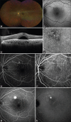
Central serous chorioretinopathy: Treatment
- PMID: 36660123
- PMCID: PMC9843567
- DOI: 10.4103/2211-5056.362040
Central serous chorioretinopathy: Treatment
Abstract
Central serous chorioretinopathy (CSC) is a pachychoroid spectrum disease characterized by serous detachment of the neurosensory retina with subretinal fluid in young and middle-aged adults. The pathogenesis of CSC is not yet fully understood. However, it is considered a multifactorial disease that is strongly associated with choroidal dysfunction or vascular engorgement. Although there is no consensus on the treatment of CSC, photodynamic therapy has been effectively used to manage serous retinal detachment (SRD) in CSC. Moreover, micropulse diode laser photocoagulation and focal laser treatment have also been used. Recently, oral medications, including mineralocorticoid receptor antagonists, have been proposed for the management of CSC. Multimodal imaging plays a significant role in the diagnosis and treatment of CSC. Optical coherence tomography angiography (OCTA) has the advantage of detecting vascular flow in the retina and choroid layer, allowing for a better understanding of the pathology, severity, prognosis, and chronicity of CSC. In addition, early detection of choroidal neovascularization in CSC is possible using OCTA. This review article aims to provide a comprehensive and updated understanding of CSC, focusing on treatment.
Keywords: Central serous chorioretinopathy; micropulse diode laser photocoagulation; mineralocorticoid receptor antagonist; photodynamic therapy.
Copyright: © 2022 Taiwan J Ophthalmol.
Conflict of interest statement
There are no conflicts of interest.
Figures

References
-
- Gass JD. Pathogenesis of disciform detachment of the neuroepithelium. Am J Ophthalmol. 1967;63:l1–139. - PubMed
-
- Wang M, Munch IC, Hasler PW, Prünte C, Larsen M. Central serous chorioretinopathy. Acta Ophthalmol. 2008;86:126–45. - PubMed
-
- Yamada K, Hayasaka S, Setogawa T. Fluorescein-angiographic patterns in patients with central serous chorioretinopathy at the initial visit. Ophthalmologica. 1992;205:69–76. - PubMed
-
- Piccolino FC, Borgia L. Central serous chorioretinopathy and indocyanine green angiography. Retina. 1994;14:231–42. - PubMed
Publication types
LinkOut - more resources
Full Text Sources
