Tracheal Rupture After Trauma: A Successful Conservative Management
- PMID: 36660502
- PMCID: PMC9846864
- DOI: 10.7759/cureus.32681
Tracheal Rupture After Trauma: A Successful Conservative Management
Abstract
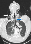
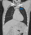
Tracheobronchial injury (TBI) is a rare life-threatening injury that can result from either penetrating or blunt trauma. Treatment may be surgical or conservative, but the evidence regarding which is the best approach is still very scarce. This case report describes the successful conservative management of a 32-year-old male with a traumatic tracheal laceration. The alarming signs and symptoms, the imaging modalities of choice, the rationale behind the treatment strategy, and the most common complications are detailed here. Through this case, the authors wish to highlight the features that should lead to the suspicion of this potentially fatal traumatic injury, as well as raise awareness on how to adequately manage these patients.
Keywords: airway trauma; conservative treatment; surgical treatment; trachea; tracheal injuries.
Copyright © 2022, Jorge et al.
Conflict of interest statement
The authors have declared that no competing interests exist.
Figures


References
-
- Acute injuries of the trachea and major bronchi: importance of early diagnosis. Cassada DC, Munyikwa MP, Moniz MP, et al. Ann Thorac Surg. 2000;69:1563–1567. - PubMed
-
- Tracheobronchial injuries. Conservative treatment. Lampl L. Interact Cardiovasc Thorac Surg. 2004;3:401–405. - PubMed
-
- Traumatic injury to the trachea and bronchus. Karmy-Jones R, Wood DE. Thorac Surg Clin. 2007;17:35–46. - PubMed
Publication types
LinkOut - more resources
Full Text Sources
