. 2023 Jun 13;7(11):2590-2605.
doi: 10.1182/bloodadvances.2022008591.

A critical role of RUNX1 in governing megakaryocyte-primed hematopoietic stem cell differentiation

Affiliations

A critical role of RUNX1 in governing megakaryocyte-primed hematopoietic stem cell differentiation

Chen Wang et al. Blood Adv. .

Abstract

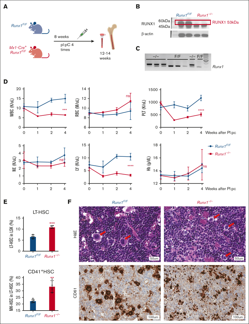
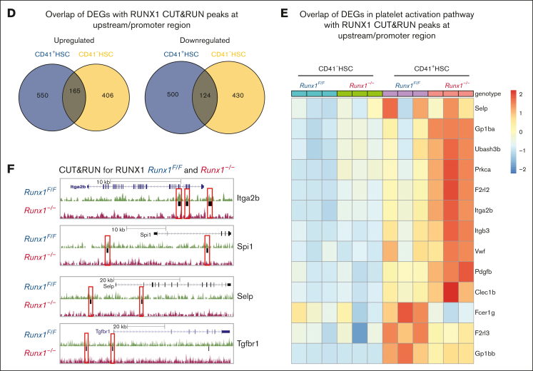
As a transcription factor in the RUNT domain core-binding factor family, RUNX1 is crucial in multiple stages of hematopoiesis, and its mutation can cause familial platelet disorder with a predisposition to acute myeloid leukemia. Previous work has established that RUNX1 is involved in the maturation of megakaryocytes (MKs) and the production of platelets. Recent studies have shown that there exists a subpopulation of hematopoietic stem cells (HSCs) with relatively high expression of von Willebrand factor and CD41 at the apex of the HSC hierarchy, termed MK-HSCs, which can give rise to MKs without going through the traditional differentiation trajectory from HSC via MPP (multipotent progenitors) and MEP (megakaryocyte-erythroid progenitor). Here, by using Runx1F/FMx1-Cre mouse model, we discovered that the MK-HSC to MK direct differentiation can occur within 1 cell division, and RUNX1 is an important regulator in the process. Runx1 knockout results in a drastic decrease in platelet counts and a severe defect in the differentiation from MK-HSCs to MKs. Single cell RNA sequencing (RNAseq) analysis shows that MK-HSCs have a distinct gene expression signature compared with non-MK-HSCs, and Runx1 deletion alters the platelet and MK-related gene expression in MK-HSCs. Furthermore, bulk RNAseq and Cut&Run analyses show that RUNX1 binds to multiple essential MK or platelet developmental genes, such as Spi1, Selp, and Itga2b and regulates their expressions in MK-HSCs. Thus, by modulating the expression of MK-related genes, RUNX1 governs the direct differentiation from MK-HSCs to MKs and platelets.


Conflict of interest statement

Conflict-of-interest disclosure: The authors declare no competing financial interests.

Figures

None
Graphical abstract
Figure 1.
Figure 1.
HSCs and CD41+ MK-HSCs are dysregulated in Runx1–/– mice and accompanied by thrombocytopenia. (A) Schematics of the mouse model of Runx1F/F;Mx1-Cre and Runx1 inducible deletion. (B) Western blotting of the knockout of RUNX1 protein in duplicate mice. (C) Genotyping of exon 4 deletion of Runx1 in Runx1F/F:Mx1-Cre mice. Triplet samples of Runx1F/F:Mx1-Cre (–/–) and Runx1F/F (F/F) are shown. (D) Peripheral blood counts of Runx1F/F;Mx1-Cre mice after pI:pC inductions. Statistics was performed using t test at 4 weeks post deletion between Runx1/ and Runx1F/F mice. The x-axis indicates the weeks after the final pI:pC injection. (E) Quantification of flow cytometry analysis of MK-HSCs (LSKCD34Flt3CD150+CD41+) in the BM at 4 weeks after pI:pC injection. The y-axis is the percentage out of their parental population. (F) hematoxylin and eosin and anti-CD61 (a MK marker) staining of BM from Runx1/ and Runx1F/F mice 4 weeks post pI:pC injections. The red arrows indicate MK cells. ∗∗∗P < .001 and ∗∗P < .01. ns, not significant.
Figure 2.
Figure 2.
RUNX1 deletion affects direct differentiation from MK-HSCs to MKs. (A) Sorted CD41+HSCs (LSKCD34Flt3CD150+CD41+), CD41HSCs (LSKCD34Flt3CD150+CD41) and MPP2 (LSK CD34+Flt3CD150+CD48+) were plated in StemSpan medium containing SCF and TPO cytokines. Cell images were taken after 2-day culture. Red circles indicate large MK-like cells in the liquid culture dish, and the cell size were measured by cell cytometer. Cells larger than 20um were indicated with green boxes. (B) Quantification of CFU assay from sorted CD41+HSC, CD41HSC and MPP2. Cells were sorted as single cell into 96 well plates with methylcellulose medium containing multiple cytokines (SCF, TPO, IL-3, IL-6, EPO, IL11, FLT3L, and GM-CSF). After a 7-day culture, colonies were counted based on morphologies. MK, granulocyte, and granulocyte/monocyte colonies were quantified for Runx1–/– and Runx1F/F group 7 days after plating. The y-axis indicates the number of colonies after normalization with Runx1F/F group. (bottom right) Representative views of the MK, G and GM colonies. (C) Sorted CD41+HSC, CD41HSC cells were cultured in collagen-based semi-solid culture, and after a 7-day culture, MK-CFUs were stained with acetylcholinesterase. Pictures show representative views of the MK-CFUs in CD41+HSC, CD41HSCs for Runx1–/– and Runx1F/F group. (D) Quantification of the MK-CFUs stained with AChE. ∗∗∗P < .001, ∗∗P < .01, and ∗∗∗∗ P < .0001.
Figure 2.
Figure 2.
RUNX1 deletion affects direct differentiation from MK-HSCs to MKs. (A) Sorted CD41+HSCs (LSKCD34Flt3CD150+CD41+), CD41HSCs (LSKCD34Flt3CD150+CD41) and MPP2 (LSK CD34+Flt3CD150+CD48+) were plated in StemSpan medium containing SCF and TPO cytokines. Cell images were taken after 2-day culture. Red circles indicate large MK-like cells in the liquid culture dish, and the cell size were measured by cell cytometer. Cells larger than 20um were indicated with green boxes. (B) Quantification of CFU assay from sorted CD41+HSC, CD41HSC and MPP2. Cells were sorted as single cell into 96 well plates with methylcellulose medium containing multiple cytokines (SCF, TPO, IL-3, IL-6, EPO, IL11, FLT3L, and GM-CSF). After a 7-day culture, colonies were counted based on morphologies. MK, granulocyte, and granulocyte/monocyte colonies were quantified for Runx1–/– and Runx1F/F group 7 days after plating. The y-axis indicates the number of colonies after normalization with Runx1F/F group. (bottom right) Representative views of the MK, G and GM colonies. (C) Sorted CD41+HSC, CD41HSC cells were cultured in collagen-based semi-solid culture, and after a 7-day culture, MK-CFUs were stained with acetylcholinesterase. Pictures show representative views of the MK-CFUs in CD41+HSC, CD41HSCs for Runx1–/– and Runx1F/F group. (D) Quantification of the MK-CFUs stained with AChE. ∗∗∗P < .001, ∗∗P < .01, and ∗∗∗∗ P < .0001.
Figure 3.
Figure 3.
MK-HSCs can directly differentiate to MK within 1 cell division. (A) 60-well Terasaki plate sorted with single cells of CD41+HSC, CD41HSCs from Runx1–/–and Runx1F/F mice after a 2-day culture in Stemspan medium containing SCF and TPO. (B) Kinetics of the plated single cell divisions. The x-axis indicates the hours after plating the cells post sorting. y-axis indicates the percentage of divided cells in total plated cells. (C) Representative images of the divided large MK-like and small non-MK–like daughter cells. (D) Quantification of the cultured cells as single large MK-like cells, single small non-MK-like cells, double large MK-like cells, and double small non-MK–like cells. (E) Heatmap of key platelet or MK-related gene expressions in large MK-like cells and small non-MK–like cells for Runx1–/–group. (F) Heatmap of key platelet/MK-related gene expressions in large MK-like cells and small non-MK–like cells for Runx1–/–group. ∗∗P < .01 and ∗P < .05.
Figure 4.
Figure 4.
scRNAseq reveals a distinct gene signature of MK-HSCs which is altered by Runx1 deficiency. (A) 10x Genomics scRNAseq was used to identify 4 distinct clusters in sorted LSK CD150+ HSCs from Runx1–/–and Runx1F/F group by Seurat program. (B) A quantification of the percentage of cells in the 4 clusters of Runx1F/F and Runx1–/–cells. (C) Heatmap of the DEGs in the 4 clusters of Runx1F/F and Runx1–/–cells. MK-HSCs has a distinct MK-biased signature, and both MK-HSCs and non-MK–HSCs have long-term HSCs signature. (D) Heatmap of the DEGs in the MK-HSCs of Runx1F/F vs Runx1–/–cells. RUNX1 deficiency alters platelet activation related gene expressions in this subset of HSCs, including Pf4, vWF, and Gp5. (E) Feature plot of marker gene Itga2b (CD41) and Gp5 (Glycoprotein V Platelet) for Runx1–/–and Runx1F/F groups.
Figure 5.
Figure 5.
RUNX1 regulates platelet and MK regulatory gene expression. (A) PCA plot of RNAseq replicates for each genotype of sorted HSCs. (B) Volcano plot of differential expressed genes in CD41+HSC vs CD41-HSCs from Runx1F/F mice and differential expressed genes in Runx1F/F vs Runx1–/–cells in CD41+HSC. (C) Overlaps of differential expressed genes between CD41+HSC vs CD41HSCs for Runx1–/–and Runx1F/F genotypes show the commonly changed gene expressions in the cell populations. (D) GSEA analysis of CD41+HSC vs CD41HSCs in Runx1F/F genotype shows major upregulated pathways in CD41+HSCs including multiple megakaryocyte/platelet related pathways. (E) GSEA analyses of CD41+HSCs for Runx1F/F and Runx1–/–groups show upregulated pathways in WT including platelet activity related pathways. (F) Heatmap of key platelet related gene expressions in CD41+HSC vs CD41HSCs for Runx1F/F genotype indicates a distinct expression pattern and general increase of platelet related genes in CD41+HSCs. (G) Heatmap of key platelet regulatory gene expression in CD41+HSCs for Runx1F/F and Runx1–/–genotypes shows that RUNX1 deletion alters the gene expression pattern of many platelets-regulatory genes.
Figure 5.
Figure 5.
RUNX1 regulates platelet and MK regulatory gene expression. (A) PCA plot of RNAseq replicates for each genotype of sorted HSCs. (B) Volcano plot of differential expressed genes in CD41+HSC vs CD41-HSCs from Runx1F/F mice and differential expressed genes in Runx1F/F vs Runx1–/–cells in CD41+HSC. (C) Overlaps of differential expressed genes between CD41+HSC vs CD41HSCs for Runx1–/–and Runx1F/F genotypes show the commonly changed gene expressions in the cell populations. (D) GSEA analysis of CD41+HSC vs CD41HSCs in Runx1F/F genotype shows major upregulated pathways in CD41+HSCs including multiple megakaryocyte/platelet related pathways. (E) GSEA analyses of CD41+HSCs for Runx1F/F and Runx1–/–groups show upregulated pathways in WT including platelet activity related pathways. (F) Heatmap of key platelet related gene expressions in CD41+HSC vs CD41HSCs for Runx1F/F genotype indicates a distinct expression pattern and general increase of platelet related genes in CD41+HSCs. (G) Heatmap of key platelet regulatory gene expression in CD41+HSCs for Runx1F/F and Runx1–/–genotypes shows that RUNX1 deletion alters the gene expression pattern of many platelets-regulatory genes.
Figure 6.
Figure 6.
Platelet and MK regulatory genes in MK-HSCs as possible RUNX1 targets. (A) RUNX1 CUT&RUN peak density map of sorted CD41+HSCs and CD41-HSCs for Runx1F/F and Runx1–/–genotypes. The map shows that there is no detectable binding of the anti-RUNX1 antibody to the genome of Runx1/ genotype HSCs. (B) Motif enrichment analysis in HOMER database by the CisBP and de novo strategy. Different motifs were found enriched in CD41+HSC and CD41-HSC subpopulations. CD41+HSCs are enriched for Ets1, Fli1 and RUNX1, while CD41HSCs are enriched for Sox4, Sox7 and Ets1. (C) RUNX1 CUT&RUN assay identifies the location of RUNX1 bounded peaks in sorted CD41+HSCs and CD41-HSCs. (D) Overlapping genes between RUNX1 bound targets identified by CUT&RUN peaks at upstream/promoter region and DEGs identified in CD41+HSCs and CD41HSCs cells of Runx1–/–genotype through RNAseq. (E) Heatmap of the enriched RUNX1 target genes that are related to platelet activation signaling. (F) Representative RUNX1 Cut&Run tracks of Itga2b, Spi1, Selp and Tgfbr1 in Runx1F/F vs Runx1–/–CD41+HSCs generated by genome browser. Red boxes label the changed peaks in the gene bodies.
Figure 6.
Figure 6.
Platelet and MK regulatory genes in MK-HSCs as possible RUNX1 targets. (A) RUNX1 CUT&RUN peak density map of sorted CD41+HSCs and CD41-HSCs for Runx1F/F and Runx1–/–genotypes. The map shows that there is no detectable binding of the anti-RUNX1 antibody to the genome of Runx1/ genotype HSCs. (B) Motif enrichment analysis in HOMER database by the CisBP and de novo strategy. Different motifs were found enriched in CD41+HSC and CD41-HSC subpopulations. CD41+HSCs are enriched for Ets1, Fli1 and RUNX1, while CD41HSCs are enriched for Sox4, Sox7 and Ets1. (C) RUNX1 CUT&RUN assay identifies the location of RUNX1 bounded peaks in sorted CD41+HSCs and CD41-HSCs. (D) Overlapping genes between RUNX1 bound targets identified by CUT&RUN peaks at upstream/promoter region and DEGs identified in CD41+HSCs and CD41HSCs cells of Runx1–/–genotype through RNAseq. (E) Heatmap of the enriched RUNX1 target genes that are related to platelet activation signaling. (F) Representative RUNX1 Cut&Run tracks of Itga2b, Spi1, Selp and Tgfbr1 in Runx1F/F vs Runx1–/–CD41+HSCs generated by genome browser. Red boxes label the changed peaks in the gene bodies.

References

    1. Sanjuan-Pla A, Macaulay IC, Jensen CT, et al. Platelet-biased stem cells reside at the apex of the haematopoietic stem-cell hierarchy. Nature. 2013;502(7470):232–236. - PubMed
    1. Benz C, Copley MR, Kent DG, et al. Hematopoietic stem cell subtypes expand differentially during development and display distinct lymphopoietic programs. Cell Stem Cell. 2012;10(3):273–283. - PubMed
    1. Rodriguez-Fraticelli AE, Weinreb C, Wang SW, et al. Single-cell lineage tracing unveils a role for TCF15 in haematopoiesis. Nature. 2020;583(7817):585–589. - PMC - PubMed
    1. Wang Q, Stacy T, Binder M, Marin-Padilla M, Sharpe AH, Speck NA. Disruption of the Cbfa2 gene causes necrosis and hemorrhaging in the central nervous system and blocks definitive hematopoiesis. Proc Natl Acad Sci U S A. 1996;93(8):3444–3449. - PMC - PubMed
    1. Song WJ, Sullivan MG, Legare RD, et al. Haploinsufficiency of CBFA2 causes familial thrombocytopenia with propensity to develop acute myelogenous leukaemia. Nat Genet. 1999;23(2):166–175. - PubMed

Publication types

MeSH terms

Substances