Review
. 2022 Dec 30;9(1):35.
doi: 10.3390/gels9010035.

Update on Chitosan-Based Hydrogels: Preparation, Characterization, and Its Antimicrobial and Antibiofilm Applications

Affiliations
Review

Update on Chitosan-Based Hydrogels: Preparation, Characterization, and Its Antimicrobial and Antibiofilm Applications

Kokila Thirupathi et al. Gels. .

Abstract

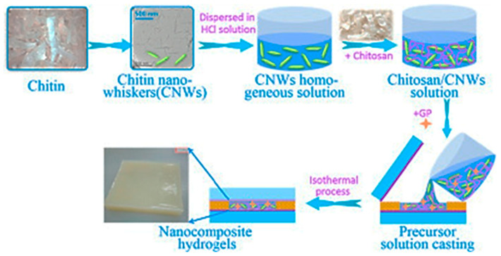
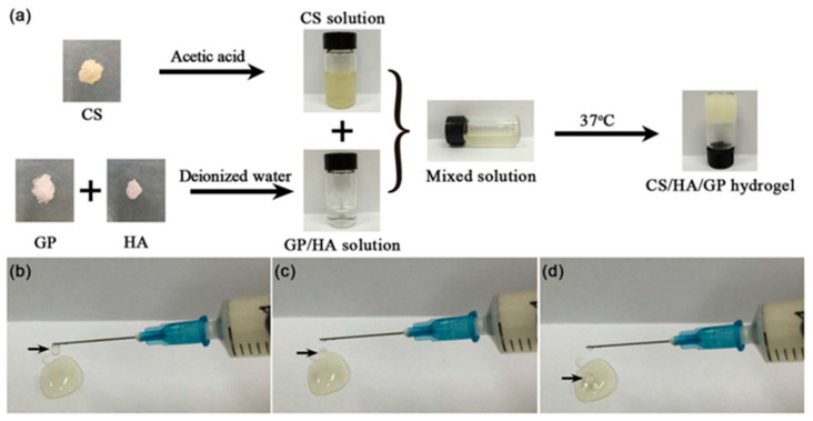
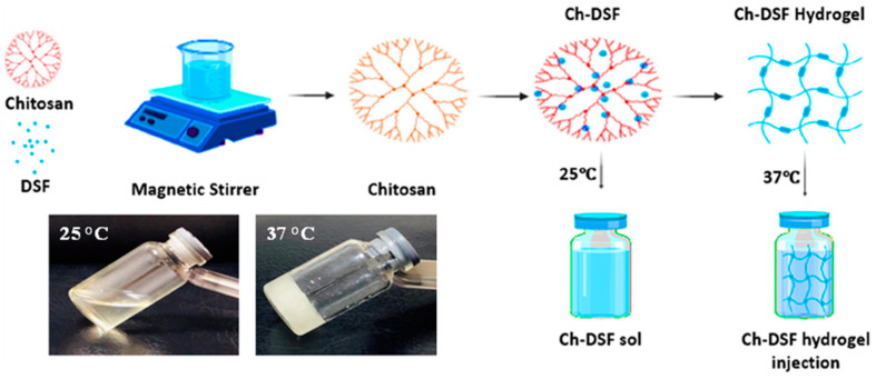
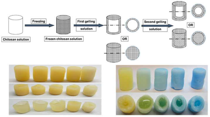
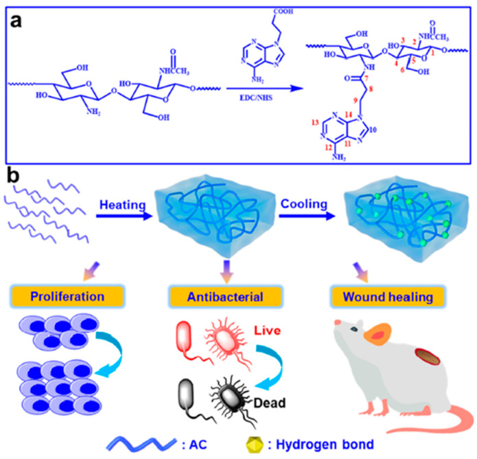
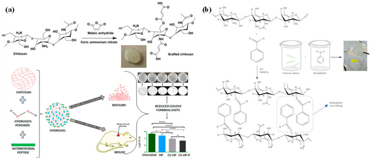
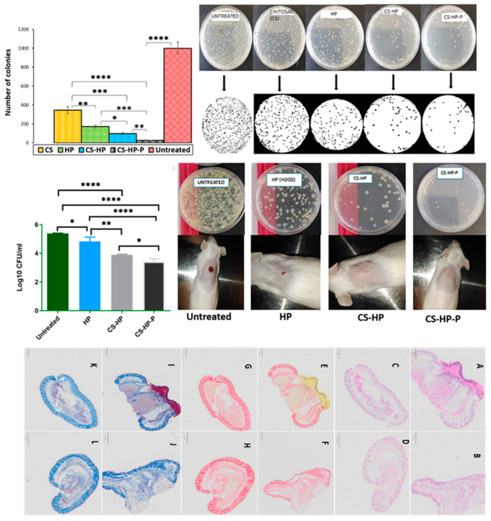
Chitosan is a prominent biopolymer in research for of its physicochemical properties and uses. Each year, the number of publications based on chitosan and its derivatives increases. Because of its comprehensive biological properties, including antibacterial, antioxidant, and tissue regeneration activities, chitosan and its derivatives can be used to prevent and treat soft tissue diseases. Furthermore, chitosan can be employed as a nanocarrier for therapeutic drug delivery. In this review, we will first discuss chitosan and chitosan-based hydrogel polymers. The structure, functionality, and physicochemical characteristics of chitosan-based hydrogels are addressed. Second, a variety of characterization approaches were used to analyze and validate the physicochemical characteristics of chitosan-based hydrogel materials. Finally, we discuss the antibacterial, antibiofilm, and antifungal uses of supramolecular chitosan-based hydrogels. This review study can be used as a base for future research into the production of various types of chitosan-based hydrogels in the antibacterial and antifungal fields.

Keywords: antibacterial activity; antibiofilm; antifungal materials; chitosan-based hydrogel; structural characterization.


Conflict of interest statement

The authors declare no conflict of interest.

Figures

Figure 12
Figure 12
TEM images of prepared chitosan caseinate PEC nanoparticles (af), and the DLS particle size analyses results (gi), adopted from open access resource [44].
Scheme 1
Scheme 1
Chemical conversion of chitin onto chitosan by deacetylation process.
Scheme 2
Scheme 2
Outline of production and chemical structure of chitosan.
Scheme 3
Scheme 3
Functional groups in chitosan polymer that can be chemically modified. M: monomer units.
Figure 1
Figure 1
(a) Chemical structure of kappa-carrageenan (k-CG) and chitosan (CS). (b) The schematic representation on the preparation of ultra-thin κ-carrageenan/chitosan hydrogel films. Reprinted with permission from [30] Copyright 2018 American Chemical Society.
Figure 2
Figure 2
The schematic representation of the preparation of CS/AM NSs hydrogel and its application towards the bacterial wound treatment on the mice, adopted from [31] with permission from Wiley through copyright clearance center.
Figure 3
Figure 3
The graphic representation on the preparation of CNWs/CS nanocomposites, adopted from [32] with permission from Elsevier through copyright clearance center.
Figure 4
Figure 4
(a) The preparation of hydrogel precursor solution (HG3) and the solution at low temperature of 4 °C and the formed hydrogel at 37 °C, (bd) the HG3 precursor solution can be readily ejected to form droplets using a syringe, adopted from [33] with permission from Elsevier through copyright clearance center. CS: Chitosan; HA: Hyaluronic acid; GP: Glycerophosphate.
Figure 5
Figure 5
The schematic representation of the preparation of chitosan-based thermosensitive hydrogels. Insert: the hydrogel at room and body temperature, adopted from open access resource [34]. DSF: disulfiram.
Figure 6
Figure 6
The schematic illustration of the preparation of the chitosan-based hydrogel; insert: photo images of the obtained hydrogel materials, adopted from [35] with permission from Elsevier through copyright clearance center.
Figure 7
Figure 7
The schematic representation with respect to the synthesis and structure of Cn nm gels. (a) The dissolution process of chitosan in alkaline solution; (c) The structure of gels with only 4r-PEG-CHO; (d) The structure of gels with 4r-PEG-CHO and 4r-PEG-NH2; (e) The molecular structure of the reactants. The SEM image on the right belongs to C2-N0 gel, the SEM images on the top left and bottom left belong to C2-N2 gel and C2-N4 gel, respectively, adopted from [36] with permission from Elsevier through copyright clearance center. PEG: Polyethyleneglycol.
Figure 8
Figure 8
(a) The synthetic route of AC derivatives. (b) Schematic diagram of AC hydrogel preparation and promotion of wound healing, adopted from [37] with permission from Elsevier through copyright clearance center. AC: Adenine-modified Chitosan.
Figure 9
Figure 9
Synthesis of norbornene-derived chitosan-based microgels, reprinted with permission from [38]. Copyright 2019 American Chemical Society.
Figure 10
Figure 10
(a) Chitosan-based hydrogel preparation using maleic anhydride towards anti-microbial applications, (b) the preparation of chitosan-based hydrogels using benzaldehyde using lyophilization method, adopted from open access resource [39,40].
Figure 11
Figure 11
The schematic depiction on the preparation of chitosan hydrogel nanocomposites, adopted from open access resource [42].
Figure 13
Figure 13
The bactericidal mechanism of DCHBSP gel (ciprofloxacin-based polymeric gel) against V. chemaguriensis strains in solid and liquid matrices. Adopted from open access resource [26].
Figure 14
Figure 14
This figure represents the application of hydrogels against infected wounds on mice model. Representational photographs of the healing progress of the wounds covered with gauze, and the sponges of chitosan, adopted from [78] with permission from Elsevier through copyright clearance center.
Figure 15
Figure 15
Number of colonies of untreated MRSA biofilms and biofilms exposed by chitosan-based hydrogels. Evaluation of MRSA burden post-treatment. Physical observation of the wound closure and bacterial burden. Photomicrographs of the untreated and treated skin sections stained with H&E, PSR, and CAB (0.5×), adopted from open access resource [39].

References

    1. Gheorghita R., Anchidin-Norocel L., Filip R., Dimian M., Covasa M. Applications of biopolymers for drugs and probiotics delivery. Polymers. 2021;13:2729. doi: 10.3390/polym13162729. - DOI - PMC - PubMed
    1. Rebitski E.P., Darder M. Chitosan and pectin core–shell beads encapsulating metformin–clay intercalation compounds for controlled delivery. New J. Chem. 2020;44:10102–10110. doi: 10.1039/C9NJ06433H. - DOI
    1. Khan A., Alamry K.A. Recent advances of emerging green chitosan-based biomaterials with potential biomedical applications: A review. Carbohydr. Res. 2021;506:108–368. doi: 10.1016/j.carres.2021.108368. - DOI - PubMed
    1. Aranaz I., Alcántara A.R., Concepción Civera M., Arias C., Elorza B., Caballero A.H., Acosta N. Chitosan: An Overview of Its Properties and Applications. Polymers. 2021;13:3256. doi: 10.3390/polym13193256. - DOI - PMC - PubMed
    1. Ardean C., Davidescu C.M., Nemeş N.S., Negrea A., Ciopec M., Duteanu N., Negrea P., Duda-Seiman D., Musta V. Factors Influencing the Antibacterial Activity of Chitosan and Chitosan Modified by Functionalization. Int. J. Mol. Sci. 2021;22:7449. doi: 10.3390/ijms22147449. - DOI - PMC - PubMed

LinkOut - more resources