. 2022 Dec 30;14(1):22.
doi: 10.3390/jfb14010022.

Improved Bone Regeneration Using Biodegradable Polybutylene Succinate Artificial Scaffold in a Rabbit Model

Affiliations

Improved Bone Regeneration Using Biodegradable Polybutylene Succinate Artificial Scaffold in a Rabbit Model

Giulio Edoardo Vigni et al. J Funct Biomater. .

Abstract

The treatment of extensive bone loss represents a great challenge for orthopaedic and reconstructive surgery. Most of the time, those treatments consist of multiple-stage surgeries over a prolonged period, pose significant infectious risks and carry the possibility of rejection. In this study, we investigated if the use of a polybutylene succinate (PBS) micro-fibrillar scaffold may improve bone regeneration in these procedures. In an in vivo rabbit model, the healing of two calvarial bone defects was studied. One defect was left to heal spontaneously while the other was treated with a PBS scaffold. Computed tomography (CT) scans, histological and immunohistochemical analyses were performed at 4, 12 and 24 weeks. CT examination showed a significantly larger area of mineralised tissue in the treated defect. Histological examination confirmed a greater presence of active osteoblasts and mineralised tissue in the scaffold-treated defect, with no evidence of inflammatory infiltrates around it. Immunohistochemical analysis was positive for CD56 at the transition point between healthy bone and the fracture zone. This study demonstrates that the use of a PBS microfibrillar scaffold in critical bone defects on a rabbit model is a potentially effective technique to improve bone regeneration.

Keywords: bone defect; bone reconstruction; bone regeneration; microfibrillar scaffold; polybutylene succinate; rabbit.


Conflict of interest statement

The authors have no relevant financial or non-financial interests to disclose.

Figures

Figure 1
Figure 1
Scaffold implantation into frontal bone defect.
Figure 2
Figure 2
Sampling of the frontal bones.
Figure 3
Figure 3
SEM image of electrospun PBS scaffold at 5000× (a) and 500× (b) magnification; microCT reconstruction of PBS scaffold (c) and photo of the planar scaffold (d).
Figure 4
Figure 4
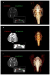
CT scan and 3D reconstruction at 4 (a), 12 (b) and 24 (c) weeks. Diameters’ comparison in coronal view of treated vs. untreated bone defects.
Figure 5
Figure 5
Comparison of the healing progression of the scaffold-treated and untreated (control) bone defect at 4, 12 and 24 weeks.
Figure 6
Figure 6
(A,B): Hematoxylin and eosin stain of untreated bone defect 4 weeks post implant (magnification: (A): 2.5×–(B): 5×). (C,D): Hematoxylin and eosin stain of treated bone defect 12 weeks post implant (magnification: (C): 2.5×–(D): 5×). Black arrows: bone tissue; black stars: fibrous tissue.
Figure 7
Figure 7
(a) Osteonecrosis in hematoxylin and eosin stain of treated bone defect 24 weeks post-implant (magnification: 20×); (b) Bone deposition and necrotic bone fragments in hematoxylin and eosin stain of treated bone defect 24 weeks post implant (magnification: 20×); (c) Periosseous tissue with embedded amorphous fragments (scaffold); (d) Bone marrow with hematopoietic stem cell niches. Black arrows: osteonecrosis areas; blue arrow: hematopoietic stem cell niches; black star: necrotic bone fragments; blue stars: amorphous fragments (scaffold); thin blue arrow: bone deposition.
Figure 8
Figure 8
Activated osteoblasts with CD56 as immunohistochemical marker 24 weeks post-implant, related to scaffold-treated specimens (a,b) and control (c,d). Magnification 10×.

References

    1. Mauffrey C., Barlow B.T., Smith W. Management of Segmental Bone Defects. J. Am. Acad. Orthop. Surg. 2015;23:143–153. doi: 10.5435/JAAOS-D-14-00018. - DOI - PubMed
    1. Hertel R., Gerber A., Schlegel U., Cordey J., Rüegsegger P., Rahn B.A. Cancellous Bone Graft for Skeletal Reconstruction. Muscular versus Periosteal Bed--Preliminary Report. Injury. 1994;25((Suppl. S1)):A59–A70. doi: 10.1016/0020-1383(94)90263-1. - DOI - PubMed
    1. Belthur M.V., Conway J.D., Jindal G., Ranade A., Herzenberg J.E. Bone Graft Harvest Using a New Intramedullary System. Clin. Orthop. Relat. Res. 2008;466:2973–2980. doi: 10.1007/s11999-008-0538-3. - DOI - PMC - PubMed
    1. Tatara A.M., Mikos A.G. Tissue Engineering in Orthopaedics. J. Bone Joint Surg. Am. 2016;98:1132. doi: 10.2106/JBJS.16.00299. - DOI - PMC - PubMed
    1. Treatment of Osteonecrosis With Autologous Bone Marrow Graft…: Clinical Orthopaedics and Related Research®. [(accessed on 9 November 2022)]. Available online: https://journals.lww.com/clinorthop/Fulltext/2002/12000/Treatment_of_Ost....