. 2023 Jan 16;9(1):19.
doi: 10.3390/jimaging9010019.

Picomolar Detection of Lead Ions (Pb2+) by Functionally Modified Fluorescent Carbon Quantum Dots from Watermelon Juice and Their Imaging in Cancer Cells

Affiliations

Picomolar Detection of Lead Ions (Pb2+) by Functionally Modified Fluorescent Carbon Quantum Dots from Watermelon Juice and Their Imaging in Cancer Cells

Kundan Singh Rawat et al. J Imaging. .

Abstract

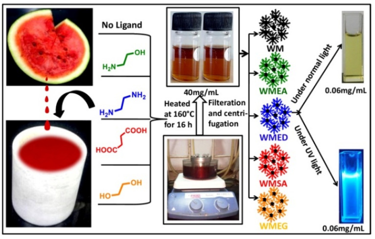
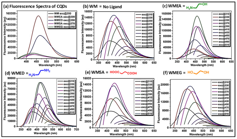
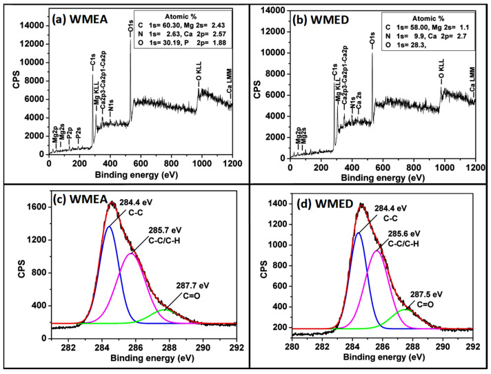
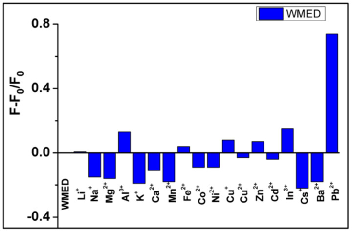
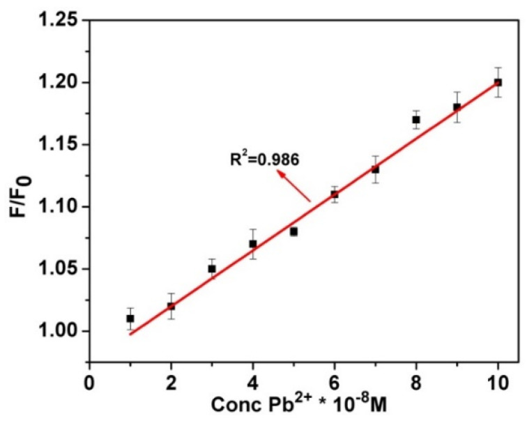
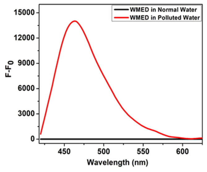
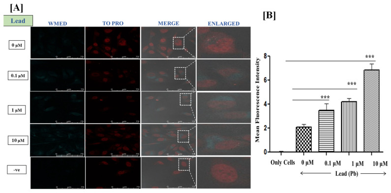
Water contamination due to the presence of lead is one of the leading causes of environmental and health hazards because of poor soil and groundwater waste management. Herein we report the synthesis of functionally modified luminescent carbon quantum dots (CQDs) obtained from watermelon juice as potential nanomaterials for the detection of toxic Pb2+ ions in polluted water and cancer cells. By introducing surface passivating ligands such as ethanolamine (EA) and ethylenediamine (ED) in watermelon juice, watermelon-ethanolamine (WMEA)-CQDs and watermelon-ethylenediamine (WMED)-CQDs exhibited a remarkable ~10-fold and ~6-fold increase in fluorescence intensity with respect to non-doped WM-CQDs. The relative fluorescence quantum yields of WMEA-CQDs and WMED-CQDs were found to be 8% and 7%, respectively, in an aqueous medium. Among various functionally-modified CQDs, only WMED-CQDs showed high selectivity towards Pb2+ ions with a remarkably good limit of detection (LoD) of 190 pM, which is less than that of the permissible limit (72 nM) in drinking water. The functionally altered WMED-CQDs detected Pb2+ metal ions in polluted water and in a human cervical cancer cell line (HeLa), thus advocating new vistas for eco-friendly nanomaterials for their use as diagnostic tools in the environment and biomedical research areas.

Keywords: bioimaging; carbon quantum dots; green synthesis; lead ion sensing; watermelon juice.


Conflict of interest statement

The authors declare no conflict of interest.

Figures

Scheme 1
Scheme 1
Graphical representation of the one-pot synthesis of CQDs from watermelon juice by adding different surface passivating agents.
Figure 1
Figure 1
(a) Emission spectra of WM-CQDs and WMEG at 350 nm excitation, emission spectra of WMEG-CQDs at 370 nm excitation, emission spectra of WMEA-CQDs and WMED-CQDs at 390 nm excitation, showing intensity difference by introducing different surface passivating agents. Excitation-dependent emission spectra of (b) WM-CQDs; (c) WMEA-CQDs; (d) WMED-CQDs; (e) WMSA-CQDs; and (f) WMEG-CQDs obtained by hydrothermal treatment at different excitation.
Figure 2
Figure 2
Time-resolved fluorescence decay of WMEA-CQDs (λex = 390 nm and λem = 460 nm) and WMED-CQDs (λex = 390 nm and λem = 470 nm) in aqueous medium.
Figure 3
Figure 3
TEM images of watermelon-CQDs (a) WMEA CQDs dispersed in water; inset shows particle size distribution and (b) WMED CQDs dispersed in water; inset shows particle size distribution; (c,d) zoomed TEM images of CQDs, red arrows showed CQDs.
Figure 4
Figure 4
Full range XPS analysis of watermelon-CQDs prepared by adding (a) ethanol amine; (b) ethylene diamine; high-resolution XPS spectra of the C1s region of watermelon-CQDs synthesized by adding (c) ethanol amine; (d) ethylene diamine.
Figure 5
Figure 5
Selectivity of the WMED to different metal ions. The concentration of Pb2+ and other metal ions was 1 × 10⁻⁴ M.
Figure 6
Figure 6
The linear variation of fluorescence intensity of WMED CQDs in triple distilled water (TDW) with increasing concentration of lead, λex = 390 nm.
Figure 7
Figure 7
Fluorescence intensity of WMED in normal water (black line) and contaminated water having Pb2+ ions (Red line).
Figure 8
Figure 8
(A) Confocal fluorescence microscopy images at 100× Magnification; Live HeLa cells were incubated with Lead ions at 0.1 μM, 1 μM, and 10 μM concentrations for 1.5 h. Staining of Live HeLa cells with 0.128 mg/mL WMED, images recorded for WMED at λex 405 nm/λem 450–550 nm. The Nucleus is stained with TO-PRO nuclear dye λex 641 nm/λem 661 nm; (B) quantification of lead ions using ImageJ software version 1.46r, *** p < 0.001.

References

    1. Flegal A.R., Smith D.R. Current Needs for Increased Accuracy and Precision in Measurements of Low Levels of Lead in Blood. Environ. Res. 1992;58:125–133. doi: 10.1016/S0013-9351(05)80209-9. - DOI - PubMed
    1. Neal A.P., Guilarte T.R. Mechanisms of lead and manganese neurotoxicity. Toxicol. Res. 2013;2:99–114. doi: 10.1039/c2tx20064c. - DOI - PMC - PubMed
    1. Fan C., He S., Liu G., Wang L., Song S. A Portable and Power-Free Microfluidic Device for Rapid and Sensitive Lead (Pb2+) Detection. Sensors. 2012;12:9467–9475. doi: 10.3390/s120709467. - DOI - PMC - PubMed
    1. Kim H.N., Ren W.X., Kim J.S., Yoon J. Fluorescent and colourimetric sensors for detecting lead, cadmium, and mercury ions. Chem. Soc. Rev. 2012;41:3210–3244. doi: 10.1039/C1CS15245A. - DOI - PubMed
    1. U.S. Environmental Protection Agency . Edition of the Drinking Water Standards and Health Advisories. Office of Water; Washington, DC, USA: 2009.

LinkOut - more resources