

The Polysaccharidic Nature of the Skeleton of Marennine as Determined by NMR Spectroscopy
- PMID: 36662215
- PMCID: PMC9865362
- DOI: 10.3390/md21010042
The Polysaccharidic Nature of the Skeleton of Marennine as Determined by NMR Spectroscopy
Abstract
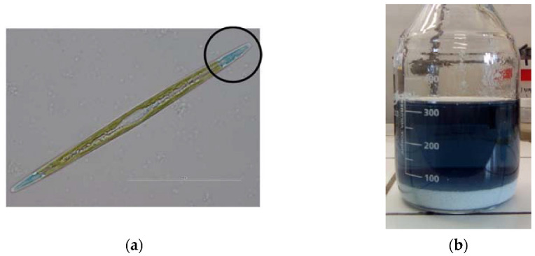
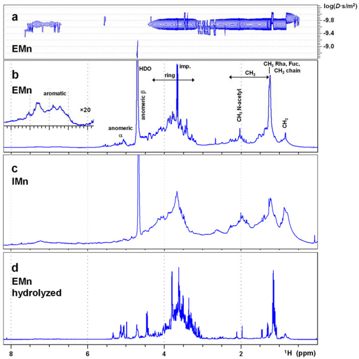
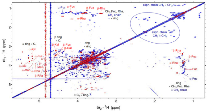
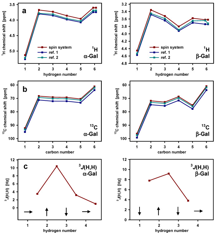
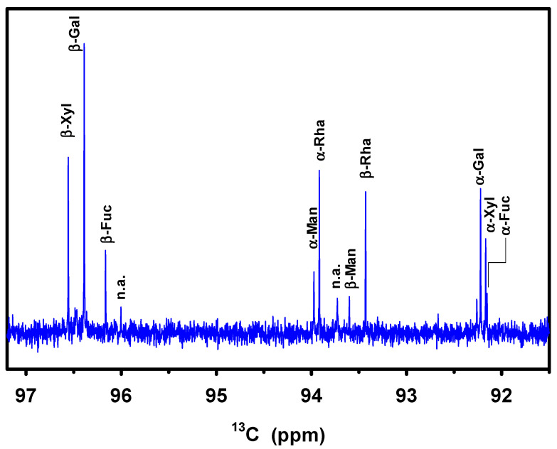
The water-soluble blue-green pigment marennine, produced and partly excreted by the diatom Haslea ostrearia, and known for a long time for its role in the greening of oysters, was isolated from the culture medium, purified, and analyzed by Nuclear Magnetic Resonance (NMR) in order to gain insight into its chemical structure. The spectra show mainly carbohydrates of a complex composition, apparently highly branched, and with a mass in the order of 10 kDa. There are, in addition, some signals of aliphatic and, much weaker, aromatic groups that present aglycons. The latter might be responsible for the color. These carbohydrates are always associated with the blue-green color and cannot be separated from it by most treatments; they are interpreted as constituting the frame of the pigment. NMR after hydrolysis identifies the most abundant monosaccharides in marennine as galactose, xylose, mannose, rhamnose, and fucose.
Keywords: Haslea ostrearia; biomolecular NMR; blue pigment; diatoms; marennine; microalgae; polysaccharides.
Conflict of interest statement
The authors declare no conflict of interest.
Figures
References
-
- Sprat T. The History of the Royal Society. Martyn; London, UK: 1667. The history of the generation and ordering green oysters, commonly called Colchester-oysters.
-
- Gaillon B. Des huîtres vertes et des causes de cette coloration. J. Phys. Chim. Hist. Nat. Arts. 1820;91:222–225.
-
- Bory de Saint-Vincent J.B. Dictionnaire Classique d’Histoire Naturelle. Rey et Gravier/Baudouin; Paris, France: 1824. Navicules; pp. 562–565.
-
- Simonsen R. The Diatom Plankton of the Indian Ocean Expedition of RV “Meteor”. Volume 19. Deutsche Forschungsgemeinschaft; Bonn, Germany: 1974. pp. 46–49.
-
- Lankester E.R. On green oysters. Q. J. Microsc. Sci. 1886;26:71–94. doi: 10.1038/052028c0. - DOI
MeSH terms
Substances
Grants and funding
LinkOut - more resources
Full Text Sources
