Review
. 2023 Jan 5;10(1):11.
doi: 10.3390/medicines10010011.

Choroidal Melanoma: A Mini Review

Affiliations
Review

Choroidal Melanoma: A Mini Review

Noha Soliman et al. Medicines (Basel). .

Abstract

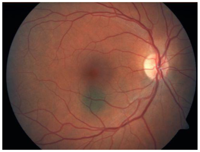
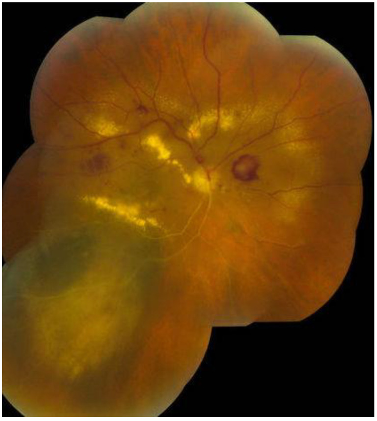
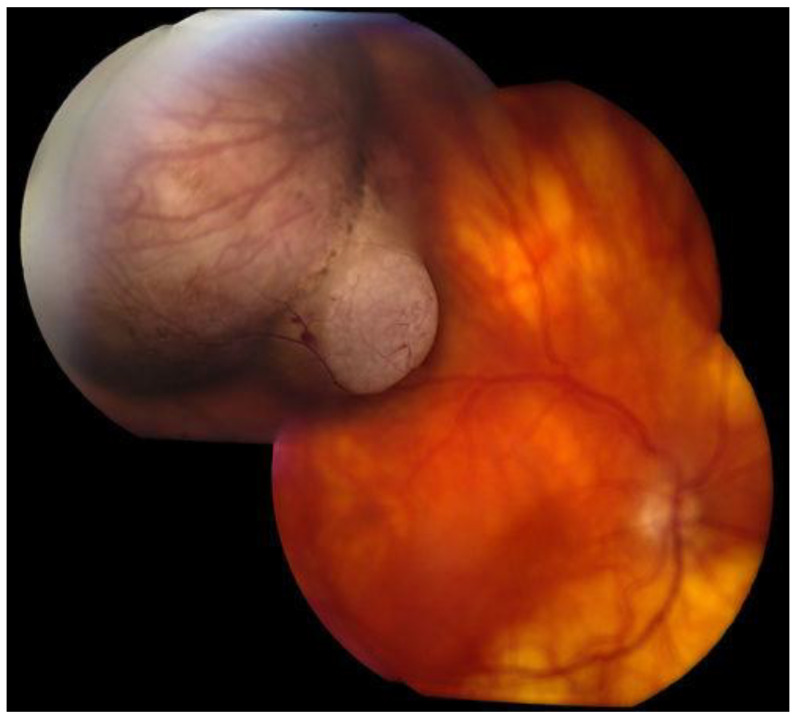
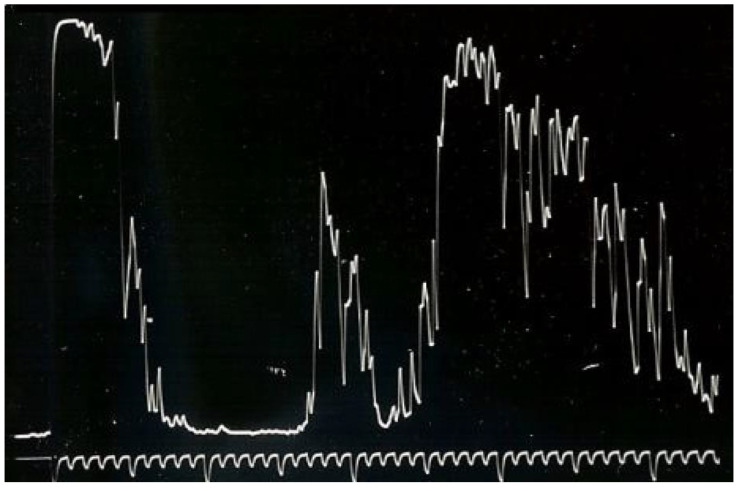
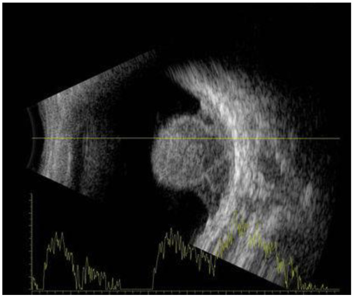
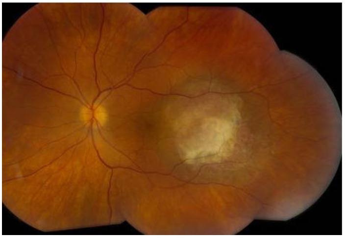
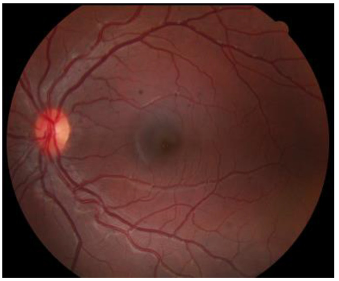
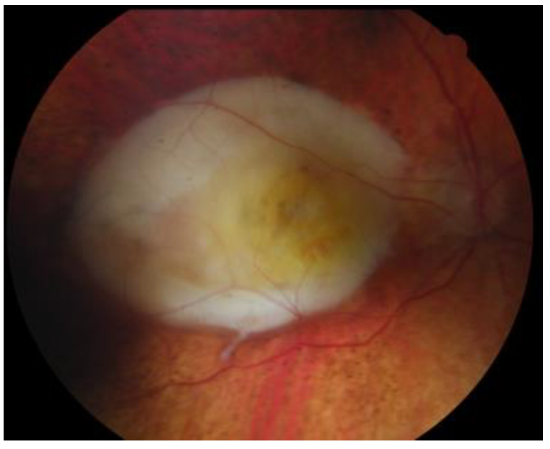
Choroidal melanoma is a rare malignant tumour, yet it is the most common primary intra-ocular neoplasm and second on the list of top ten most malignant melanoma sites in the body. Clinical presentation can be non-specific and includes photopsia, floaters, progressive visual field loss, and blurry vision. The tumour is quite often diagnosed clinically during fundus examination; however, the most valued diagnostic tests are A- and B-scan ultrasonography (US). Several factors affect prognosis, including the patient's age, tumour size, histological features, and presence of metastases. Still, with primary treatment and tight surveillance, around 50% of choroidal melanoma patients metastasise.

Keywords: choroidal melanoma; eye cancer; management; prognosis.


Conflict of interest statement

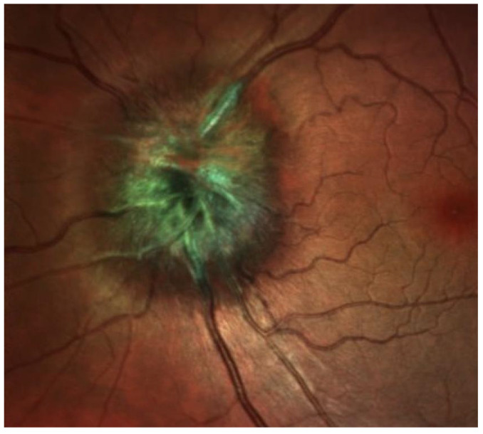
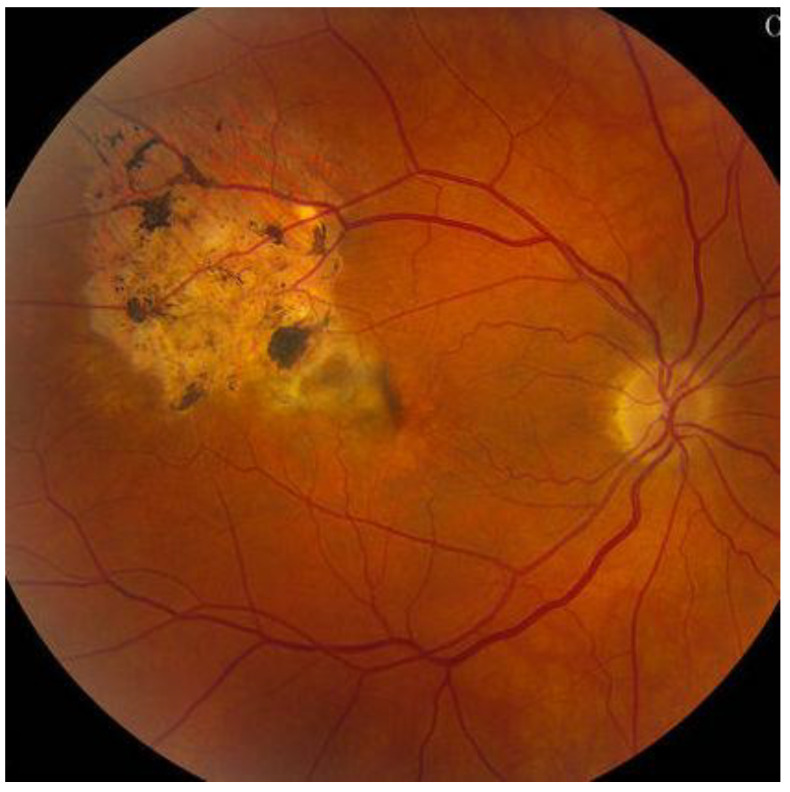
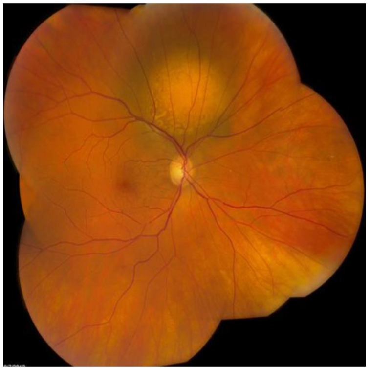
The authors declare no conflict of interest. Figures: All figures illustrated in this review were retrieved from the Retina Gallery via https://www.retinagallery.com/index.php (accessed on 21 November 2022). Retina Gallery is a library of free and non-copyrighted Retinal images. The authors declare no conflict of interest with Retina Gallery.

Figures

Figure 1
Figure 1
Choroidal nevus.
Figure 2
Figure 2
Darkly pigmented melanoma.
Figure 3
Figure 3
Amelanotic choroidal melanoma.
Figure 4
Figure 4
A-scan US reveals medium to low internal reflectivity with vascular pulsations.
Figure 5
Figure 5
B-scan US shows a mushroom or dome-shaped tumour. The mass is acoustically silent, indicating a dark appearance inside the tumour.
Figure 6
Figure 6
Choroidal haemangiomas.
Figure 7
Figure 7
Retinal pigment epithelium hypertrophy.
Figure 8
Figure 8
Disciform scar.
Figure 9
Figure 9
Melanocytoma.
Figure 10
Figure 10
Choroidal osteoma.
Figure 11
Figure 11
Choroidal metastasis.

References

    1. Singh P., Singh A. Choroidal melanoma. Oman J. Ophthalmol. 2012;5:3. doi: 10.4103/0974-620X.94718. - DOI - PMC - PubMed
    1. Yang J., Manson D.K., Marr B.P., Carvajal R.D. Treatment of uveal melanoma: Where are we now? Ther. Adv. Med. Oncol. 2018;10:1758834018757175. doi: 10.1177/1758834018757175. - DOI - PMC - PubMed
    1. Violanti S.S., Bononi I., Gallenga C.E., Martini F., Tognon M., Perri P. New insights into molecular oncogenesis and therapy of uveal melanoma. Cancers. 2019;11:694. doi: 10.3390/cancers11050694. - DOI - PMC - PubMed
    1. Krantz B.A., Dave N., Komatsubara K.M., Marr B.P., Carvajal R.D. Uveal melanoma: Epidemiology, etiology, and treatment of primary disease. Clin. Ophthalmol. 2017;11:279. doi: 10.2147/OPTH.S89591. - DOI - PMC - PubMed
    1. Terzidou C., Trivli A., Dalianis G., Apessou D., Spandidos D.A., Goulielmos G.N. Advanced choroidal melanoma with a desirable aesthetic outcome after enucleation: A case report. Oncol. Lett. 2018;16:511–514. doi: 10.3892/ol.2018.8661. - DOI - PMC - PubMed

LinkOut - more resources