. 2023 Jan 20;18(1):e0277442.
doi: 10.1371/journal.pone.0277442. eCollection 2023.

Promoting early neovascularization by allotransplanted adipose-derived Muse cells in an ovine model of acute myocardial infarction

Affiliations

Promoting early neovascularization by allotransplanted adipose-derived Muse cells in an ovine model of acute myocardial infarction

Martha G Castillo et al. PLoS One. .

Abstract

Background: Recent preclinical studies have demonstrated that bone marrow (BM)-derived Muse cells have a homing mechanism to reach damaged cardiac tissue while also being able to reduce myocardial infarct size and improve cardiac function; however, the potential of BM-Muse cells to foster new blood-vessel formation has not been fully assessed. Up to date, adipose tissue (AT)-derived Muse cells remain to be studied in acute myocardial infarction (AMI). The aim of the present study was to analyze in vitro and in vivo the neovascularization capacity of AT-Muse cells while exploring their biodistribution and differentiation potential in a translational ovine model of AMI.

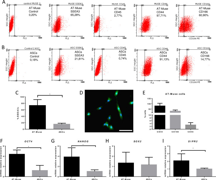
Methods and results: AT-Muse cells were successfully isolated from ovine adipose tissue. In adult sheep, one or more diagonal branches of the left anterior descending coronary artery were permanently ligated for thirty minutes. Sheep were randomized in two groups and treated with intramyocardial injections: Vehicle (PBS, n = 4) and AT-Muse (2x10⁷ AT-Muse cells labeled with PKH26 Red Fluorescent Dye, n = 4). Molecular characterization showed higher expression of angiogenic genes (VEGF, PGF and ANG) and increased number of tube formation in AT-Muse cells group compared to Adipose-derived mesenchymal stromal cells (ASCs) group. At 7 days post-IAM, the AT-Muse group showed significantly more arterioles and capillaries than the Vehicle group. Co-localization of PKH26+ cells with desmin, sarcomeric actin and troponin T implied the differentiation of Muse cells to a cardiac fate; moreover, PKH26+ cells also co-localized with a lectin marker, suggesting a possible differentiation to a vascular lineage.

Conclusion: Intramyocardially administered AT-Muse cells displayed a significant neovascularization activity and survival capacity in an ovine model of AMI.


Conflict of interest statement

The authors have declared that no competing interests exist.

Figures

Fig 1
Fig 1. Flow cytometry and gene expression analysis.
Labeling of A: AT-Muse cells and B: ASCs with antibodies anti- SSEA3, CD45, CD44, CD166 and controls. C: The % of SSEA3+ cells was higher in the AT-Muse group than in the ASCs group. D: Immunofluorescence image confirming the labeling of SSEA3 (green) and DAPI (blue) in AT-Muse cells at P4 in vitro. Scale bar = 100 μm. E: AT-Muse cells were CD166+, CD44+ and CD45- confirming their immunophenotype. F-I: The AT-Muse group showed higher expression of OCT4, NANOG and S1PR2 than the ASCs group. Results are means ± SD. *p<0.05, Mann-Whitney U test.
Fig 2
Fig 2. Angiogenic potential of AT-Muse cells.
A-C: VEGF, ANG, and PGF gene expression was higher in the AT-Muse group than in the ASCs group. D: Tube formation assay: the AT-Muse group showed an increased number of rings per area (mm2) compared to the ASCs group. E-F: Representative images of rings formed in the AT-Muse and ASCs groups. G-H: VEGF protein expression was higher in the AT-Muse group than in the ASCs group. Scale bar 1000 μm. Results are mean ± SD. ** p < 0.01; *p < 0.05, Mann-Whitney U test.
Fig 3
Fig 3. Differentiation of PKH26+ cells.
A-F: Fluorescence microscopy analysis revealed the presence of PKH26+ cells at 7 days post-AMI in the infarct border zone of AT-Muse group. A: PKH26+ Muse cells (red), Desmin (green), and DAPI (blue) are merged. B: PKH26+ Muse cells (red), Sarcomeric actin (green), and DAPI (blue) are merged. C: PKH26+ Muse cells (red), Troponin T (green), and DAPI (blue) are merged. D: PKH26+ Muse cells (red), Connexin-43 (green), and DAPI (blue) are merged. E: PKH26+ Muse cells (red), Lectin (green), and DAPI (blue) are merged. F: PKH26+ Muse cells (red), Smooth muscle, actin (green) and DAPI (blue) are merged. One cell is enlarged in the top left box (white arrow). Bars = 50 μm.
Fig 4
Fig 4. Microvascular density at 7 days post-AMI.
A-B: Arteriolar and capillary densities were significantly higher in the AT-Muse group than in the Vehicle group. C-D: Representative slices of the infarct border zone for the AT-Muse and Vehicle groups. Arterioles were stained with an anti-smooth muscle actin antibody and counterstained with hematoxylin (bars = 100 μm). E-F: Representative slices of the infarct border zone for the AT-Muse and Vehicle groups showing capillaries stained with a biotinylated lectin and counterstained with hematoxylin (bars = 100 μm). Results are means ± SD. *p<0.05, Mann-Whitney U test.
Fig 5
Fig 5. Cycling cells and protein expression of angiogenic markers at 7 days post-AMI.
A: The number of Ki67+ cells in the AT-Muse group was significantly higher than in the Vehicle group. B: Representative image of Ki67+ nuclei (brown) counterstained with sarcomeric actin (violet) in the AT-Muse group (bar = 20 μm). C-D: The VEGF had higher gene and protein expression in the infarct border zones of the hearts of AT-Muse group in comparison with the Vehicle group, respectively. E: The CD34 protein expression was higher than in the Vehicle group. F: representative western blot images for VEGF and CD34. Results are means ± SD. *p<0.05, Mann-Whitney U test.
Fig 6
Fig 6. Biodistribution analysis of AT-Muse cells.
A: High number of PKH26-labeled ovine AT-Muse cells were found in the infarct border cross sections. Some Muse cells were found in the lungs although no Muse cells were found in spleen, liver, kidney, retina, gonad or in the Vehicle group. Results are means ± SD; ****p<0.0001, two-way ANOVA with Tukey’s post-hoc test for multiple comparisons. B: Representative microscopic merged image of infarct border zone of heart injected with Muse, PKH26+ cells (red) and DAPI (blue). C: Representative microscopic merged image from lung tissue of AT-Muse group, PKH26+ cells (red) and DAPI (blue).

References

    1. Roth G, Mensah G, Johnson C, Addolorato G, Ammirati E, Baddour L, et al.. Global Burden of Cardiovascular Diseases and Risk Factors, 1990–2019. Journal of the American College of Cardiology. 2020;76(25):2982–3021. - PMC - PubMed
    1. Hashimoto H, Olson E, Bassel-Duby R. Therapeutic approaches for cardiac regeneration and repair. Nature Reviews Cardiology. 2018;15(10):585–600. doi: 10.1038/s41569-018-0036-6 - DOI - PMC - PubMed
    1. Karantalis V, Hare J. Use of Mesenchymal Stem Cells for Therapy of Cardiac Disease. Circulation Research. 2015;116(8):1413–1430. doi: 10.1161/CIRCRESAHA.116.303614 - DOI - PMC - PubMed
    1. Bagno L, Hatzistergos K, Balkan W, Hare J. Mesenchymal Stem Cell-Based Therapy for Cardiovascular Disease: Progress and Challenges. Molecular Therapy. 2018;26(7):1610–1623. doi: 10.1016/j.ymthe.2018.05.009 - DOI - PMC - PubMed
    1. Golpanian S, Wolf A, Hatzistergos K, Hare J. Rebuilding the Damaged Heart: Mesenchymal Stem Cells, Cell-Based Therapy, and Engineered Heart Tissue. Physiological Reviews. 2016;96(3):1127–1168. doi: 10.1152/physrev.00019.2015 - DOI - PMC - PubMed

Publication types