The Effect of Ascorbic Acid and Nicotinamide on Panton-Valentine Leukocidin Cytotoxicity: An Ex Vivo Study
- PMID: 36668859
- PMCID: PMC9865643
- DOI: 10.3390/toxins15010038
The Effect of Ascorbic Acid and Nicotinamide on Panton-Valentine Leukocidin Cytotoxicity: An Ex Vivo Study
Abstract
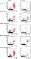
Background: Panton−Valentine Leukocidin sustains a strong cytotoxic activity, targeting immune cells and, consequently, perforating the plasma membrane and inducing cell death. The present study is aimed to examine the individual effect of ascorbic acid and nicotinamide on PVL cytotoxicity ex vivo, as well as their effect on granulocytes viability when treated with PVL. Materials and Methods: The PVL cytotoxicity assay was performed in triplicates using the commercial Cytotoxicity Detection Kit PLUS (LDH). LDH release was measured to determine cell damage and cell viability was measured via flow cytometry. Results and discussion: A clear reduction in PVL cytotoxicity was demonstrated (p < 0.001). Treatment with ascorbic acid at 5 mg/mL has shown a 3-fold reduction in PVL cytotoxicity; likewise, nicotinamide illustrated a 4-fold reduction in PVL cytotoxicity. Moreover, granulocytes’ viability after PVL treatment was maintained when incubated with 5 mg/mL of ascorbic acid and nicotinamide. Conclusions: our findings illustrated that ascorbic acid and nicotinamide exhibit an inhibitory effect on PVL cytotoxicity and promote cell viability, as the cytotoxic effect of the toxin is postulated to be neutralized by antioxidant incubation. Further investigations are needed to assess whether these antioxidants may be viable options in PVL cytotoxicity attenuation in PVL-associated diseases.
Keywords: PVL; antioxidant; ascorbic acid; nicotinamide; vitamin.
Conflict of interest statement
The authors declare that there are no conflict of interest.
Figures


Similar articles
-
Reduction of Panton-Valentine Leukocidin Production in the Staphylococcal Strain USA300 After In Vitro Ascorbic Acid and Nicotinamide Treatment.Cureus. 2023 Oct 24;15(10):e47588. doi: 10.7759/cureus.47588. eCollection 2023 Oct. Cureus. 2023. PMID: 38022293 Free PMC article.
-
α-Defensins partially protect human neutrophils against Panton-Valentine leukocidin produced by Staphylococcus aureus.Lett Appl Microbiol. 2015 Aug;61(2):158-64. doi: 10.1111/lam.12438. Epub 2015 Jun 1. Lett Appl Microbiol. 2015. PMID: 25963798
-
Accumulation of staphylococcal Panton-Valentine leukocidin in the detergent-resistant membrane microdomains on the target cells is essential for its cytotoxicity.FEMS Immunol Med Microbiol. 2012 Dec;66(3):343-52. doi: 10.1111/j.1574-695X.2012.01027.x. Epub 2012 Sep 12. FEMS Immunol Med Microbiol. 2012. PMID: 22924956
-
Panton-Valentine Leukocidin Induces Cytokine Release and Cytotoxicity Mediated by the C5a Receptor on Rabbit Alveolar Macrophages.Jpn J Infect Dis. 2021 Jul 21;74(4):352-358. doi: 10.7883/yoken.JJID.2020.657. Epub 2021 Jan 29. Jpn J Infect Dis. 2021. PMID: 33518621
-
The staphylococcal toxin Panton-Valentine Leukocidin targets human C5a receptors.Cell Host Microbe. 2013 May 15;13(5):584-594. doi: 10.1016/j.chom.2013.04.006. Cell Host Microbe. 2013. PMID: 23684309
Cited by
-
The impact of aging and oxidative stress in metabolic and nervous system disorders: programmed cell death and molecular signal transduction crosstalk.Front Immunol. 2023 Nov 8;14:1273570. doi: 10.3389/fimmu.2023.1273570. eCollection 2023. Front Immunol. 2023. PMID: 38022638 Free PMC article. Review.
-
Reduction of Panton-Valentine Leukocidin Production in the Staphylococcal Strain USA300 After In Vitro Ascorbic Acid and Nicotinamide Treatment.Cureus. 2023 Oct 24;15(10):e47588. doi: 10.7759/cureus.47588. eCollection 2023 Oct. Cureus. 2023. PMID: 38022293 Free PMC article.
-
Cognitive Impairment in Multiple Sclerosis.Bioengineering (Basel). 2023 Jul 23;10(7):871. doi: 10.3390/bioengineering10070871. Bioengineering (Basel). 2023. PMID: 37508898 Free PMC article. Review.
-
Cornerstone Cellular Pathways for Metabolic Disorders and Diabetes Mellitus: Non-Coding RNAs, Wnt Signaling, and AMPK.Cells. 2023 Nov 9;12(22):2595. doi: 10.3390/cells12222595. Cells. 2023. PMID: 37998330 Free PMC article. Review.
-
Innovative therapeutic strategies for cardiovascular disease.EXCLI J. 2023 Jul 26;22:690-715. doi: 10.17179/excli2023-6306. eCollection 2023. EXCLI J. 2023. PMID: 37593239 Free PMC article. Review.
References
-
- Panton P.N., Valentine F.C.O. Staphylococcal Toxin. Lancet. 1932;219:506–508. doi: 10.1016/S0140-6736(01)24468-7. - DOI
-
- Diep B.A., Chan L., Tattevin P., Kajikawa O., Martin T.R., Basuino L., Mai T.T., Marbach H., Braughton K.R., Whitney A.R., et al. Polymorphonuclear leukocytes mediate Staphylococcus aureus Panton-Valentine leukocidin-induced lung inflammation and injury. Proc. Natl. Acad. Sci. USA. 2010;107:5587–5592. doi: 10.1073/pnas.0912403107. - DOI - PMC - PubMed
-
- Lipinska U., Hermans K., Meulemans L., Dumitrescu O., Badiou C., Duchateau L., Haesebrouck F., Etienne J., Lina G. Panton-Valentine leukocidin does play a role in the early stage of Staphylococcus aureus skin infections: A rabbit model. PLoS ONE. 2011;6:e22864. doi: 10.1371/journal.pone.0022864. - DOI - PMC - PubMed
MeSH terms
Substances
LinkOut - more resources
Full Text Sources
Medical

