. 2023 Jan 12;15(1):70.
doi: 10.3390/toxins15010070.

The Bi-Functional Paxilline Enriched in Skin Secretion of Tree Frogs (Hyla japonica) Targets the KCNK18 and BKCa Channels

Affiliations

The Bi-Functional Paxilline Enriched in Skin Secretion of Tree Frogs (Hyla japonica) Targets the KCNK18 and BKCa Channels

Chuanling Yin et al. Toxins (Basel). .

Abstract

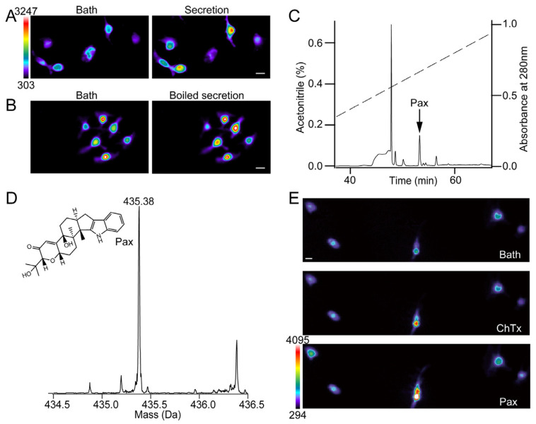
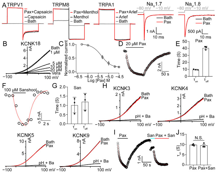
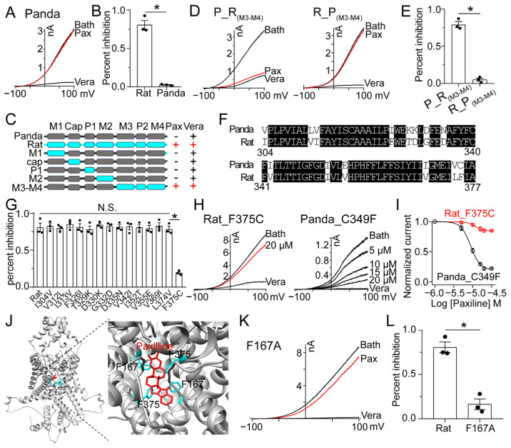
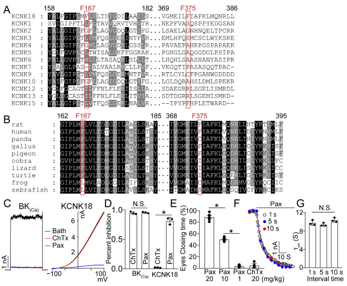
The skin secretion of tree frogs contains a vast array of bioactive chemicals for repelling predators, but their structural and functional diversity is not fully understood. Paxilline (PAX), a compound synthesized by Penicillium paxilli, has been known as a specific antagonist of large conductance Ca2+-activated K+ Channels (BKCa). Here, we report the presence of PAX in the secretions of tree frogs (Hyla japonica) and that this compound has a novel function of inhibiting the potassium channel subfamily K member 18 (KCNK18) channels of their predators. The PAX-induced KCNK18 inhibition is sufficient to evoke Ca2+ influx in charybdotoxin-insensitive DRG neurons of rats. By forming π-π stacking interactions, four phenylalanines located in the central pore of KCNK18 stabilize PAX to block the ion permeation. For PAX-mediated toxicity, our results from animal assays suggest that the inhibition of KCNK18 likely acts synergistically with that of BKCa to elicit tingling and buzzing sensations in predators or competitors. These results not only show the molecular mechanism of PAX-KCNK18 interaction, but also provide insights into the defensive effects of the enriched PAX.

Keywords: KCNK18; defensive strategy; paxilline; skin secretion; tree frogs.


Conflict of interest statement

The authors declare no conflict of interest.

Figures

Figure 1
Figure 1
PAX induces Ca2+ signals on ChTx-insensitive neurons. (A,B) Representative calcium increase of rat DRG neurons in the presence of intact (A) or boiled (B) skin secretions of tree frogs. Scale bar, 50 μm (horizontal), 303 to 3247 AU (vertical). (C) Isolation of native PAX (black arrow) from frog skin secretions by a C18 RP-HPLC column. (D) The chemical structure and molecular weight of PAX. (E) Calcium imaging of rat DRG neurons in the presence of 100 nM ChTx and 20 μM PAX. Scale bar, 50 μm (horizontal), 294 to 4095 AU (vertical).
Figure 2
Figure 2
PAX selectively inhibits rat KCNK18. (A) 100 μM PAX had no inhibitory effect on TRPV1, TRPM8, TRPA1, NaV1.7 and NaV1.8. (B) Representative KCNK18 currents inhibited by PAX at different concentrations. 100 μM verapamil (vera) was used as a positive control. (C) Dose–response relationship of PAX inhibiting KCNK18. Data were fitted to a Hill equation (average ± SEM; n = 3 for each data point). (D) Representative wash-in and wash-out time course of 20 μM PAX on KCNK18 recorded at 100 mV, superimposed with fittings of a single-exponential function (red dotted curves). (E) The associated and dissociated time of 20 μM PAX on KCNK18 (average ± SEM; n = 3). (F) Representative wash-in and wash-out time course of 100 μM sanshool on KCNK18 recorded at 100 mV, superimposed with fittings of a single-exponential function (red dotted curves). (G) The associated and dissociated time of 100 μM sanshool on KCNK18 (average ± SEM; n = 3). (H) 100 μM PAX had no inhibitory effect on KCNK3, KCNK4, KCNK5 and KCNK9. (I) Representative wash-in time course of 20 μM PAX on KCNK18 recorded at 100 mV in the absence and presence of 50 μM sanshool, superimposed with fittings of a single-exponential function (red dotted curves). The solid black line indicates the presence of 20 μM PAX. The solid blue line indicates the presence of 50 μM sanshool. (J) The associated time of 20 μM PAX on KCNK18 in the absence and presence of 50 μM sanshool (average ± SEM; n = 3; N.S., no significance).
Figure 3
Figure 3
The binding pocket of PAX to KCNK18. (A) 20 μM PAX had no inhibitory effect on the panda KCNK18 channel. (B) The inhibitory rate of 20 μM PAX on rat and panda KCNK18 (average ± SEM; n = 3; * p < 0.01). (C) Schematic representation of the chimeras between panda (grey) and rat (cyan) KCNK18. The responses of wild-type and chimeric KCNK18 channels to 20 μM PAX and 100 μM vera are given. (D) Representative currents of chimeric KCNK18 inhibited by 20 μM PAX and 100 μM vera. P_R(M3-M4) represents that the M3-M4 region of panda KCNK18, was replaced by the homologous region of rat KCNK18, and vice versa R_P(M3-M4). (E) The inhibitory rate of 20 μM PAX on chimeric KCNK18 channels (average ± SEM; n = 3; * p < 0.01). (F) The sequence alignment of M3-M4 domain of panda and rat KCNK18. (G) The inhibitory rate of 20 μM PAX on rat KCNK18 mutants (average ± SEM; n = 3; N.S., no significance; * p < 0.01). (H) Representative currents of KCNK18 mutants inhibited by PAX and 100 μM vera. (I) Dose–response relationship of PAX inhibiting KCNK18 mutants. Data were fitted to a Hill equation (average ± SEM; n = 3). (J) The side view of PAX docking to rat KCNK18 model (left). The binding pocket of PAX was enlarged (right), and four phenylalanine are located near PAX. (K) Representative currents of rat KCNK18 mutants inhibited by 20 μM PAX and 100 μM vera. (L) The inhibitory rate of 20 μM PAX on rat KCNK18 and F167A mutant (average ± SEM; n = 3; * p < 0.01).
Figure 4
Figure 4
The sequential and functional comparison of KCNK families. (A) The sequence alignment of rat KCNK families. (B) The sequence alignment of KCNK18 orthologs. (C) Representative currents of BK(Ca) and KCNK18 inhibited by 1 μM ChTx (red) and 20 μM Pax (blue). (D) The inhibitory rate of 1 μM ChTx and 10 μM Pax on BK(Ca) and KCNK18 (average ± SEM; n = 3; * p < 0.01). (E) Eye closing response of rat after intravenous tail injection of PAX or ChTx. The time of both eyes closing was calculated (average ± SEM; n = 6; * p < 0.01). (F) Representative wash-in time course of 20 μM PAX on rat KCNK18. The perfusion of 20 μM PAX was constantly applied (indicated by a black bar). For evaluation of the inhibitory effect of PAX, the current was evoked from the holding potential (−80 mV) by the test pulse at +100 mV. The interval between each sweep was 1, 5 or 10 s, respectively. The currents were superimposed with fittings of a single-exponential function. (G) The associated time of 20 μM PAX on KCNK18 at different intervals of stimuli (average ± SEM; n = 3; N.S., no significance).

Similar articles

Cited by

References

    1. Daly J.W., Myers C.W. Toxicity of Panamanian poison frogs (Dendrobates): Some biological and chemical aspects. Science. 1967;156:970–973. doi: 10.1126/science.156.3777.970. - DOI - PubMed
    1. Daly J.W., Martin Garraffo H., Spande T.F., Jaramillo C., Stanley Rand A. Dietary source for skin alkaloids of poison frogs (Dendrobatidae)? J. Chem. Ecol. 1994;20:943–955. doi: 10.1007/BF02059589. - DOI - PubMed
    1. Clarke B.T. The natural history of amphibian skin secretions, their normal functioning and potential medical applications. Biol. Rev. Camb. Philos. Soc. 1997;72:365–379. doi: 10.1017/S0006323197005045. - DOI - PubMed
    1. Daly J.W., Myers C.W., Whittaker N. Further classification of skin alkaloids from neotropical poison frogs (Dendrobatidae), with a general survey of toxic/noxious substances in the amphibia. Toxicon. 1987;25:1023–1095. doi: 10.1016/0041-0101(87)90265-0. - DOI - PubMed
    1. Lazarus L.H., Attila M. The toad, ugly and venomous, wears yet a precious jewel in his skin. Prog. Neurobiol. 1993;41:473–507. doi: 10.1016/0301-0082(93)90027-P. - DOI - PubMed

Publication types