Case Reports
. 2022 Dec 29;10(1):23.
doi: 10.3390/vetsci10010023.

A Case of Congenital Pulmonary Vein Stenosis with Secondary Post-Capillary Pulmonary Hypertension and Left Sided Congestive Heart Failure in a Cat

Affiliations
Case Reports

A Case of Congenital Pulmonary Vein Stenosis with Secondary Post-Capillary Pulmonary Hypertension and Left Sided Congestive Heart Failure in a Cat

Karin Kriström et al. Vet Sci. .

Abstract

A five-month-old, 3.8 kg intact male Maine coon cat presented for dyspnea characterized by increased respiratory effort in addition to open-mouth breathing. Thoracic radiographs showed pectus excavatum, enlarged cardiac silhouette, and generalized interstitial patterns. Echocardiography revealed normal left atrial (LA) and left ventricular dimensions. A large tubular structure, suspected to be a distended pulmonary vein (PV), was identified as draining into the LA. Severe eccentric and concentric right ventricular hypertrophy and paradoxical septal motion were noted. Based on Doppler echocardiography, both pulmonary venous and pulmonary artery pressure was severely elevated. Clinical, radiographic, and echocardiographic abnormalities were hypothesized to result from pulmonary vein stenosis (PVS), causing severely elevated pulmonary venous pressures and resulting in clinical signs of left-sided congestive heart failure (L-CHF) and severe post-capillary pulmonary hypertension (Pc-PH). The prognosis for good quality of life was assessed as poor, and the owner elected euthanasia. Necropsy confirmed the presence of PVS with severe dilation of the PVs draining all but the left cranial lung lobe. All lung lobes except the left cranial lobe had increased tissue density and a mottled cut surface. This case report shows that, in rare cases, both L-CHF and Pc-PH may be present without LA enlargement. To the authors' knowledge, this is the first report on PVS in veterinary medicine.

Keywords: congenital heart disease; feline; pulmonary veins; stenosis.


Conflict of interest statement

The authors declare no conflict of interest.

Figures
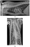
Figure 1
Figure 1
(a,b) Left lateral thoracic radiographs showing a generalized, unstructured interstitial pattern. The cat also had pectus excavatum, and the caudal sternum was dorsally deviating, displacing the cardiac silhouette to the left.
Figure 2
Figure 2
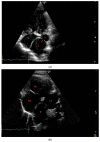
(a,b) Transthoracic left apical window modified 4-chamber optimized for showing the dilated PV (red *) opening into the LA through a narrow ostium at the PV-LA junction (red arrow). LA: left atrium, PV: pulmonary vein, RA: right atrium, RV: right ventricle.
Figure 3
Figure 3
Transthoracic left apical window modified 4–chamber optimized for the right heart. Doppler imaging shows a tricuspid regurgitation with a peak blood flow velocity of 5 m/s (red stripe), representing an estimated RV pressure of at least 100 mmHg.
Figure 4
Figure 4
Transthoracic left apical window modified 4–chamber view optimized for the right ventricular outflow tract. Doppler (color and spectral) examination shows a pulmonary artery profile with type II profile but normal peak blood flow velocity, suggesting increased pulmonary artery pressures.
Figure 5
Figure 5
(ac) Transthoracic left apical window modified 4–chamber optimized for showing the dilated PV (red *) emptying into the LA through a narrow ostium at the PV-LA junction. The anatomy and clinical condition of the cat did not allow for standard windows and views. (a) Color Doppler examination showing a turbulent blood flow between the dilated PV (red arrow) and the LA.; (b) Spectral Doppler examination performed with pulsed wave gate placed at what was believed to be the PV inlet. This view shows a continuous flow in at least one cycle. The angle is suboptimal, and therefore velocity is underestimated in the image.; (c) Spectral Doppler examination performed with pulsed wave gate placed at what was believed to be the PV inlet. The image shows a peak blood flow velocity of 2.6 m/s representing an estimated pressure gradient of 27 mmHg between the dilated PV and the LA and, therefore, an estimated PV pressure of at least 32 mmHg. The angle is suboptimal, and therefore continuous flow could not be shown. LA: left atrium, PV: pulmonary vein.
Figure 5
Figure 5
(ac) Transthoracic left apical window modified 4–chamber optimized for showing the dilated PV (red *) emptying into the LA through a narrow ostium at the PV-LA junction. The anatomy and clinical condition of the cat did not allow for standard windows and views. (a) Color Doppler examination showing a turbulent blood flow between the dilated PV (red arrow) and the LA.; (b) Spectral Doppler examination performed with pulsed wave gate placed at what was believed to be the PV inlet. This view shows a continuous flow in at least one cycle. The angle is suboptimal, and therefore velocity is underestimated in the image.; (c) Spectral Doppler examination performed with pulsed wave gate placed at what was believed to be the PV inlet. The image shows a peak blood flow velocity of 2.6 m/s representing an estimated pressure gradient of 27 mmHg between the dilated PV and the LA and, therefore, an estimated PV pressure of at least 32 mmHg. The angle is suboptimal, and therefore continuous flow could not be shown. LA: left atrium, PV: pulmonary vein.
Figure 6
Figure 6
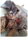
Necropsy image showing the anomalous junction of the pulmonary veins into the LA. Two normal PVs are seen, draining the cranial and caudal part of the left cranial lobe (marked with black arrows), opening in a normal ostium in the LA left wall. Three severely dilated PVs draining other lung lobes (red arrows) merge into a sac-like, dilated formation located in direct proximity to the LA (red star). LA:left atrium, PV: pulmonary vein.
Figure 7
Figure 7
Diagram showing the collecting dilated structure attached to the LA (red *) and the PVs draining the left caudal, accessory, right caudal, and the right cranial lung lobe emptying into the dilated common structure. The normal PV draining the left cranial lung lobe (LcPV) empty via the ostium in the left LA-wall. LA: left atrium, LcPV: Left cranial lung lobe, PV: pulmonary vein.

References

    1. Thomas W.P., Gaber C.E., Jacobs G.J., Kaplan P.M., Lombard C.W., Vet M., Moise N.S., Moses B.L. Recommendations for standards in transthoracic two-dimensional echocardiography in the dog and cat. J. Vet. Intern. Med. 1993;7:247–252. doi: 10.1111/j.1939-1676.1993.tb01015.x. - DOI - PubMed
    1. Latson L.A., Prieto L.R. Congenital and acquired pulmonary vein stenosis. Circulation. 2007;115:103–108. doi: 10.1161/CIRCULATIONAHA.106.646166. - DOI - PubMed
    1. Sun C.-C.J., Doyle T., Ringel R.E. Pulmonary vein stenosis. Hum. Pathol. 1995;26:880–886. doi: 10.1016/0046-8177(95)90011-X. - DOI - PubMed
    1. Allen H.D., Driscoll D.J., Shaddy R.E., Feltes T.F. Moss & Adams’ Heart Disease in Infants, Children, and Adolescents: Including the Fetus and Young Adult. Lippincott Williams & Wilkins; Philadelphia, PA, USA: 2013.
    1. Saad E.B., Rossillo A., Saad C.P., Martin D.O., Bhargava M., Erciyes D., Bash D., Williams-Andrews M., Beheiry S., Marrouche N.F. Pulmonary vein stenosis after radiofrequency ablation of atrial fibrillation: Functional characterization, evolution, and influence of the ablation strategy. Circulation. 2003;108:3102–3107. doi: 10.1161/01.CIR.0000104569.96907.7F. - DOI - PubMed

Publication types