. 2023 Feb 6;20(2):1230-1246.
doi: 10.1021/acs.molpharmaceut.2c00902. Epub 2023 Jan 20.

Development of In Vitro and Ex Vivo Biofilm Models for the Assessment of Antibacterial Fibrous Electrospun Wound Dressings

Affiliations

Development of In Vitro and Ex Vivo Biofilm Models for the Assessment of Antibacterial Fibrous Electrospun Wound Dressings

Kairi Lorenz et al. Mol Pharm. .

Abstract

Increasing evidence suggests that the chronicity of wounds is associated with the presence of bacterial biofilms. Therefore, novel wound care products are being developed, which can inhibit biofilm formation and/or treat already formed biofilms. A lack of standardized assays for the analysis of such novel antibacterial drug delivery systems enhances the need for appropriate tools and models for their characterization. Herein, we demonstrate that optimized and biorelevant in vitro and ex vivo wound infection and biofilm models offer a convenient approach for the testing of novel antibacterial wound dressings for their antibacterial and antibiofilm properties, allowing one to obtain qualitative and quantitative results. The in vitro model was developed using an electrospun (ES) thermally crosslinked gelatin-glucose (GEL-Glu) matrix and an ex vivo wound infection model using pig ear skin. Wound pathogens were used for colonization and biofilm development on the GEL-Glu matrix or pig skin with superficial burn wounds. The in vitro model allowed us to obtain more reproducible results compared with the ex vivo model, whereas the ex vivo model had the advantage that several pathogens preferred to form a biofilm on pig skin compared with the GEL-Glu matrix. The in vitro model functioned poorly for Staphylococcus epidermidis biofilm formation, but it worked well for Escherichia coli and Staphylococcus aureus, which were able to use the GEL-Glu matrix as a nutrient source and not only as a surface for biofilm growth. On the other hand, all tested pathogens were equally able to produce a biofilm on the surface of pig skin. The developed biofilm models enabled us to compare different ES dressings [pristine and chloramphenicol-loaded polycaprolactone (PCL) and PCL-poly(ethylene oxide) (PEO) (PCL/PEO) dressings] and understand their biofilm inhibition and treatment properties on various pathogens. Furthermore, we show that biofilms were formed on the wound surface as well as on a wound dressing, indicating that the demonstrated methods mimic well the in vivo situation. Colony forming unit (CFU) counting and live biofilm matrix as well as bacterial DNA staining together with microscopic imaging were performed for biofilm quantification and visualization, respectively. The results showed that both wound biofilm models (in vitro and ex vivo) enabled the evaluation of the desired antibiofilm properties, thus facilitating the design and development of more effective wound care products and screening of various formulations and active substances.

Keywords: antibacterial; antibiofilm; electrospinning; ex vivo biofilm model; in vitro biofilm model; skin wound infection; wound dressings.


Conflict of interest statement

The authors declare no competing financial interest.

Figures

Figure 1
Figure 1
Schematics of the experimental work conducted for the development and validation of the biofilm models. Key: CAM, chloramphenicol; ES, electrospinning; GEL, gelatin; Glu, glucose; PCL, polycaprolactone; PEO, polyethylenoxide.
Figure 2
Figure 2
(A) Micrographs of cross-sections of heat-treated pig ear skin (with superficial burn wounds) with and without γ-irradiation and glycerol treatment; H&E stained. Scale bar: 500 μm. (B) AFM micrographs of ES fibrous wound dressings. (C) European Pharmacopoeia 10.0 sterility test results after 14 days of incubation of γ-irradiated (50 kGy) samples (ES pristine fibrous wound dressings and infection model substrates) under aerobic conditions at room temperature (RT) in the dark. Positive bacterial control (E. coli). Key: GEL, gelatin; Glu, glucose; PCL, polycaprolactone; PEO, polyethylenoxide. Scale bar 1 cm.
Figure 3
Figure 3
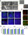
(A) SEM micrographs of biofilm formation after 24 h on top of the substrates—gelatin–glucose matrix (GEL-Glu matrix) and pig skin. Arrows point out the bacteria. Three different wound bacteria were used: E. coli DSM 1103, S. aureus DSM 2569, and S. epidermidis DSM 28319. The scale bar is 3 μm for the overview image of the pig skin surface, and it is 10 μm for all other SEM micrographs. (B) CFM micrographs of biofilm formation (24 and 48 h) on top of the GEL-Glu matrix in the in vitro model. Biofilm matrix visualized using EbbaBiolight 630 is in pink; ES gelatin–glucose matrix (GEL-Glu matrix) fibers visualized by autofluorescence are in blue; and bacteria stained using SYTO-9 are in green. Three different wound bacteria were used: E. coli DSM 1103, S. aureus DSM 2569, and S. epidermidis DSM 28319. Scale bar: 10 μm. The sample depth shown is 14.4 μm. (C) CFM micrographs of biofilm formation (24 h) in the ex vivo model. Pig skin had nonspecific red autofluorescence; bacteria were stained with SYTO-9 in green. Two different wound bacteria were used: E. coli DSM 1103 and S. aureus DSM 2569. Scale bar: 10 μm. (D) Biofilm formation after 24 and 48 h on top of the substrates—gelatin–glucose matrix (GEL-Glu matrix) and pig skin. Three different wound bacteria were used: E. coli DSM 1103, S. aureus DSM 2569, and S. epidermidis DSM 28319. Results are shown in the logarithmic scale as the number of colony-forming units (CFUs), with standard deviation bars (n = 3). Statistical significance is shown as follows: *p < 0.05; **p < 0.01; and ***p < 0.001. Experiments were performed using at least three technical replicates. Key: GEL, gelatin; Glu, glucose.
Figure 4
Figure 4
(A) Use of the gelatin–glucose matrix (GEL-Glu matrix) as a nutritional substrate in HEPES buffer. Bacteria were inoculated into 10 mM HEPES buffer alone and/or together with the GEL-Glu matrix for up to 96 h. S. aureus DSM 2569 and S. epidermidis DSM 28319 were used. The number of colony-forming units (CFUs), with standard deviation bars (n = 3), are shown in the logarithmic scale. Experiments were performed using at least three technical replicates. The detection limit of the assay is 2 log10 CFU/cm2. (B) S. epidermidis DSM 28319 biofilm formation on top of a single-layered gelatin–glucose matrix (1 × GEL-Glu) and triple-layered gelatin–glucose matrices (3× GEL-Glu matrix) in HEPES-buffered DMEM/F-12 growth media at different time points (24, 48, and 72 h). The number of colony-forming units (CFUs), with standard deviation bars (n = 3), are shown in the logarithmic scale. Statistical significance is shown as **P < 0.01. Experiments were performed using three technical replicates. Key: GEL, gelatin; Glu, glucose.
Figure 5
Figure 5
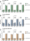
Model validation using electrospun (ES) wound dressings. For the in vitro model, the GEL-Glu matrix was used as artificial skin, and for the ex vivo model, pig skin was used. Three different bacteria were used: (A) E. coli DSM 1103, (B) S. aureus DSM 2569, and (C) S. epidermidis DSM 28319. Results are shown in the logarithmic scale as the number of colony-forming units (CFUs) cm2, with standard deviation bars (n = 3). The detection limit of the assay is 2 log10 CFU/cm2. CAM-loaded ES wound dressings were compared with pristine control wound dressings. A comparison between the PCL vs PCL/PEO formulations is also presented. Statistical significance is shown as follows: *P < 0.05; **P < 0.01; and ***P < 0.001. Key: CAM, chloramphenicol; PCL, polycaprolactone; and PEO, polyethylenoxide.
Figure 6
Figure 6
Bacterial biofilm adhesion on top of the substrate−gelatin−glucose matrix (GEL-Glu matrix) or pig skin or onto the covering wound dressing (ES pristine wound dressings). Substrates were covered with pristine wound dressings and incubated for 24 h, after which the substrate and wound dressing were separated, and the biofilm formed on top of each part was studied independently. Three different bacteria were used: (A) E. coli DSM 1103, (B) S. aureus DSM 2569, and (C) S. epidermidis DSM 28319. Two different formulations were tested—PCL wound dressing and PCL/PEO wound dressing (control dressings); 100% is shown to mark the arithmetic mean of biofilm formation on top of the different uncovered substrates. Key: GEL, gelatin; Glu, glucose; PCL, pristine wound dressings made from polycaprolactone; PCL/PEO, pristine wound dressings made from polycaprolactone and polyethylenoxide.
Figure 7
Figure 7
Biofilm treatment model. Effect of the model CAM-loaded wound dressings (CAM-PCL and CAM-PCL/PEO dressings) on already formed E. coli DSM 1103 biofilms (24 h). A comparison is made with pristine wound dressings and uncovered substrates. The in vitro setup was created on top of the ES GEL-Glu matrix and the ex vivo setup was created on top of pig skin. The number of biofilm-forming bacteria before treatment was 108 CFU/cm2. Results are shown in a linear scale as the number of colony-forming units (CFUs), with standard deviation bars (n = 3). Changes in the threshold value (108 CFU/cm2) are presented. Statistical significance is shown as follows: *P < 0.05; **P < 0.01; ***P < 0.001. Key: CAM, chloramphenicol-loaded wound dressings; PCL, pristine wound dressings made from polycaprolactone; PCL/PEO, pristine wound dressings made from polycaprolactone and polyethylenoxide.

Similar articles

Cited by

References

    1. Omar A.; Wright J.; Schultz G.; Burrell R.; Nadworny P. Microbial Biofilms and. Chronic Wounds. Microorganisms 2017, 5, 9.10.3390/microorganisms5010009. - DOI - PMC - PubMed
    1. Stuermer E. K.; Besser M.; Brill F.; Geffken M.; Plattfaut I.; Severing A. L.; Wiencke V.; Rembe J. D.; Naumova E. A.; Kampe A.; Debus S.; Smeets R. Comparative Analysis of Biofilm Models to Determine the Efficacy of Antimicrobials. Int. J. Hyg. Environ. Health 2021, 234, 11374410.1016/j.ijheh.2021.113744. - DOI - PubMed
    1. Stoffel J. J.; Kohler Riedi P. L.; Hadj Romdhane B. A Multimodel Regime for Evaluating Effectiveness of Antimicrobial Wound Care Products in Microbial Biofilms. Wound Repair Regen. 2020, 28, 438–447. 10.1111/wrr.12806. - DOI - PMC - PubMed
    1. Wu Y. K.; Cheng N. C.; Cheng C. M. Biofilms in Chronic Wounds: Pathogenesis and Diagnosis. Trends Biotechnol. 2019, 37, 505–517. 10.1016/j.tibtech.2018.10.011. - DOI - PubMed
    1. Jannesari M.; Varshosaz J.; Morshed M.; Zamani M. Composite Poly(Vinyl Alcohol)/Poly(Vinyl Acetate) Electrospun Nanofibrous Mats as a Novel Wound Dressing Matrix for Controlled Release of Drugs. Int. J. Nanomed. 2011, 6, 993–1003. 10.2147/IJN.S17595. - DOI - PMC - PubMed

Publication types

LinkOut - more resources