. 2023 Jan 24;120(4):e2209964120.
doi: 10.1073/pnas.2209964120. Epub 2023 Jan 20.

TMEM161B regulates cerebral cortical gyration, Sonic Hedgehog signaling, and ciliary structure in the developing central nervous system

Affiliations

TMEM161B regulates cerebral cortical gyration, Sonic Hedgehog signaling, and ciliary structure in the developing central nervous system

Shyam K Akula et al. Proc Natl Acad Sci U S A. .

Abstract

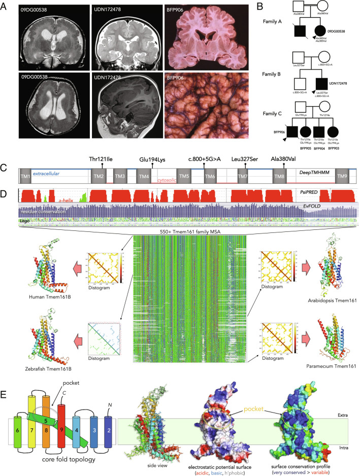
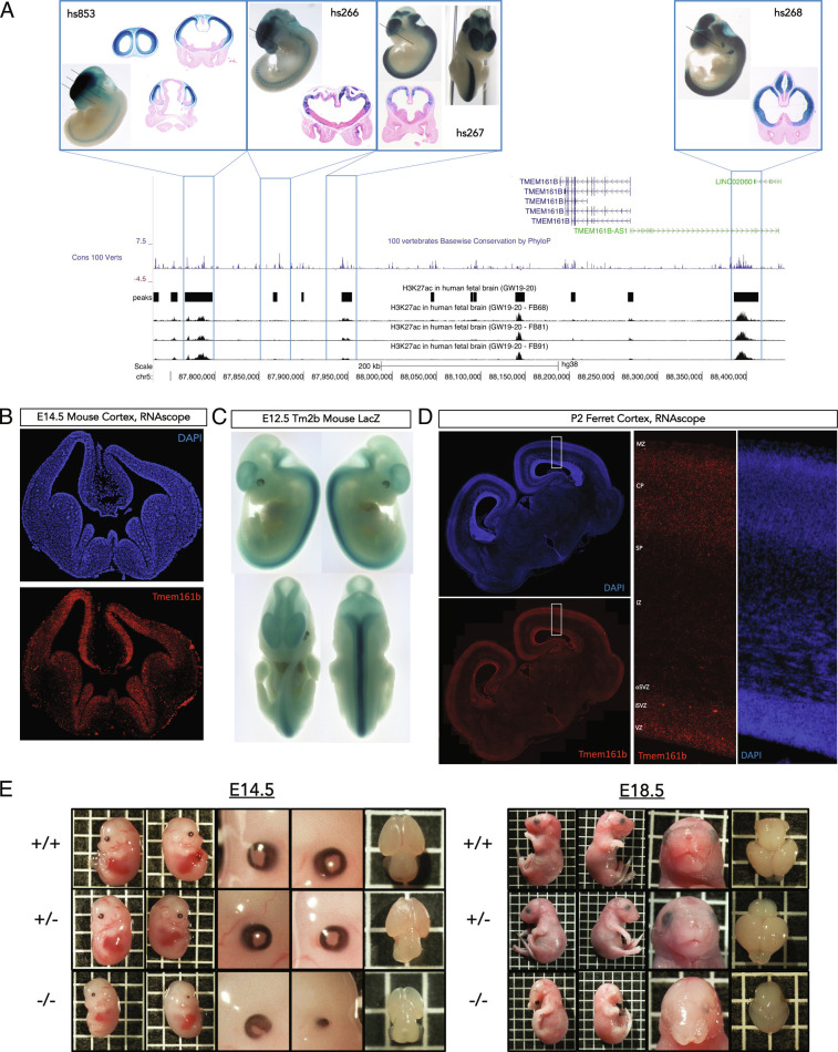
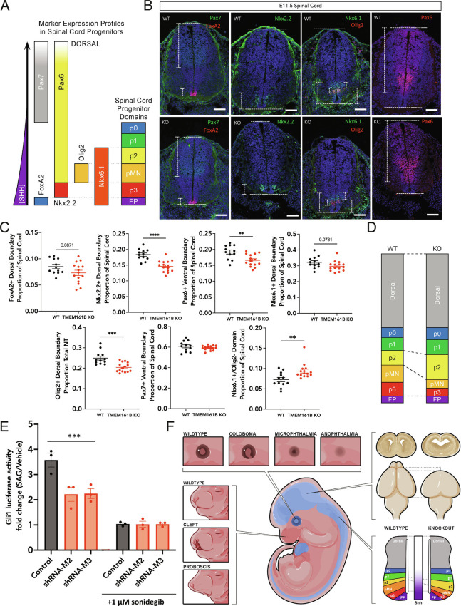
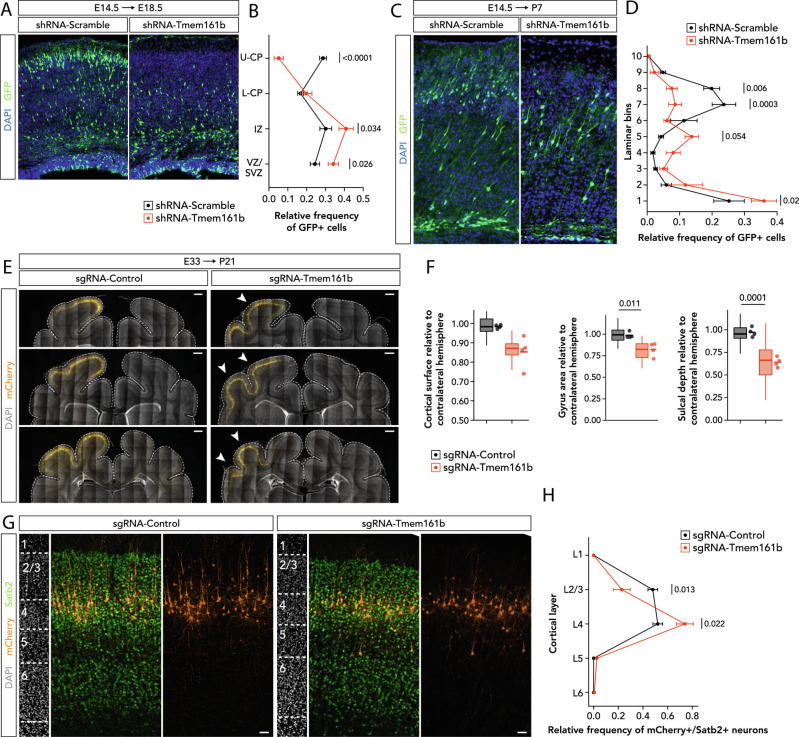
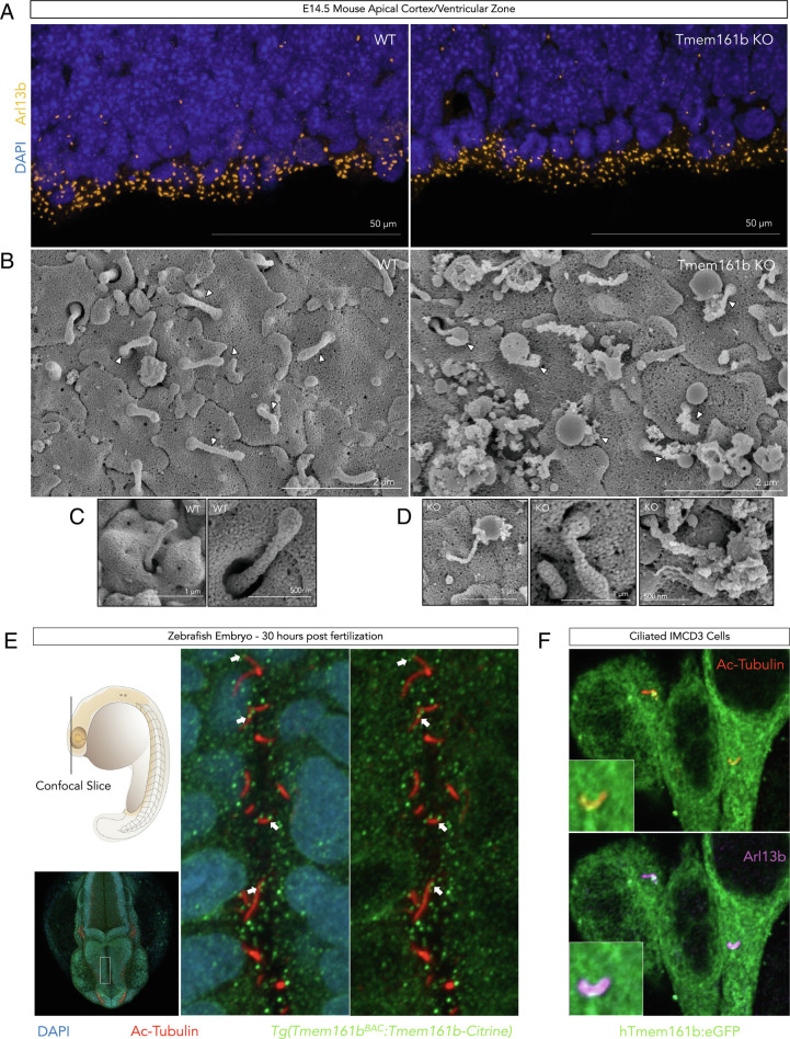
Sonic hedgehog signaling regulates processes of embryonic development across multiple tissues, yet factors regulating context-specific Shh signaling remain poorly understood. Exome sequencing of families with polymicrogyria (disordered cortical folding) revealed multiple individuals with biallelic deleterious variants in TMEM161B, which encodes a multi-pass transmembrane protein of unknown function. Tmem161b null mice demonstrated holoprosencephaly, craniofacial midline defects, eye defects, and spinal cord patterning changes consistent with impaired Shh signaling, but were without limb defects, suggesting a CNS-specific role of Tmem161b. Tmem161b depletion impaired the response to Smoothened activation in vitro and disrupted cortical histogenesis in vivo in both mouse and ferret models, including leading to abnormal gyration in the ferret model. Tmem161b localizes non-exclusively to the primary cilium, and scanning electron microscopy revealed shortened, dysmorphic, and ballooned ventricular zone cilia in the Tmem161b null mouse, suggesting that the Shh-related phenotypes may reflect ciliary dysfunction. Our data identify TMEM161B as a regulator of cerebral cortical gyration, as involved in primary ciliary structure, as a regulator of Shh signaling, and further implicate Shh signaling in human gyral development.

Keywords: Sonic Hedgehog; cortical gyration; holoprosencephaly; polymicrogyria; primary cilia.


Conflict of interest statement

The authors declare no competing interest.

Figures

Fig. 1.
Fig. 1.
Missense variants in TMEM161B, a highly conserved 9-transmembrane domain protein, are associated with cortical malformation. (A) Brain MRIs and pathology of affected individuals display diffuse polymicrogyria. MRI images of 09DG00538 are at 2 y of age, MRI images of UDN172478 are at 3 y of age, and images of BFP906 are from postmortem examination at age 32 y. All three show a similar coronal brain contour and diffuse PMG across the entire cortex bilaterally. (B) Pedigrees of affected families. TMEM161B (NM_153354.4) genotypes are labeled where they were available and confirmed, black shading indicates affected or presumed affected individuals, tissue for BFP906 was unavailable for sequencing. The recessive inheritance and segregation of TMEM161B variants with disease supports a loss-of-function disease model. (C) Distribution of identified variants in TMEM161B. The missense variants identified are distributed throughout TMEM161B in highly conserved residues ( SI Appendix, Fig. S1C ), marked atop the TM helix prediction by DeepTMHMM (https://dtu.biolib.com/DeepTMHMM). (D) Multisequence alignment of TMEM161B orthologs across domains of eukaryotic life reveals deep conservation of orthologs across its 9TM chain architecture, suggesting a fundamental biological role. Structural models build with TrRosetta and Tfold (with attendant predicted distance matrices or distograms that highly resemble each other)––that agree well with more recent AlphaFold2 predictions––are drawn for the human TMEM161B as well as its zebrafish ortholog. More distant plant and amoeba TMEM161s likewise share the 9TM helix fold. (E) Modeling of the predicted structure of TMEM161B integrating evolutionary conservation data. TM1 could serve as an atypical signal peptide, for a net 8-TM domains in the mature protein. TMEM161s have eight predicted TM helices with an unusual, highly conserved, non-symmetric core fold topology that defines the TMEM161 superfamily. The most conserved feature is a shallow extracellular pocket bordered by TM helix extensions that just above the membrane, as labeled on the figure in both an electrostatic surface profile, and surface residue conservation profile of human TMEM161B.
Fig. 2.
Fig. 2.
TMEM161B shows spatiotemporal expression enrichment for the developing CNS, and Tmem161b KO mouse embryos show gross abnormalities. (A) Multiple conserved active/open enhancers in developing CNS flank TMEM161B. Alignment of human fetal H3K27ac data with TMEM161B locus, with overlays of representative LacZ stains from the VISTA Enhancer Browser (24) demonstrating that several local H3K27ac peaks correspond to highly conserved enhancers expressed in the developing CNS. (B) RNAscope for Tmem161b in developing mouse brain. In situ hybridization using RNAscope against Tmem161b at E14.5 shows diffuse Tmem161b expression in developing brain with a bias towards ventricular zones in both ventral and dorsal forebrain, this expression pattern is concordant with the enhancer LacZ staining in Fig. 2A . (C) LacZ staining in Tmem161b tm2b/+ embryos reveals enriched Tmem161b expression in the developing CNS. E12.5 embryos reveal high concentration of Tmem161b-driven LacZ expression in the developing CNS on top of diffuse low-level expression. (D) RNAscope for Tmem161b in P2 ferret brain. During neurogenesis and migration in the gyrencephalic ferret, Tmem161b is expressed and enriched slightly in the ventricular zone, but also shows expression across cortical plate. (E) Tmem161b null embryos display numerous anatomical abnormalities: KO mice were smaller, had a range of eye defects from coloboma to anophthalmia, and craniofacial defects. KO brains were smaller and demonstrated variable holoprosencephaly (more examples of all phenotypes in the supplement). Heterozygous mice were indistinguishable from WT mice. One box = 0.5 cm side length.
Fig. 3.
Fig. 3.
Tmem161b KO embryos show multiple sequalae of Shh disruption in the developing CNS. (A) Schematic of spinal cord markers examined in Tmem161b null embryos; in combination these markers define spinal cord domains in development patterned by response to a ventrally sourced Shh gradient. (BD) Tmem161b KO mice show decreased ventral domains in the developing spinal cord. E11.5 control (n = 4) and Tmem161b KO (n = 5) embryos were harvested, and spinal cords including rostral and thoracic sections were prepared for IF for markers corresponding to ventral and dorsal progenitor domains in the spinal cord, which are dependent on a gradient of Shh signaling. (CE) Tmem161b KO embryos showed a lower dorsal boundary of the Nkx2.2 domain/span of Nkx2.2 relative to the length of the spinal cord, with a correspondingly increased Pax6 domain, which is mutually exclusive with Nkx2.2 (BH corrected multiple unpaired tests of imaged fields, < 0.0001 for Nkx2.2 and P = 0.0027 for Pax6 between WT and KO). There was a decrease in both dorsal boundary (P < 0.0001) and the span of the Olig2+ domain relative to total spinal cord length (P = 0.0324). Both FoxA2 and Nkx6.1 showed marginal decreases in their span in KOs relative to WTs (P = 0.0871 and 0.0781 respectively, BH multiple testing corrected t tests), but the Nkx6.1+/Olig2− domain was increased in the KO relative to the WT (unpaired t test, P = 0.0064). There was no difference between Pax7+ domains in the WT vs. KO. Measurements of FoxA2, Nkx2.2, Pax6, Olig2, Pax7 and Nkx6.1 domains were performed by experimenters blinded to genotype. (E) Tmem161b supports sensitivity to Shh. SL2 cells (modified NIH3T3s with Gli1 luciferase reporter activity) were transfected with control or Tmem161b shRNAs and then treated with SAG for 48 h. Luciferase content was measured at the end of incubation to determine the relative amount of Gli1 transcriptional activity during the assay. Each data point represents an independent experimental replicate (separate passages of cells, between two batches of independently prepped plasmid and drug, and n = 3 technical replicates per condition/experiment). Tmem161b shRNA treated cells showed ~40% less Gli1 luciferase activity in response to SAG compared to controls (2- way ANOVA shows effect of shRNAs, P = 0.0048, effect of sonidegib, a smoothened antagonist, P < 0.0001, and interaction effect, P = 0.0054. Sidak post-hoc comparison, corrected, demonstrates Tmem161b shRNA treated conditions show less Gli1 luciferase activity relative to control shRNA, P = 0.0005 for shRNA-M2, and P = 0.0006 for shRNA-M3), and this effect was eliminated with the addition of sonidegib, suggesting that Tmem161b’s role in supporting Shh signaling is secondary to canonical Smoothened activation. (F) Summary of Shh related phenotypes observed across >50 Tmem161b KO embryos: eye defects ranging from coloboma to anophthalmia, craniofacial defects from minor cleft palate to failure of midline formation leading to proboscis, holoprosencephaly, and spinal cord patterning shifts. No limb abnormalities were noted across all nulls examined.
Fig. 4.
Fig. 4.
In vivo disruption of Tmem161b causes abnormal cortical histogenesis. (AD) Knockdown of Tmem161b in the developing cortex causes cell autonomous neuronal positioning defects. (AB) CD1 embryos were electroporated at E14.5 with either a control plasmid expressing a non-targeting shRNA (n = 6), or an shRNA targeting mouse Tmem161b (n = 6); both plasmids had second ORF containing a GFP tag to visualize electroporated cells. Cells electroporated with an shRNA targeting Tmem161b showed a cell autonomous lamination defect, concentrating more in the VZ/SVZ and IZ than in the cortical plate after the 4-d period between electroporation and harvest; of the Tmem161b depleted cells that did enter the cortical plate, virtually none migrated into the upper cortical plate by the analysis timepoint. 2-way ANOVA of plasmid condition X cortical region showed an interaction effect, P < 0.001, with post Sidak multiple comparison tests, corrected for multiple testing, demonstrating more knockdown cells found in the VZ/SVZ (P = 0.0255) and IZ (P = 0.0336), and more control cells found in the upper cortical plate (P < 0.0001). >200 GFP+ cells per brain were analyzed. (C, D) IUE performed as described above, but electroporation at E14.5, and analysis at P7 (i.e., longer survival), where final distribution of GFP+ cells was quantified across 10 laminar bins of the P7 cortex divided evenly from pial surface to bottom of cortex. Knockdown of Tmem161b at E14.5 led to an altered distribution of GFP+ cells at P7 (2-way ANOVA of plasmid condition X cortical region showed an interaction effect, P < 0.001, with post Sidak multiple comparison tests, corrected for multiple testing). (EH) Tmem161b knockdown via IUE of pyramidal cell progenitors in the developing ferret cortex at E33.5 using sgRNA targeting Tmem161b results in reduced cortical surface, reduced gyrus size and sulcal depth, and altered neuronal positioning in the postnatal ferret cortex at P21. (E) Coronal sections of through the P21 ferret cortex processed for immunohistochemistry against mCherry (orange) counterstained with DAPI (gray) in control and Tmem161b-knockdown ferrets. Dotted lines indicate the dorsal surface of the cerebral cortex. Arrows indicate the reduced size of gyri and sulci in somatosensory cortical areas with neurons electroporated with the sgRNA-Tmem161b plasmid. (F) Quantification of the cortical surface, gyrus size, and sulcal depth in control and Tmem161b-knockdown ferret brains. For each gross anatomical variable, the ratio between the region with electroporated cells and the corresponding region in the contralateral, non-electroporated hemisphere was calculated. Four ferret brains per experimental condition and >10 coronal sections per brain were analyzed for each variable (n = 52 sections in sgRNA-Control, n = 52 sections for sgRNA-Tmem161b). Two-tailed Student’s t test: P = 0.020 for cortical surface, P = 0.011 for gyrus size, and P = 0.0001 for sulcal depth. (G) Coronal sections through the somatosensory cortex processed for immunohistochemistry against Satb2 (green) and mCherry (orange) counterstained with DAPI (gray) in control and Tmem161b-knockdown ferrets at P21. Confocal images illustrating the distribution of mCherry+/Satb2+ neurons and quantified across cortical layers. Note the presence of some mCherry+/Satb+ neurons in L5, which were rarely seen in the control brains. (H) Relative frequency of the distribution of mCherry+/Satb2+ neurons quantified across cortical layers of P21 ferret cortex. Knockdown of Tmem161b leads to an altered distribution of mCherry+/Satb2+ cells in L2/3 and L4 (two-way ANOVA, P < 0.001, with post Sidak multiple comparison tests). Four ferret brains per experimental condition and >200 cells per brain were analyzed. Data are shown as mean ± SEM. Data shown as box plots represent the distributions of values from all coronal sections quantified per condition, and the adjacent data points and lines represent the averages per ferret and averaged mean per condition, respectively. (Scale bars, 1 mm (E) and 50 μm (G).)
Fig. 5.
Fig. 5.
Tmem161b null embryos have abnormal primary cilia in the developing CNS, and TMEM161B protein localizes to primary cilia. (A) Tmem161b KO embryos show normal numbers of apical primary cilia in the cortex. N = 7 WT and N = 8 Tmem161b KO embryos had cortices stained with Arl13b, and number of apical puncta were counted. There was no difference in the number of puncta between genotypes, but a qualitative appearance of more rounded/abnormally shaped puncta was noted leading to follow-up SEM. (B) Tmem161b KO apical primary cilia show structural abnormalities. SEM images of the apical cortical surface of Tmem161b KO mice and control with some primary cilia labeled with white arrows. WT primary cilia display normal morphology, while the KO surface shows increased vesicular debris, and primary cilia with ballooned tips and disrupted stalks. (C, D) Examples of Tmem161b KO primary cilia. Typical examples of the abnormalities found in Tmem161b KO primary cilia – ciliary tip ballooning or shortened cilia with abnormal contour. (E) Tmem161b localizes to primary cilia in zebrafish hindbrain. Confocal imaging of transgenic zebrafish that express a citrine tagged Tmem161b at 30 h post fertilization shows co-localization with citrine puncta and primary cilia marker Acetylated tubulin in the hindbrain ventricular zone; see also Movie S1. (F) TMEM161B localizes to primary cilia in IMCD3 cells. GFP tagged human TMEM161B was expressed in IMCD3 cells, which were then ciliated and confocal imaging performed. GFP signal showed clear co-localization with primary cilia markers Arl13b and Acetylated tubulin, see also Movie S2.

References

    1. Traiffort E., Angot E., Ruat M., Sonic Hedgehog signaling in the mammalian brain. J. Neurochem. 113 , 576–590 (2010). - PubMed
    1. Yabut O. R., Pleasure S. J., Sonic Hedgehog signaling rises to the surface: Emerging roles in neocortical development. Brain Plast. 3 , 119–128 (2018). - PMC - PubMed
    1. Andreu-Cervera A., Catala M., Schneider-Maunoury S., Cilia, ciliopathies and hedgehog-related forebrain developmental disorders. Neurobiol. Dis. 150 , 105236 (2021). - PubMed
    1. Cavodeassi F., Creuzet S., Etchevers H. C., The Hedgehog pathway and ocular developmental anomalies. Hum. Genet. 138 , 917–936 (2019). - PMC - PubMed
    1. Roessler E., Muenke M., The molecular genetics of holoprosencephaly. Am. J. Med. Genet. C Semin. Med. Genet. 154C , 52–61 (2010). - PMC - PubMed

Publication types